. 2019 Jun:24:101215.
doi: 10.1016/j.redox.2019.101215. Epub 2019 May 13.

STING-IRF3 contributes to lipopolysaccharide-induced cardiac dysfunction, inflammation, apoptosis and pyroptosis by activating NLRP3

Affiliations

STING-IRF3 contributes to lipopolysaccharide-induced cardiac dysfunction, inflammation, apoptosis and pyroptosis by activating NLRP3

Ning Li et al. Redox Biol. 2019 Jun.

Abstract

Mountainous evidence suggests that inflammation, cardiomyocyte apoptosis and pyroptosis are involved in the development of sepsis and sepsis-induced cardiomyopathy (SIC). Stimulator of interferon genes (STING) is an indispensable molecule that could regulate inflammation and immune response in multiple diseases. However, the role of STING in cardiovascular disease, especially SIC remains unclear. This study was designed to investigate the potential molecular mechanisms of STING in lipopolysaccharide (LPS)-induced cardiac injury using STING global knockout mice. In wild type mice and cardiomyocytes, LPS stimulation triggered the perinuclear translocation of STING, which further bound to Type-I interferons (IFN) regulatory factor 3 (IRF3) and phosphorylated IRF3. Phosphorylated (P-) IRF3 subsequently translocated into nucleus and increased the expression of NOD-like receptor protein 3 (NLRP3). Knockout of STING in mice significantly improved survival rate and cardiac function, apart from suppressing myocardial and serum inflammatory cytokines, apoptosis, as well as cardiomyocyte pyroptosis. In vitro experiments revealed that NLRP3 overexpression by adenovirus could offset protective effects of STING knockdown in LPS-induced cardiomyocytes. Additionally, LPS stimulation also promoted the production of intracellular reactive oxygen (ROS), which further induced the NLRP3 translocation to the cytoplasm from the nucleus. Dissociative TXNIP could directly interact with cytoplasmic NLRP3 and form inflammasome, eventually triggering cardiomyocyte injury. Collectively, our findings disclose that STING deficiency could alleviate LPS-induced SIC in mice. Hence, targeting STING in cardiomyocytes may be a promising therapeutic strategy for preventing SIC.

Keywords: Apoptosis; NLRP3 inflammasome; Pyroptosis; STING-IRF3; Sepsis-induced cardiomyopathy.


Figures

Fig. 1
Fig. 1
The expression level of STING and IRF3 in SIC. A . The protein levels of STING, P-IRF3 and T-IRF3 in mouse heart stimulated by different doses (ranging from 0 to 15 mg/kg) of LPS for 12 h in mice (n = 6, *P<0.05 vs. 0 mg/kg group). B. The protein levels of STING, P-IRF3 and T-IRF3 in mouse heart stimulated by LPS (10 mg/kg) for 0, 4, 8, 12, and 24 h in mice (n = 6, *P<0.05 vs. 0 h group). C. The protein levels of STING, P-IRF3 and T-IRF3 in NRCMs after LPS stimulation (1 μg/ml for 6 h) (n = 6, *P<0.05 vs. PBS group).
Fig. 2
Fig. 2
LPS affected the intracellular location of STING and IRF3. A-B. NRCMs or H9c2 cells were stimulated by LPS for 6 h. A. Representative images of immunofluorescence of STING in NRCMs and H9c2 cells (n = 6). B. Representative images of immunofluorescence of IRF3 in NRCMs and H9c2 cells (n = 6). C. The cytoplasm protein and nucleus protein levels of STING in NRCMs stimulated by LPS for 6 h (n = 6, *P<0.05 vs. indicated group, NS, no significance). D. The cytoplasm protein and nucleus protein levels of T-IRF3 in NRCMs stimulated by LPS for 6 h (n = 6, *P<0.05 vs. indicated group). E. Representative images of IP of STING and IRF3 in NRCMs stimulated by LPS for 6 h (n = 6, *P<0.05 vs. PBS group).
Fig. 3
Fig. 3
STING knockdown inhibited LPS-induced phosphorylation and nuclear translocation of IRF3. A . Representative images of immunofluorescence of IRF3 in NRCMs or H9c2 cells transfected with STING siRNA and then stimulated by LPS for 6 h. B. The protein levels of STING, P-IRF3 and T-IRF3 in NRCMs stimulated by LPS for 6 h (n = 6, *P<0.05 vs. indicated group).
Fig. 4
Fig. 4
STING deficiency improved survival and cardiac function of LPS-treated mice. A . Effect of STING deficiency on the 7-day survival rate after LPS injection (n = 10). B. Effect of STING deficiency on LPS-induced LDH release (n = 6). C. Effect of STING deficiency on LPS-induced CK MB release (n = 6). D. Representative echocardiographic images of each group (n = 6). E. Effect of STING deficiency on left ventricle ejection fraction and left ventricle fractional shortening of each group after Sham or LPS injection (n = 6). *P < 0.05 vs. Sham + WT, #P < 0.05 vs. LPS + WT.
Fig. 5
Fig. 5
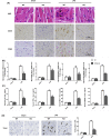
STING deficiency suppressed cardiac inflammation, apoptosis and pyroptosis of LPS-treated mice. A . Representative images of the morphological analysis and inflammatory cells infiltration as reflected by the H&E staining, and immunohistochemistry staining for CD45 and CD68 protein (n = 6, 10 + fields per heart). The arrow showed inflammatory cells. B. The levels of inflammatory cytokines including IL-1β, TNF-α, MCP-1 and HMGB1 in myocardial tissues of each group after Sham or LPS injection (n = 6). C. The levels of inflammatory cytokines including IL-1β, TNF-α, MCP-1 and HMGB1 in serum of each group after Sham or LPS injection (n = 6). D. TUNEL staining of each group after Sham or LPS injection (n = 6, 10 + fields per heart). The arrow indicated Tunel positive cells. E. The protein levels of C-Caspase3, Caspase3, BAX and BCL-2 of each group after Sham or LPS injection (n = 6). F. Representative images of the immunohistochemistry staining for Caspase1 (n = 6, 10 + fields per heart). E. The protein levels of Caspase1, IL-1β and IL-18 of each group after Sham or LPS injection (n = 6). *P < 0.05 vs. Sham + WT, #P < 0.05 vs. LPS + WT.
Fig. 5
Fig. 5
STING deficiency suppressed cardiac inflammation, apoptosis and pyroptosis of LPS-treated mice. A . Representative images of the morphological analysis and inflammatory cells infiltration as reflected by the H&E staining, and immunohistochemistry staining for CD45 and CD68 protein (n = 6, 10 + fields per heart). The arrow showed inflammatory cells. B. The levels of inflammatory cytokines including IL-1β, TNF-α, MCP-1 and HMGB1 in myocardial tissues of each group after Sham or LPS injection (n = 6). C. The levels of inflammatory cytokines including IL-1β, TNF-α, MCP-1 and HMGB1 in serum of each group after Sham or LPS injection (n = 6). D. TUNEL staining of each group after Sham or LPS injection (n = 6, 10 + fields per heart). The arrow indicated Tunel positive cells. E. The protein levels of C-Caspase3, Caspase3, BAX and BCL-2 of each group after Sham or LPS injection (n = 6). F. Representative images of the immunohistochemistry staining for Caspase1 (n = 6, 10 + fields per heart). E. The protein levels of Caspase1, IL-1β and IL-18 of each group after Sham or LPS injection (n = 6). *P < 0.05 vs. Sham + WT, #P < 0.05 vs. LPS + WT.
Fig. 6
Fig. 6
STING triggered the activation of NLRP3 inflammasome in heart of LPS-treated mice. A . The protein levels of TXNIP, NLRP3 and ASC of each group after Sham or LPS injection (n = 6). B. Representative images of IP of NLPR3, TXNIP and Trx in each group after Sham or LPS injection (n = 6). *P < 0.05 vs. Sham + WT, #P < 0.05 vs. LPS + WT.
Fig. 7
Fig. 7
STING activated NLRP3 in an IRF3-dependent manner in LPS-induced NRCMs. A . The protein levels of NLRP3 in indicated groups that were treated with IRF3 siRNA and (or) Ad-STING and then stimulated with LPS for 6 h (n = 6). B . IRF3 agonist, KIN1148 increased the protein expression level of NLRP3 (n = 6). *P<0.05 vs. indicated group.
Fig. 8
Fig. 8
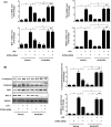
STING induced inflammation, apoptosis and pyroptosis in LPS-induced cardiomyocytes by activating NLRP3. A . The mRNA levels of IL-1β, TNF-α, MCP-1 and HMGB1 in indicated groups that were treated with STING siRNA and (or) Ad-IRF3and then stimulated with LPS for 6 h (n = 6). B. The protein levels of C-Caspase3, Caspase3, BAX and BCL-2 in indicated groups (n = 6). C. Apoptosis analysis by flow cytometry in indicated groups (n = 6). D. The protein levels of Caspase1, IL-1β and IL-18 in indicated groups (n = 6). E. Cell viability was detected by an CCK8 assay in indicated group (n = 6). *P<0.05 vs. indicated group.
Fig. 8
Fig. 8
STING induced inflammation, apoptosis and pyroptosis in LPS-induced cardiomyocytes by activating NLRP3. A . The mRNA levels of IL-1β, TNF-α, MCP-1 and HMGB1 in indicated groups that were treated with STING siRNA and (or) Ad-IRF3and then stimulated with LPS for 6 h (n = 6). B. The protein levels of C-Caspase3, Caspase3, BAX and BCL-2 in indicated groups (n = 6). C. Apoptosis analysis by flow cytometry in indicated groups (n = 6). D. The protein levels of Caspase1, IL-1β and IL-18 in indicated groups (n = 6). E. Cell viability was detected by an CCK8 assay in indicated group (n = 6). *P<0.05 vs. indicated group.
Fig. 9
Fig. 9
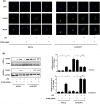
ROS is essential for cytoplasmic translocation of NLRP3 in LPS-induced NRCMs. A . Representative images of immunofluorescence of NLRP3 in NRCMs in indicated groups (n = 6). B. The cytoplasm protein and nucleus protein levels of NLPR3 in NRCMs in indicated groups (n = 6). C. The ROS levels of NRCMs in indicated groups (n = 6). D. ROS scavenger, Nac blocked the cytoplasmic translocation of NLRP3 (n = 6). E. The cytoplasm protein and nucleus protein levels of NLPR3 in NRCMs in NRCMs treated with Nac (n = 6). *P<0.05 vs. indicated group, NS, no significance.
Fig. 9
Fig. 9
ROS is essential for cytoplasmic translocation of NLRP3 in LPS-induced NRCMs. A . Representative images of immunofluorescence of NLRP3 in NRCMs in indicated groups (n = 6). B. The cytoplasm protein and nucleus protein levels of NLPR3 in NRCMs in indicated groups (n = 6). C. The ROS levels of NRCMs in indicated groups (n = 6). D. ROS scavenger, Nac blocked the cytoplasmic translocation of NLRP3 (n = 6). E. The cytoplasm protein and nucleus protein levels of NLPR3 in NRCMs in NRCMs treated with Nac (n = 6). *P<0.05 vs. indicated group, NS, no significance.
Fig. 10
Fig. 10
A putative scheme illustrating the mechanism by which STING-IRF3 contributes to inflammation, pyroptosis and apoptosis of cardiomyocytes in LPS-induced cardiac injury.
figs1
figs1

Similar articles

Cited by

References

    1. Deutschman C.S., Tracey K.J. Sepsis: current dogma and new perspectives. Immunity. 2014;40:463–475. - PubMed
    2. Deutschman, C. S. & Tracey, K. J. Sepsis: current dogma and new perspectives. Immunity 40, 463-475 (2014). - PubMed
    1. Van T.E., Wiersinga W.J., Scicluna B.P., Van T.D.P. Biomarkers in sepsis. Crit. Care Clin. 2018;34:139. - PubMed
    2. Van, T. E., Wiersinga, W. J., Scicluna, B. P. & Van, T. D. P. Biomarkers in Sepsis. Critical Care Clinics 34, 139 (2018). - PubMed
    1. Sato R., Nasu M. A review of sepsis-induced cardiomyopathy. J. Intensive Care. 2015;3:1–7. - PMC - PubMed
    2. Sato, R. & Nasu, M. A review of sepsis-induced cardiomyopathy. Journal of Intensive Care 3, 1-7 (2015). - PMC - PubMed
    1. Liu Y.C., Yu M.M., Shou S.T., Chai Y.F. Sepsis-induced cardiomyopathy: mechanisms and treatments. Front. Immunol. 2017;8:1021. - PMC - PubMed
    2. Liu, Y. C., Yu, M. M., Shou, S. T. & Chai, Y. F. Sepsis-Induced Cardiomyopathy: Mechanisms and Treatments. Frontiers in Immunology 8, 1021- (2017). - PMC - PubMed
    1. Wang Y. β 1 -adrenoceptor stimulation promotes LPS-induced cardiomyocyte apoptosis through activating PKA and enhancing CaMKII and IκBα phosphorylation. Crit. Care. 2015;19:76. - PMC - PubMed
    2. Wang, Y. et al. β 1 -adrenoceptor stimulation promotes LPS-induced cardiomyocyte apoptosis through activating PKA and enhancing CaMKII and IκBα phosphorylation. Critical Care 19, 76 (2015). - PMC - PubMed

Publication types

MeSH terms