. 2019 May 21;10(1):2260.
doi: 10.1038/s41467-019-09892-6.

The extracellular gate shapes the energy profile of an ABC exporter

Affiliations

The extracellular gate shapes the energy profile of an ABC exporter

Cedric A J Hutter et al. Nat Commun. .

Abstract

ABC exporters harness the energy of ATP to pump substrates across membranes. Extracellular gate opening and closure are key steps of the transport cycle, but the underlying mechanism is poorly understood. Here, we generated a synthetic single domain antibody (sybody) that recognizes the heterodimeric ABC exporter TM287/288 exclusively in the presence of ATP, which was essential to solve a 3.2 Å crystal structure of the outward-facing transporter. The sybody binds to an extracellular wing and strongly inhibits ATPase activity by shifting the transporter's conformational equilibrium towards the outward-facing state, as shown by double electron-electron resonance (DEER). Mutations that facilitate extracellular gate opening result in a comparable equilibrium shift and strongly reduce ATPase activity and drug transport. Using the sybody as conformational probe, we demonstrate that efficient extracellular gate closure is required to dissociate the NBD dimer after ATP hydrolysis to reset the transporter back to its inward-facing state.


Conflict of interest statement

The authors declare no competing interests.

Figures

Fig. 1
Fig. 1
Three outward-facing structures of TM287/288 solved in complex with single domain antibody fragments. The transporters are viewed along the membrane plane (indicated as gray rectangle). a 3.2 Å structure of TM287/288(EtoA) in complex with ATPγS-Mg and state-specific sybody Sb_TM#35. b 3.5 Å structure of TM287/288(2xDtoA/EtoA) in complex with ATPγS-Mg and state-specific nanobody Nb_TM#1. c 4.2 Å crystal structure of TM287/288(2xDtoA/EtoA) in complex with ATPγS-Mg and state-unspecific nanobody Nb_TM#2. d SPR analyses in the absence (upper panel) and presence (lower panel) of ATP using immobilized TM287/288(EtoQ) as ligand and Sb_TM#35, Nb_TM#1 and Nb_TM#2 as analytes. Injected concentrations of Sb_TM#35: 0, 9, 27, 81, 243, 729 nM; Nb_TM#1: 0, 1, 3, 9, 27, 81 nM; Nb_TM#2: 0, 0.9, 2.7, 8.1, 24.3, 72.9 nM. Kinetic analysis is shown in Supplementary Table 2
Fig. 2
Fig. 2
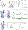
The sybody traps TM287/288 in its OF state. a Sybody Sb_TM#35 is shown as cartoon in gray with the CDR1, 2 and 3 highlighted in yellow, orange and red, respectively. Four aromatic residues (Y33, W52, Y59, and W113) that wedge between TMs 1 and 2 of TM287 (teal) and TMs 5′ and 6′ of TM288 (magenta) are highlighted as sticks. b Inhibition of TM287/288’s ATP hydrolysis by Sb_TM#35, Nb_TM#1 and Nb_TM#2. A non-randomized sybody served as control. The data were fitted with a hyperbolic decay function to determine IC50 values, as well as residual activities. The error bars are standard deviations of technical triplicates. c, d DEER analyses of spin-label pairs introduced to probe the extracellular and intracellular TMDs and the NBDs (c), as well as sybody binding to the transporter (d). DEER traces were recorded in the presence of ATP-EDTA with or without unlabeled Sb_TM#35 (c) or in the presence of ATP-EDTA and spin-labeled Sb_TM#35 (d). The graphs show experimental distance distributions, and vertical dotted lines shown in c highlight changes in the mean distances
Fig. 3
Fig. 3
Structural analysis of the closed NBD dimer. a The fully closed NBD dimer (NBD287 in teal and NBD288 in magenta) sandwiches two ATPγS-Mg molecules (shown as sticks with corresponding electron density) between Walker A motif (red) and the opposite ABC signature motif (green) at the degenerate and the consensus site in a highly symmetric manner. Residues involved in ATP binding and hydrolysis are shown as sticks. b Superimposition of the consensus ATP binding site of the previously solved IF structure (PDB: 4Q4A, light pink) and the OF structure (magenta). Distortions of the catalytic dyad (E517TM288 and H548TM288) are relaxed during NBD closure to adopt a hydrolysis-competent arrangement. The side chain of E517TM288 was modeled into the TM287/288(EtoA) structure. c Slice-cut through the two nucleotide binding sites reveals two possible Pi exit tunnels at the consensus site which are not present at the degenerate site. ATPγS (partially clipped) is shown as yellow sticks
Fig. 4
Fig. 4
The extracellular gate is sealed by two conserved aspartates. a Structure of TM287/288’s extracellular gate in the IF (left, PDB: 4Q4H) and OF (right) state shown as cartoon. D41TM287and D65TM288 are shown as sticks and establish hydrogen bonds (dashed yellow lines) with the peptide backbone (shown as sticks) of neighboring helices TM6 and TM6' that are broken during IF–OF transition. b Sequence alignment of bacterial ABC exporters in the region containing the conserved extracellular gate aspartates. c ATPase activities of single mutants D41ATM287 and D65ATM288 and the corresponding double mutant (2xDtoA) relative to wild-type TM287/288 determined in detergent. d Drug stimulated ATPase activities of wild-type EfrEF, the single mutants D41AEfrE and D50AEfrF and the corresponding double mutant (2xDtoA) reconstituted into proteoliposomes determined in the absence (basal activity) or in the presence of ethidium at the concentrations indicated. Data were normalized to the basal ATPase activity of the respective mutant. The error bars are standard deviations of technical triplicates. e Ethidium accumulation of Lactococcus lactis cells expressing wild-type EfrEF, the inactive Walker B mutant E515QEfrF or the extracellular gate mutants D41AEfrE and D50AEfrF or the corresponding double mutant (2xDtoA). f DEER analyses probing the extracellular and intracellular TMDs and the NBDs (same positions as in Fig. 2c). DEER traces were recorded in the presence of ATP-EDTA for the wild-type transporter and for TM287/288(2xDtoA). g Relative ATPase activities of the 2xDtoA mutant in the presence of increasing concentrations of Sb_TM#35, Nb_TM#1 and Nb_TM#2. A non-randomized sybody served as control. The data were fitted with a hyperbolic decay function to determine IC50 values as well as residual activities. The error bars are standard deviations of technical triplicates
Fig. 5
Fig. 5
Probing the OF–IF transition using state-specific binders. a Exemplary raw data of SPR traces based on which conformational probing was detected. At time point zero, immobilized TM287/288(EtoA) was charged with 1 mM ATP (red arrow) and binders were injected (gray arrows) at saturating concentrations (Sb_TM#35, 1 µM; Nb_TM#1, 500 nM; Nb_TM#2, 100 nM) to obtain maximal response unit values (RUmax) at the indicated time points. b RUmax values for wild-type and mutant TM287/288 were obtained as shown in a by charging the transporter with 1 mM ATP either in presence of Mg2+ (upper panel) or EDTA (lower panel). For state-specific binders Sb_TM#35 and Nb_TM#1, data were fitted using a one phase decay function to determine half-life values (t1/2) of the OF state. State-unspecific Nb_TM#2 was used as a control
Fig. 6
Fig. 6
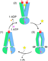
Role of the extracellular gate in the transport cycle of TM287/288. Substrate (yellow star) binds to the IF transporter (1) with high affinity, while the extracellular gate is sealed by two aspartates (closed D-lock). Binding and occlusion of two ATP-Mg molecules at the NBD interface drives the transition to the OF state (2). The extracellular gate opens (open D-lock) and substrate is released. The extracellular gate of the empty outward-oriented cavity closes (3) and thereby may trigger ATP hydrolysis at the consensus site. The mechanical force of the firmly sealed extracellular gate (closed D-lock) is required to dissociate the NBDs after ATP hydrolysis in order to reset the transporter to its IF state

Similar articles

Cited by

References

    1. Locher KP. Mechanistic diversity in ATP-binding cassette (ABC) transporters. Nat. Struct. Mol. Biol. 2016;23:487–493. doi: 10.1038/nsmb.3216. - DOI - PubMed
    1. Ramachandra M, et al. Human P-glycoprotein exhibits reduced affinity for substrates during a catalytic transition state. Biochemistry. 1998;37:5010–5019. doi: 10.1021/bi973045u. - DOI - PubMed
    1. Dawson RJ, Locher KP. Structure of a bacterial multidrug ABC transporter. Nature. 2006;443:180–185. doi: 10.1038/nature05155. - DOI - PubMed
    1. Choudhury HG, et al. Structure of an antibacterial peptide ATP-binding cassette transporter in a novel outward occluded state. Proc. Natl Acad. Sci. USA. 2014;111:9145–9150. doi: 10.1073/pnas.1320506111. - DOI - PMC - PubMed
    1. George AM, Jones PM. Perspectives on the structure-function of ABC transporters: the switch and constant contact models. Prog. Biophys. Mol. Biol. 2012;109:95–107. doi: 10.1016/j.pbiomolbio.2012.06.003. - DOI - PubMed

Publication types

MeSH terms

Substances