. 2019 Jun 3;29(11):1735-1745.e4.
doi: 10.1016/j.cub.2019.04.034. Epub 2019 May 16.

Pheromones and Nutritional Signals Regulate the Developmental Reliance on let-7 Family MicroRNAs in C. elegans

Affiliations

Pheromones and Nutritional Signals Regulate the Developmental Reliance on let-7 Family MicroRNAs in C. elegans

Orkan Ilbay et al. Curr Biol. .

Abstract

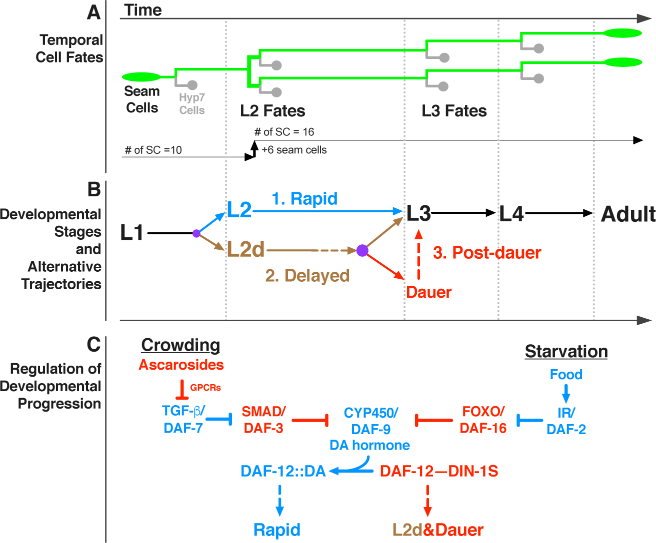
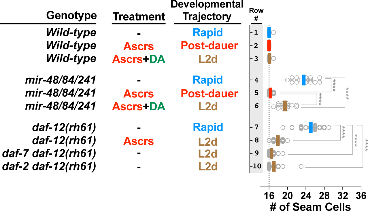
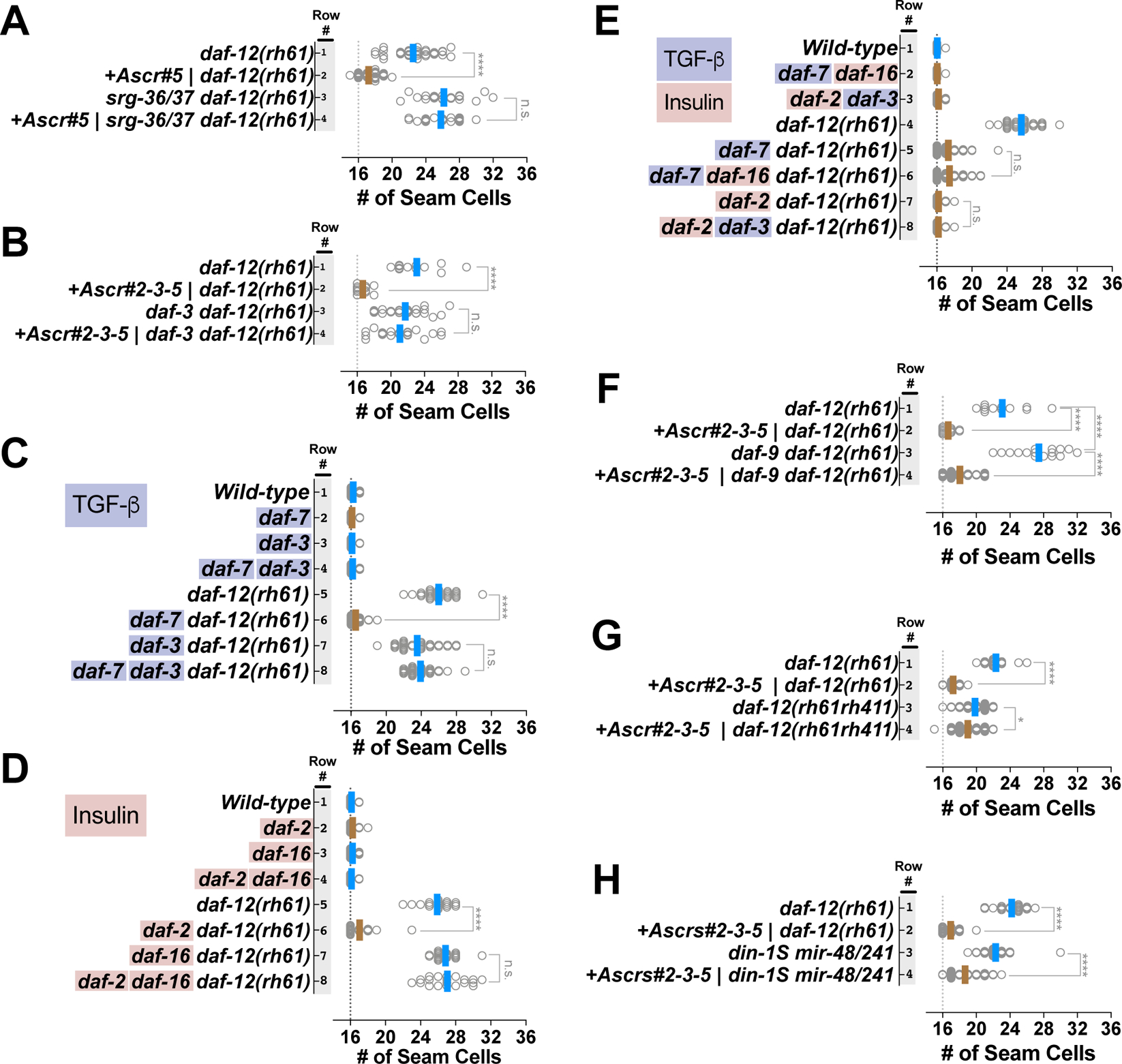
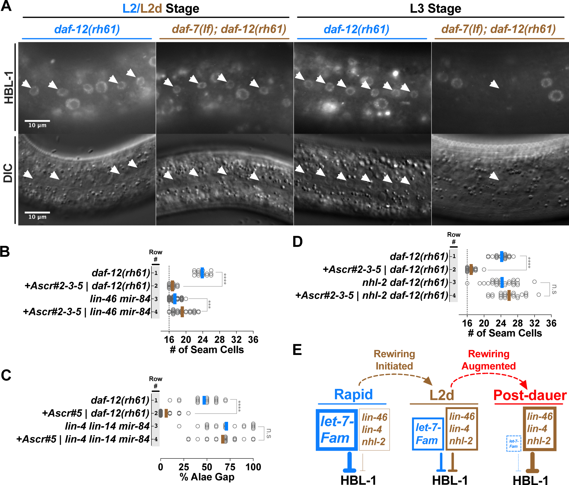
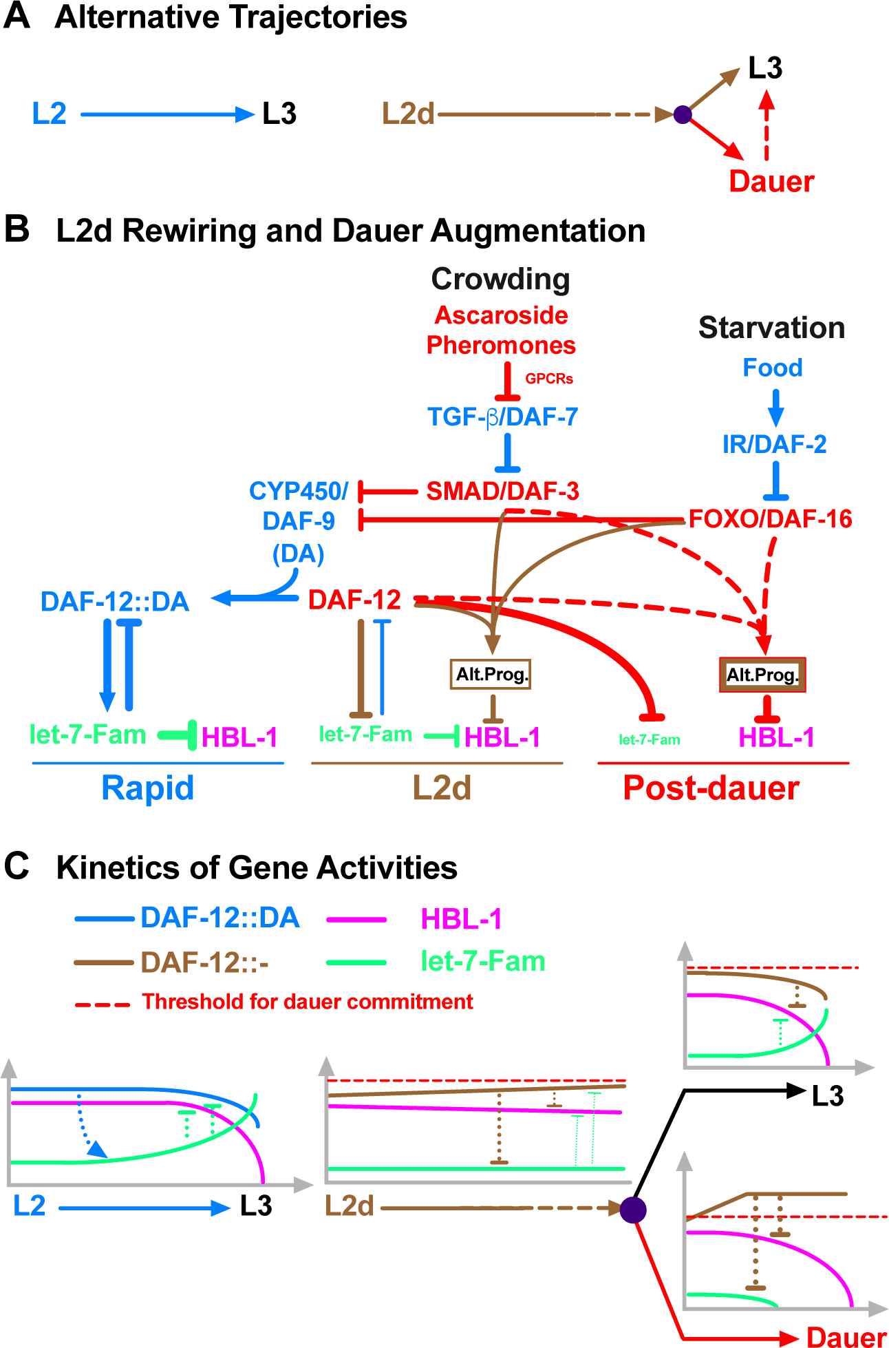
Adverse environmental conditions can affect rates of animal developmental progression and lead to temporary developmental quiescence (diapause), exemplified by the dauer larva stage of the nematode Caenorhabditis elegans (C. elegans). Remarkably, patterns of cell division and temporal cell-fate progression in C. elegans larvae are not affected by changes in developmental trajectory. However, the underlying physiological and gene regulatory mechanisms that ensure robust developmental patterning despite substantial plasticity in developmental progression are largely unknown. Here, we report that diapause-inducing pheromones correct heterochronic developmental cell lineage defects caused by insufficient expression of let-7 family microRNAs in C. elegans. Moreover, two conserved endocrine signaling pathways, DAF-7/TGF-β and DAF-2/Insulin, that confer on the larva diapause and non-diapause alternative developmental trajectories interact with the nuclear hormone receptor, DAF-12, to initiate and regulate a rewiring of the genetic circuitry controlling temporal cell fates. This rewiring includes engagement of certain heterochronic genes, lin-46, lin-4, and nhl-2, that are previously associated with an altered genetic program in post-diapause animals, in combination with a novel ligand-independent DAF-12 activity, to downregulate the critical let-7 family target Hunchback-like-1 (HBL-1). Our results show how pheromone or endocrine signaling pathways can coordinately regulate both developmental progression and cell-fate transitions in C. elegans larvae under stress so that the developmental schedule of cell fates remains unaffected by changes in developmental trajectory.

Keywords: ascarosides; dauer larva; developmental robustness; endocrine signaling; heterochronic genes; let-7; microRNAs; pheromones; reprogramming; stem cell.


Conflict of interest statement

DECLARATION OF INTERESTS

The authors declare no competing interests.

Figures

Figure 1.
Figure 1.. Temporal fates of hypodermal seam cells are robust against changes in developmental trajectory induced by crowding or starvation.
(A) Lineage diagram showing temporal (stage-specific) hypodermal seam cell fates. Seam cells (green) divide asymmetrically at each larval stage, renewing themselves while giving rise to hyp7 cells (gray). At the L2 stage, seam cells undergo a single round of symmetric cell division, resulting in an increase in their number. Note that only six out of ten seam cells undergo symmetric cell division which increases the total number of seam cells on each side of the worm from ten to sixteen. (B) Developmental stages and three distinct developmental trajectories: 1. Continuous, unipotent, and rapid progression define the L2 trajectory (blue); 2. Continuous but bipotent and delayed progression define the L2d trajectory (brown), 3. Developmental progression is interrupted by a diapause in the dauer-interrupted/post-dauer trajectory (brown followed by red). Time axis indicates the order of events in time (not proportional to absolute time). Lateral dotted lines indicate the molts between stages. Purple dots represent decision points between different trajectory options. (C) Regulation of developmental progression. Under favorable conditions dafachronic acid (DA) hormone is abundant and DA-bound DAF-12 promotes rapid development. Crowding or starvation induces L2d and dauer formation by repressing TGF-β/DAF-7 signals or insulin signaling (IR/DAF-2: insulin receptor), respectively. Activated effectors of these signaling pathways (DAF-3 or DAF-16) inhibit the biosynthesis of DA; and the unliganded DAF-12 interacts with DIN-1S, which together promote L2d and dauer formation.
Figure 2.
Figure 2.. Sensitized genetic backgrounds reveal that L2d-inducing environmental and endocrine signals impact the regulation of temporal cell fates.
Genotypes are indicated in the first column; treatments and corresponding developmental trajectories are indicated in the second and third columns, respectively. Each dot in the plots to the right shows the number of seam cells on one side (left side or right side, observed interchangeably) of a single young adult animal, and solid lines (color code matching the developmental trajectory) indicate the average seam cell number of the animals scored for each condition. Wild-type animals have sixteen seam cells per side (vertical dotted line), regardless of developmental trajectory (lines 1–3). Experiments involving temperature sensitive alleles of daf-2 and daf-7 (lines 6 and 7) are performed at a permissive temperature (20°C) that allows continuous (L2d-to-L3 without dauer arrest) development. The student’s t-test is used to calculate statistical significance (p): n.s.(not significant) p>0.05, *p<0.05, **p<0.01, ***p<0.001, ****p<0.0001. See also Figure S1 and S2 and Table S1.
Figure 3.
Figure 3.. DAF-7/TGF-β and DAF-2/Insulin signaling pathways act in parallel to modulate a ligand-independent activity of DAF-12 that is responsible for correcting heterochronic phenotypes caused by insufficient expression of let-7 family microRNAs.
Number of seam cells in young adult animals of various mutants cultured on ascaroside or control plates (A, B and F. G), or on standard NGM plates (C-E). Each dot in the plots shows the number of seam cells of a single young adult animal, and solid lines indicate the average seam cell number of the animals scored for each condition (blue lines: rapid trajectory; brown lines: L2d trajectory). (A) Ascarosides suppress daf-12(rh61) via srg-36/37-encoded GPCR signaling upstream of DAF-7/TGF-β-DAF-3 signaling. (B) daf-3 activity is required for suppression of daf-12(rh61) by ascarosides. (C-E) DAF-7/TGF-β and DAF-2/Insulin signaling pathway act in parallel to mediate the suppression of daf-12(rh61). (F, G) Ligand-independent activity of daf-12 is required for the ascaroside-mediated L2d rewiring of the pathways regulating temporal cell fates. (H) The DAF-12 corepressor DIN-1S is not required for the ascaroside-mediated suppression of heterochronic phenotypes caused by insufficient expression of let-7 family microRNAs. Suppression of extra seam cell phenotype of daf-12(rh61) is shown as a measure of the strength of the ascaroside conditions tested for din-1S(lf); mir-48/241(lf) animals. The student’s t-test is used to calculate statistical significance (p): n.s.(not significant) p>0.05, *p<0.05, **p<0.01, ***p<0.001, ****p<0.0001. See also Figure S3 and Table S1.
Figure 4.
Figure 4.. Heterochronic genes associated with the altered HBL-1 down-regulation program in post-dauer animals are required for the L2d suppression of heterochronic phenotypes caused by insufficient expression of let-7 family microRNAs.
(A) Upper row: fluorescent images showing HBL-1 expression in L2/L2d and L3 stage hypodermal seam (white arrowheads) and hyp7 (all other) nuclei in daf-12(rh61) and daf-7(lf); daf-12(rh61) animals. Lower row: corresponding DIC images of the hypodermis. It should be noted that, consistent with the variability in the extra seam cell phenotype of daf-12(rh61) animals, HBL-1 expression at the L3 stage daf-12(rh61) animals displays variability across seam cells of individual worms. For example, HBL-1 expression may be present and absent in two neighboring seam cells, which presumably expresses L2 and L3 cell fates, respectively. (B) Ascaroside conditions that suppress the extra seam cell phenotype of daf-12(rh61) enhance the extra seam cell phenotype of larvae lacking lin-46 and mir-84. (C) Ascaroside conditions that suppress the gapped alae (a consequence of retarded seam cell development that is manifested in young adults) phenotype do not suppress the gapped alae phenotype of lin-4; lin-14; mir-84 animals. (D) nhl-2 activity is required for ascaroside-mediated suppression of daf-12(rh61) (E) A model for the L2d rewiring and its potential augmentation during dauer arrest. Under L2d-inducing conditions, let-7 family microRNAs are downregulated and also become less important. The reduction in the let-7 level and importance is coupled to enhanced roles for the heterochronic genes previously associated with the altered HBL-1 downregulation program in post-dauer animals, involving lin-46, lin-4, and nhl-2. This shift in the reliance on the let-7 family microRNAs to the reliance on the alternative program for proper HBL-1 down-regulation (hence for proper L2-to-L3 cell fate progression) constitutes the L2d rewiring. In post-dauer animals, consistent with an augmentation of the L2d rewiring program, the reliance on the altered HBL-1 down-regulation program further increases while the let-7 family microRNAs become dispensable for proper HBL-1 down-regulation. It should be noted that we do not know the mechanisms (e.g. elevated levels vs enhanced activities) of increased roles for lin-46, nhl-2, or lin-4 during L2d or post-dauer development. The student’s t-test is used to calculate statistical significance (p): n.s.(not significant) p>0.05, *p<0.05, **p<0.01, ***p<0.001, ****p<0.0001. See also Table S1.
Figure 5.
Figure 5.. Coordinate regulation of developmental progression and cell fate transitions in C. elegans larvae.
(A) Alternative trajectories. The L2-to-L3 transition is rapid and deterministic (once committed to the L2 stage, the larvae do not have the dauer option), whereas the L2d-to-L3 transition is slower and bipotential. In both cases, HBL-1 is present throughout the L2/L2d stage but it is downregulated by the beginning of the L3 stage (Figure S4). (B) Pheromone and endocrine signals engage DAF-12 to initiate and regulate the rewiring of the HBL-1 down-regulation. In response to crowding and starvation, TGF-β and insulin signaling pathways, respectively, modulate the ligand-dependent DAF-12 activity to repress the transcription of let-7 family microRNAs, and, at the same time, cooperate with DAF-12 in a ligand independent manner to activate the alternative HBL-1 downregulation program (Alt. Prog.). The alternative program of L2d and dauer-interrupted trajectories are similar, but the alternative program of dauer-interrupted trajectory is stronger either due to an enhancement of the alternative program of L2d (depicted as thicker brown border line) and/or due to employment of additional factors (depicted as red border line) after the L2d larvae commit to dauer formation. (C) DAF-12 ensures properly delayed but robust HBL-1 down-regulation during L2d-to-L3 transition by coordinating the repression of let-7 family microRNAs with the activation of the alternative HBL-1 downregulation program. During rapid, L2 development, DAF-12 activates the transcription of let-7 family microRNAs, which in turn, negatively regulate DAF-12, eliminating the dauer option. During slow, bipotential, L2d development, DAF-12 represses let-7 family microRNAs, which otherwise prevent the accumulation of DAF-12. If the unliganded DAF-12 reaches the threshold, larvae commit to dauer formation, if not, larvae commit to continuous development. While mediating this decision, which necessitates the repression of let-7 family microRNAs (for maintaining the dauer option) and delaying the downregulation of HBL-1 (for postponing L3 cell fates), DAF-12 cooperates with DAF-3 or DAF-16 to activate the alternative HBL-1 downregulation program (Alt. Prog) to ensure robust HBL-1 downregulation during the L2d-to-L3 transition. See also Figure S4.

Comment in

Similar articles

Cited by

References

    1. Sulston JE, and Horvitz HR (1977). Post-embryonic cell lineages of the nematode, Caenorhabditis elegans. Dev. Biol 56, 110–156. - PubMed
    1. Ambros V, and Horvitz HR (1984). Heterochronic mutants of the nematode Caenorhabditis elegans. Science 226, 409–16. - PubMed
    1. Ambros V (2011). MicroRNAs and developmental timing. Curr. Opin. Genet. Dev 21, 511–7. - PMC - PubMed
    1. Lin SY, Johnson SM, Abraham M, Vella MC, Pasquinelli A, Gamberi C, Gottlieb E, and Slack FJ (2003). The C. elegans hunchback homolog, hbl-1, controls temporal patterning and is a probable MicroRNA target. Dev. Cell 4, 639–650. - PubMed
    1. Abrahante JE, Daul AL, Li M, Volk ML, Tennessen JM, Miller EA, and Rougvie AE (2003). The Caenorhabditis elegans hunchback-like gene lin-57/hbl-1 controls developmental time and is regulated by microRNAs. Dev. Cell 4, 625–37. - PubMed

Publication types