. 2019 May 17;15(5):e1008169.
doi: 10.1371/journal.pgen.1008169. eCollection 2019 May.

The processivity factor Pol32 mediates nuclear localization of DNA polymerase delta and prevents chromosomal fragile site formation in Drosophila development

Affiliations

The processivity factor Pol32 mediates nuclear localization of DNA polymerase delta and prevents chromosomal fragile site formation in Drosophila development

Jingyun Ji et al. PLoS Genet. .

Abstract

The Pol32 protein is one of the universal subunits of DNA polymerase δ (Pol δ), which is responsible for genome replication in eukaryotic cells. Although the role of Pol32 in DNA repair has been well-characterized, its exact function in genome replication remains obscure as studies in single cell systems have not established an essential role for Pol32 in the process. Here we characterize Pol32 in the context of Drosophila melanogaster development. In the rapidly dividing embryonic cells, loss of Pol32 halts genome replication as it specifically disrupts Pol δ localization to the nucleus. This function of Pol32 in facilitating the nuclear import of Pol δ would be similar to that of accessory subunits of DNA polymerases from mammalian Herpes viruses. In post-embryonic cells, loss of Pol32 reveals mitotic fragile sites in the Drosophila genome, a defect more consistent with Pol32's role as a polymerase processivity factor. Interestingly, these fragile sites do not favor repetitive sequences in heterochromatin, with the rDNA locus being a striking exception. Our study uncovers a possibly universal function for DNA polymerase ancillary factors and establishes a powerful system for the study of chromosomal fragile sites in a non-mammalian organism.


Conflict of interest statement

The authors have declared that no competing interests exist.

Figures

Fig 1
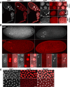
Fig 1. Loss of Pol32 impedes genomic replication.
A. DAPI stained embryos of wild-type and pol32 mutants. These are embryos collected at 0-2hr after egg laying. Two areas of enlargement marked with rectangles are shown in the middle and the right panels, with the middle ones showing the polar bodies and the right ones showing zygotic nuclei. B. Agarose gel showing total embryonic DNA from mutant (pol32) and wild-type (wt) embryos either uncut (left two lanes) or cut with EcoRI (right two lanes). The markers in the leftmost lane, from top to bottom, have sizes in kb of: 15, 10, 7.5, 5, 2.5, 1, 0.25. C. Summary results from whole genome sequencing showing DNA from pol32 mutant embryos primarily consists of mitochondrial DNA. Scale bars indicate 40μm.
Fig 2
Fig 2. Pol32 localization during normal development.
A separate image is provided for the DAPI signal (in white), the anti-Pol32 signal (in red), and the merged product of the two channels. A. Pol32 in ovarian tissue. A series of egg chambers (left three panels) are shown with three enlarged areas included in the right three columns of images. The nucleus of the oocyte is marked with an arrowhead. In Aa, Pol32 can be seen in the nuclei of nurse cells. In Ab, Pol32 can be seen in the nucleoplasm of the oocyte with chromosomes marked with an arrowhead. In Ac, Pol32 can be seen in nuclei of follicle cells. B. Pol32 in 0-2hr old embryos. The top left panels show images of an embryo in interphase, and the top right panels showing an embryo with condensed chromosomes. The five haploid nuclei (three polar body nuclei and the parental pronuclei) are shown underneath as enlarged images for each embryo. Ba and Bb show interphase nuclei. Bc and Bd show metaphase nuclei. C. Pol32 in syncytial cell cycles. The top row shows interphase cells with nuclear Pol32 signals. The bottom row shows mitotic cells with no Pol32 in the nucleus. Scale bars in red indicate 40μm, and 10μm in white.
Fig 3
Fig 3. Pol δ complex is absent in embryonic nuclei of pol32 mutants.
Representative confocal images of antibody staining of the Pol δ complex (PolD, Pol31 and Pol32), PCNA and Polα in wild-type and pol32-mutant 0-2hr old embryos. For each antibody, 50 wild-type embryos were imaged. The number of pol32-mutant embryos imaged for each antibody are: 70 for PolD, 71 for Pol31, 52 for PCNA, and 59 for Polα. Scale bars indicate 10μm.
Fig 4
Fig 4. Interactions within the Pol δ complex.
A. The level of Pol δ in wild-type and pol32 mutant embryonic extracts. Different amount of extracts (3 or 6μl) were used for Western blot analyses with Tubulin as a general loading control. Genotypes are on top and antibodies are listed to the left of the images. B. Co-IP experiments to detect interactions among Pol δ subunits. For each panel, the genotypes are indicated at the bottom, and the antibodies used for IP on top of the images. “preIM” indicates that the corresponding pre-Immune serum was used for IP. The antibodies used for Western blot detection (IB) are listed to the left of the panels. In the bottom panel, “input” marks the lane with 1/20 of the input extract loaded. “flow” marks the lane with a sample of “flow through” in the IP experiments loaded. “wash” denotes the lane with a sample of “wash” in the IP experiments.
Fig 5
Fig 5. PolD remains nuclear localized in post-embryonic cells in pol32 mutants.
Confocal images showing PolD and Pol32 localizations in a larval salivary gland (A) and an egg chamber from adult ovary (B). The genotypes are indicated to the left. In the “merged” image in B, the nucleus of the oocyte is demarcated with a rectangular box, which is shown as enlarged images to the right with chromosomes marked with an arrowhead. Note the lack of PolD signals in the mutant. Scale bars indicate 40μm.
Fig 6
Fig 6. Pol32-Pol31 interaction is essential for Pol32 function.
A. Constructs used in rescuing experiments and their effects on the mutant phenotypes. The 431aa Pol32 protein is denoted as a rectangular box with domains of interest labelled in black. The names of the domain are on top of the boxes with the range in amino acids in parentheses. For the Polα-interacting DPIM and the PCNA-interacting PIP domain, the amino acid sequences of the domain are listed underneath with the conserved residues in Bold and a larger Font. For constructs with a deletion, the deleted range in amino acids is shown under the name of the mutant construct and proceeded by a “Δ”. For constructs with residues changed to Alanine, the mutant composition of the mutated domain is shown underneath and the mutated domain denoted with an “*”. The constructs were tested for their abilities to rescue two mutant phenotypes. “Bristle” indicates shorten or missing bristles in pol32 adults. “Fertility” indicates female sterility of pol32 mutants. “+” indicates that a construct can recue the phenotype when introduced into a pol32 mutant background, while “-” cannot. B. The levels of Pol32 mutant proteins. Western blots using total extracts from either ovaries or embryos were probed with antibodies indicated to the left of the images. The animals are pol32 homozygotes with a rescuing construct listed on top of the images and shown in A. Tubulin was used as a general loading control. For the two wHTH mutants, the level of PolD was also measured (right panels in B). C. The effect of Pol31 RNAi on the level of Pol δ. Two transgenes carrying different RNAi hairpins were used to knockdown Pol31 in larval tissues. The levels of Pol32 and PolD were assayed by Western blotting. “+” indicates samples from animals with the hairpin construct, while “-” from control animals without the construct.
Fig 7
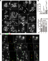
Fig 7. Loss of Pol32 induces chromosome breaks in post-embryonic cells.
A. Mitotic figures showing the different classes of DSBs in the mutant pol32L27/L30; polDl10/+. In the two wild-type nuclei chromosomes are individually identified. In the mutant nuclei, only the chromosome with a DSB of interest is denoted and the approximate location of the DSB marked with either an arrowhead (centric DSB) or an arrow (non-centric DSB). Aa, Ab: two wild-type nuclei with no broken chromosomes. Ac: a non-centric X chromosome break. Ad: a DSB at the centromere-proximal rDNA region on X. Ae: a DSB at the distal rDNA region. Af: a DSB at the DAPI-bright block of X. Ag: a non-centric DSB on chromosome 2. Ah: a centric DSB of 2. Note the rest of chromosome 2 is missing in this nucleus. Ai: a non-centric DSB on chromosome 3. Aj, Ak: two nuclei each with a centric DSB on 3. Al; a DSB at the rDNA region of the Y chromosome. Am: multiple DSBs in a nucleus. B. DSB frequencies. The top chart quantifies the percentage of nuclei with at least one chromosome break in three different genetic backgrounds. The bottom chart quantifies the two classes of breaks on each major chromosome from neuroblasts of the genotype pol32L27/L30; polDl10/+, with the numbers indicating the percentages of centric DSBs. C. FISH identifies broken sites in X peri-centromeric region. In each triplet of images, to the left is a DAPI-stained chromosome figure; in the middle is FISH image of a nucleus double labelled with rDNA (green) and 359 satellite (red) probes; and to the right is the merged product. Ca, a normal female nucleus. Cb, a nucleus with a DSB at the distal region of rDNA where the sister chromatids remain synapsed. Cc, a nucleus with a DSB splitting the rDNA arrays into approximate halves. Cd, a nucleus with a DSB splitting the DAPI-bright 359 satellite arrays into approximate halves. Ce, severe rDNA instability. Numerous acentric chromosomal fragments are visible with rDNA at one end (arrow), and rDNA fragments are present that do not appear to be attached to any major chromosomes (arrowhead). Cf, a possible case of rDNA expansion. An X chromosome with a greatly expanded rDNA array (arrow), when compared with another X chromosome with a normal appearance. In the same nucleus, the 359 satellite is attached to an aberrant chromosome proximally (arrowhead). Scale bars indicate 10μm.
Fig 8
Fig 8. Genetic interactions between pol32 and other DNA replication and repair factors.
A. Pictures of adult flies (three females at the top and three males at the bottom) showing etching of the abdominal region. Genotypes are given at the top. B. Quantification of the “etched abdomen” phenotype. The specific mutant alleles (m) are as followed: pol32m = pol32L27/L30, spnAm = spnA1, spnADf = Df(3R)X3F, mus309m = mus309D2, mus309Df = Df(3R)P-21, polαm = polαG13925, poldm = poldl10, polαDf = Df(3R)Exel6186, pol31m = pol31G16501, pol31Df = Df(3L)Bsc122.
Fig 9
Fig 9. A speculative model for the dual function of Drosophila Pol32.
The normal Pol δ complex consists of PolD (black box), Pol31 (grey box) and Pol32 (white triangle). In normal cells, the cytoplasmically assembled Pol δ enters the nucleus efficiently, and possesses normal polymerase processivity leading to efficient DNA synthesis (long strands of newly synthesized DNA). In pol32-mutant post-embryonic cells, Pol δ might enter the nucleus at a reduced efficiency (smaller arrow and question mark), accompanied by the partial loss of processivity (shortened strands of new DNA). This leads to the formation of chromosome breaks. In pol32-mutant embryonic cells, however, Pol δ is inhibited from entering the nucleus halting genome replication and embryonic development.

Similar articles

Cited by

References

    1. Stodola JL, Stith CM, Burgers PM. Proficient Replication of the Yeast Genome by a Viral DNA Polymerase. J Biol Chem. 2016. May 27;291(22):11698–705. 10.1074/jbc.M116.728741 - DOI - PMC - PubMed
    1. Tomasetti C, Vogelstein B. Cancer etiology. Variation in cancer risk among tissues can be explained by the number of stem cell divisions. Science. 2015. January 2;347(6217):78–81. 10.1126/science.1260825 - DOI - PMC - PubMed
    1. Tomasetti C, Li L, Vogelstein B. Stem cell divisions, somatic mutations, cancer etiology, and cancer prevention. Science. 2017. March 24;355(6331):1330–1334. 10.1126/science.aaf9011 - DOI - PMC - PubMed
    1. Durkin SG, Glover TW. Chromosome fragile sites. Annu Rev Genet. 2007;41:169–92. 10.1146/annurev.genet.41.042007.165900 - DOI - PubMed
    1. Zlotorynski E, Rahat A, Skaug J, Ben-Porat N, Ozeri E, Hershberg R, Levi A, Scherer SW, Margalit H, Kerem B. Molecular basis for expression of common and rare fragile sites. Mol Cell Biol. 2003. October;23(20):7143–51. 10.1128/MCB.23.20.7143-7151.2003 - DOI - PMC - PubMed

Publication types

MeSH terms

Substances