. 2019 May 10;11(5):431.
doi: 10.3390/v11050431.

Histone Deacetylase Inhibitors Enhance Cell Killing and Block Interferon-Beta Synthesis Elicited by Infection with an Oncolytic Parainfluenza Virus

Affiliations

Histone Deacetylase Inhibitors Enhance Cell Killing and Block Interferon-Beta Synthesis Elicited by Infection with an Oncolytic Parainfluenza Virus

Candace R Fox et al. Viruses. .

Abstract

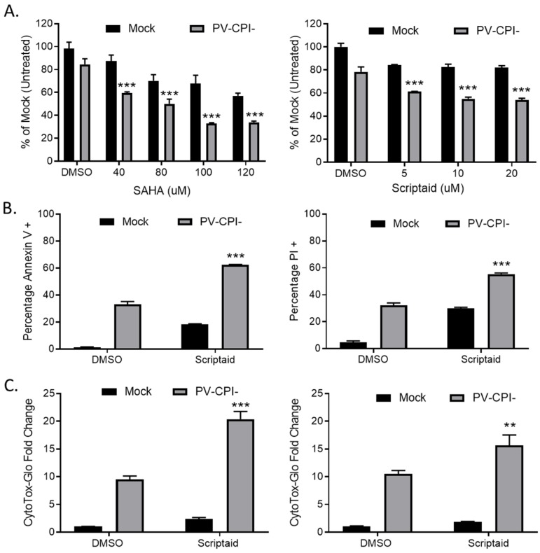
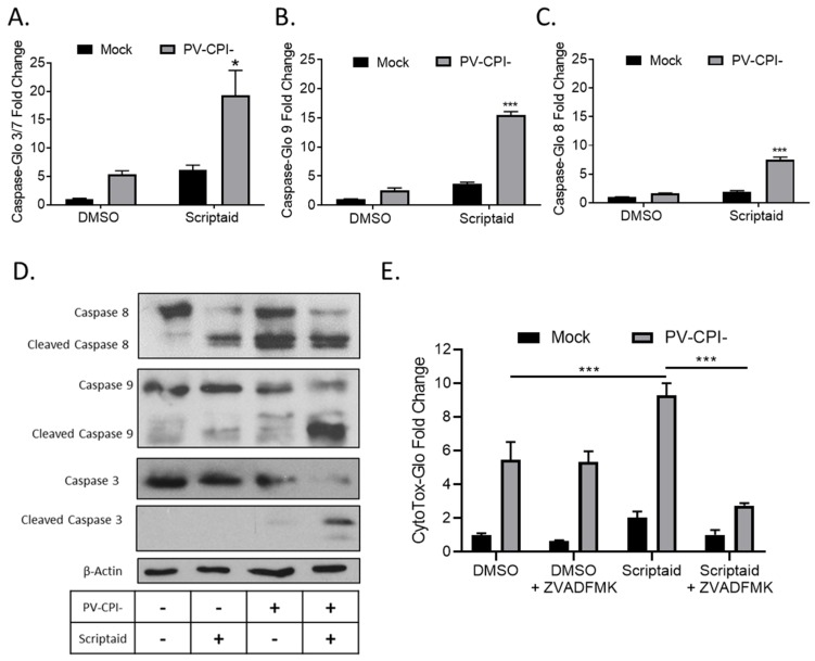
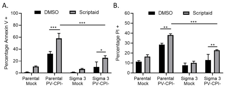
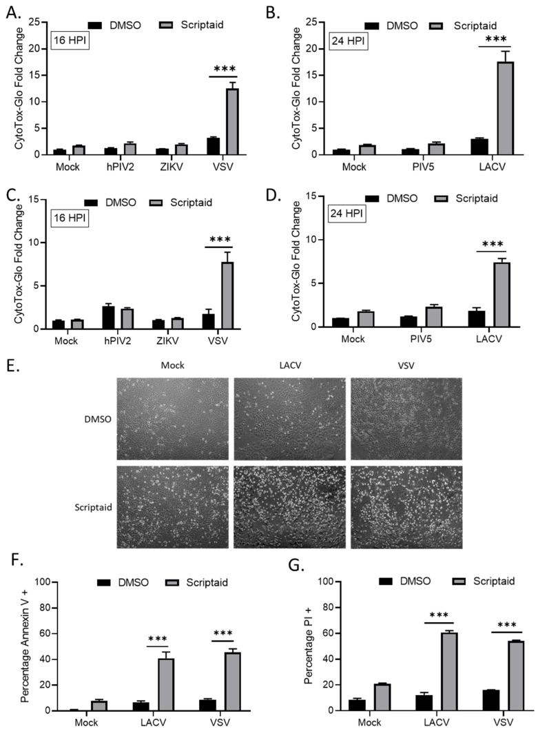
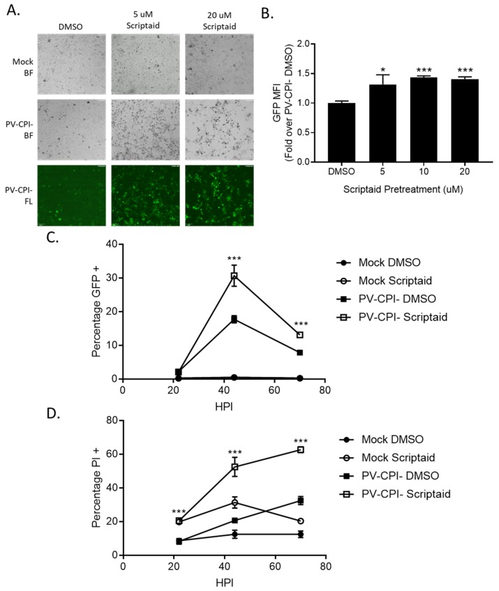
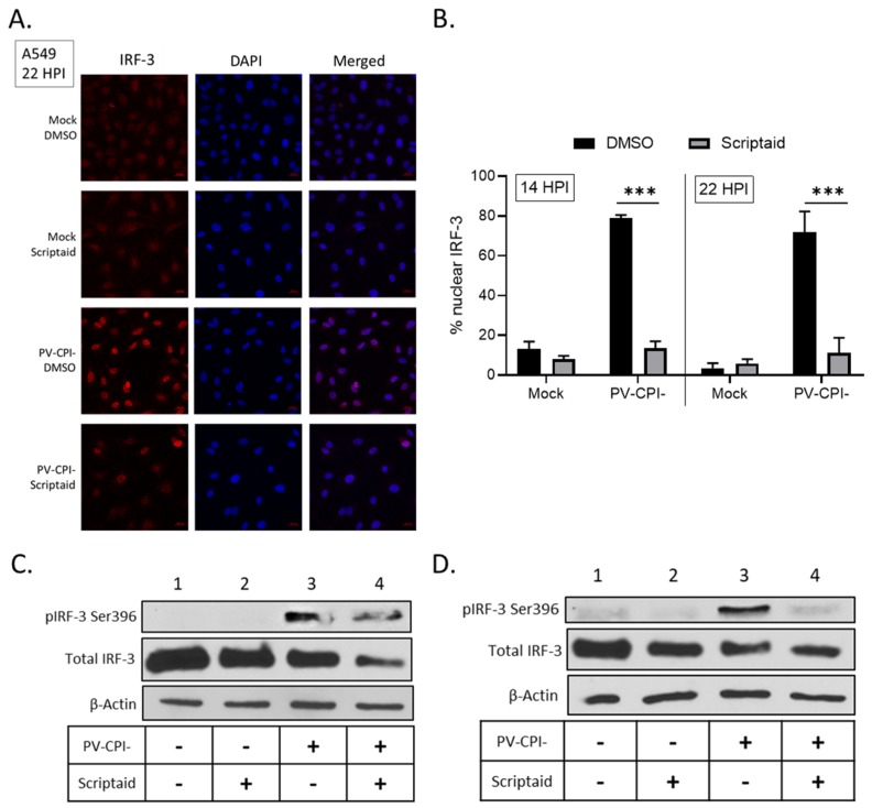
Previous results have shown that infection with the cytoplasmic-replicating parainfluenza virus 5 mutant P/V-CPI- sensitizes cells to DNA damaging agents, resulting in the enhanced killing of airway cancer cells. Here, we have tested the hypothesis that histone deacetylase (HDAC) inhibitors can also act with P/V-CPI- infection to enhance cancer cell killing. Using human small cell lung cancer and laryngeal cancer cell lines, 10 HDAC inhibitors were tested for their effect on viability of P/V-CPI- infected cells. HDAC inhibitors such as scriptaid enhanced caspase-3/7, -8 and -9 activity induced by P/V-CPI- and overall cell toxicity. Scriptaid-mediated enhanced killing was eliminated in lung cancer cells that were engineered to express a protein which sequesters double stranded RNA. Scriptaid also enhanced cancer cell killing by two other negative strand RNA viruses - the La Crosse virus and vesicular stomatitis virus. Scriptaid treatment enhanced the spread of the P/V-CPI- virus through a population of cancer cells, and suppressed interferon-beta induction through blocking phosphorylation and nuclear translocation of Interferon Regulatory Factor 3 (IRF-3). Taken together, these data support a role for combinations of a cytoplasmic-replicating RNA virus such as the P/V-CPI- mutant along with chemotherapeutic agents.

Keywords: histone deacetylase inhibitors; oncolytic virus; parainfluenza virus.


Conflict of interest statement

The authors declare no conflict of interest.

Figures

Figure 1
Figure 1
Pretreatment with HDAC inhibitors enhances cell death by the P/V-CPI- mutant virus. (A) H1299 cells were pretreated with the indicated concentrations of SAHA (left panel), scriptaid (right panel), or DMSO as a vehicle control for 12 h. Cells were mock infected or infected with the P/V-CPI- mutant at an MOI of 10, and cultured in the presence of the indicated concentration of drugs. After 24 h, cell viability was determined by MTT assay as described in Methods section. (B) A549 cells were pretreated with 5 µM scriptaid or DMSO as a vehicle control for 12 h. Cells were mock infected or infected with the P/V-CPI- mutant at an MOI of 10, and cultured in the presence of 5 µM scriptaid or DMSO. Cell viability was determined by annexin V (left panel) and PI staining (right panel) at 24 hpi. (C) H1299 (left panel) or A549 (right panel) cells were pretreated with 20 µM scriptaid or DMSO for 12 h before mock infection or P/V-CPI- infection at an MOI of 10. After culturing in the presence of 1 µM scriptaid or DMSO for 24 h, cytotoxicity was determined by CytoTox-Glo assay. Values in all panels are the mean of three replicates normalized to DMSO pretreated mock infected samples with error bars indicating standard deviation. ** and *** indicates p-value of <0.01 and <0.001 comparing DMSO versus HDAC inhibitor pretreated infected samples.
Figure 2
Figure 2
HDAC inhibitor enhances caspase activation in P/V-CPI- infected cells. (AC) H1299 cells were pretreated with 20 µM Scriptaid or DMSO as a control for 12 h. Cells were then mock infected or infected with P/V-CPI- at an MOI of 10, and cultured for 24 h in media containing either 1 µM Scriptaid or DMSO. At 24 hpi, caspase activity was determined by Caspase-Glo-3/7 (A), -9 (B) or -8 (C) assays. Values are the mean of three replicates normalized to DMSO pretreated mock infected samples with * and *** indicating p-values of <0.05 and <0.001, respectively, comparing DMSO versus HDAC inhibitor pretreated infected samples. (D) A549 cells were treated as described for panels A-C. At 24 hpi, cells were harvested, and lysates were analyzed by western blotting for β-actin or for caspase-8, -9, and -3 cleavage products. (E) H1299 cells were pretreated with 20 µM scriptaid or DMSO for 12 h, and then mock infected or infected with P/V-CPI- at an MOI of 10. Cells were then cultured in media containing either 1 µM scriptaid or DMSO, with and without 50 µM of the pan-caspase inhibitor Z-VAD-FMK. At 24 hpi, cytotoxicity was determined by CytoTox-Glo assay. Values are the mean of three replicates normalized to DMSO pretreated mock infected samples with error bars indicating standard deviation and *** indicating p-values of <0.001.
Figure 3
Figure 3
dsRNA contributes to enhanced death in cells treated with HDAC inhibitor and P/V-CPI- infection. (A, B) Parental A549 cells and A549 cells expressing reovirus sigma 3 protein were pretreated with 20 µM Scriptaid or DMSO for 12 h. Cells were mock infected or infected with P/V-CPI- virus at an MOI of 10, and cultured in the presence of 1 µM Scriptaid or DMSO control. Cell viability was determined by Annexin V (A) and PI (B) staining at 24 hpi. Values are the mean of three replicates with error bars indicating standard deviation. ** and *** indicating p-values of <0.01 and <0.001, respectively.
Figure 4
Figure 4
HDAC inhibitor pretreatment enhances cell death by other RNA viruses. (AD) H1299 (A,B) or A549 (C,D) cells were pretreated with 20 µM scriptaid or DMSO for 12 h. Cells were then either mock infected or infected with the indicated viruses at an MOI of 10. After culturing in the presence of 1 µM scriptaid or DMSO for either 16 (A,C) or 24 (B,D) h, cytotoxicity was determined by CytoTox-Glo assay. hPIV2, human parainfluenza virus 2; ZIKV, Zika virus; VSV, vesicular stomatitis virus. LACV, La Crosse virus. Values are the mean of three replicates normalized to DMSO pretreated mock infected samples, with error bars indicating standard deviation. ** and *** indicating p-values of <0.01 and <0.001, respectively. (EG) A549 cells were pretreated with 20 µM scriptaid or DMSO and either mock infected or infected with LACV or VSV at MOIs of 10. After culturing in the presence of 1 µM scriptaid or DMSO for 16 h, cells were imaged at 10× magnification, and representative bright field images are shown (E). Cell viability was determined by annexin V (F) and PI (G) staining.
Figure 5
Figure 5
Pretreatment of cells with an HDAC inhibitor increases P/V-CPI- GFP expression and promotes low MOI virus spread. (A,B) H1299 cells were pretreated with the indicated concentrations of scriptaid or control DMSO for 12 h. Cells were then mock infected or infected with P/V-CPI- at an MOI of 10. After 24 h of culture in media containing either 1 µM scriptaid or DMSO, 20× bright field (BF) or fluorescence (FL) images of cells were captured (A). Alternatively, cells were analyzed by flow cytometry for GFP expression (B). Values are expressed as fold change over DMSO treated virus infected cells set at 1.0. (C,D). A549 cells were pretreated with 20 µM scriptaid or DMSO for 12 h. Cells were then mock infected or infected with the P/V-CPI- mutant at an MOI of 0.05, and cultured in media containing 1 µM scriptaid or DMSO. At the indicated hpi, cells were harvested and analyzed by flow cytometry for GFP expression (C) or PI staining (D). For all panels, error bars indicate standard deviation. * and *** indicates p-values of <0.05 and <0.001, respectively.
Figure 6
Figure 6
HDAC inhibitor pretreatment decreases P/V-CPI- induction of IFN-β and ISG expression. (AG) H1299 (A,C,E) or A549 (B,D,E) cells were pretreated with 20 µM scriptaid or DMSO for 12 h. Cells were then mock infected or infected with P/V-CPI- at an MOI of 10 and cultured in media containing either 1 µM scriptaid or DMSO. At 24 hpi, media was collected and analyzed for IFN-β amounts by ELISA (A,B). Total cellular RNA was extracted and evaluated for IFIT1 (C,D) and OAS2 (E,F) expression levels by RT-qPCR. (G) H1299 cell lysates were analyzed for levels of IFIT protein by western blotting. (HJ) H1299 cells were pretreated with 20 µM scriptaid or DMSO for 12 h. Cells were then either mock treated or treated with 100 or 1000 U/mL of universal type 1 IFN. At 24 hpi, total cellular RNA was extracted and evaluated for IFIT1 (H), OAS2 (I), and TLR3 (J) expression levels by RT-qPCR. For all panels, error bars indicate standard deviation. ** and *** indicates p-values of <0.01 and <0.001, respectively.
Figure 7
Figure 7
Effect of scriptaid treatment on P/V-CPI-induced IRF-3 nuclear localization and phosphorylation. (A,B) A549 cells were pretreated with 20 µM scriptaid or DMSO for 12 h. Cells were then mock infected or infected with P/V-CPI- at an MOI of 10 and cultured in media containing either 1 µM scriptaid or DMSO. IRF-3 immunostaining and DAPI nuclear staining was performed at 22 hpi and imaged at 40× magnification (A). Samples from the experiment displayed in panel A were used to determine the number of cells displaying intense nuclear staining as a percentage of the population (B). For each sample, five random fields were counted and averaged, with error bars denoting standard deviations. (C,D) A549 (C) and H1299 (D) cells were treated as described in panel A. At 24 hpi, cell lysates were evaluated for IRF-3 phosphorylated at Ser396, total IRF-3 and β-actin by Western blotting.
Figure 8
Figure 8
Differential effect of various HDAC inhibitors on killing of mock infected and P/V-CPI- infected human laryngeal cancer cells. (A,B) HEp-2 cells were mock infected or infected with P/V-CPI- at an MOI of 10 PFU/cell. At 12 hpi, cells were treated with indicated concentrations of HDAC inhibitors or solvent control. At 24 h post treatment, cell viability was determined by MTT assay. Graphs were organized based if HDAC inhibitors reduced viability of mock infected cells (panel A) or if the inhibitors did not reduce viability of mock infected cells (panel B). Values are the mean of three replicates with ** and *** indicating p-values of <0.01 and <0.001, respectively, comparing DMSO versus HDAC inhibitor treated infected samples. Error bars indicate standard deviation.

Similar articles

Cited by

References

    1. Kaufman H.L., Kohlhapp F.J., Zloza A. Oncolytic viruses: A new class of immunotherapy drugs. Nat. Rev. Drug Discov. 2015;14:642–662. doi: 10.1038/nrd4663. - DOI - PMC - PubMed
    1. Fukuhara H., Ino Y., Todo T. Oncolytic virus therapy: A new era of cancer treatment at dawn. Cancer Sci. 2016;107:1373–1379. doi: 10.1111/cas.13027. - DOI - PMC - PubMed
    1. Russell S.J., Peng K.W., Bell J.C. Oncolytic virotherapy. Nat. Biotechnol. 2012;30:658–670. doi: 10.1038/nbt.2287. - DOI - PMC - PubMed
    1. Elankumaran S., Rockemann D., Samal S.K. Newcastle disease virus exerts oncolysis by both intrinsic and extrinsic caspase-dependent pathways of cell death. J. Virol. 2006;80:7522–7534. doi: 10.1128/JVI.00241-06. - DOI - PMC - PubMed
    1. Kinoh H., Inoue M., Washizawa K., Yamamoto T., Fujikawa S., Tokusumi Y., Iida A., Nagai Y., Hasegawa M. Generation of a recombinant Sendai virus that is selectively activated and lyses human tumor cells expressing matrix metalloproteinases. Gene Ther. 2004;11:1137–1145. doi: 10.1038/sj.gt.3302272. - DOI - PubMed

Publication types

MeSH terms