Review
. 2019 Jun;18(11):1199-1211.
doi: 10.1080/15384101.2019.1617008. Epub 2019 May 20.

Where is the right path heading from the centromere to spindle microtubules?

Affiliations
Review

Where is the right path heading from the centromere to spindle microtubules?

Masatoshi Hara et al. Cell Cycle. 2019 Jun.

Abstract

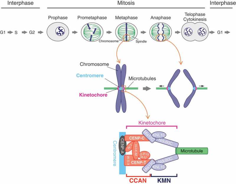
The kinetochore is a large protein complex that ensures accurate chromosome segregation during mitosis by connecting the centromere and spindle microtubules. One of the kinetochore sub-complexes, the constitutive centromere-associated network (CCAN), associates with the centromere and recruits another sub-complex, the KMN (KNL1, Mis12, and Ndc80 complexes) network (KMN), which binds to spindle microtubules. The CCAN-KMN interaction is mediated by two parallel pathways (CENP-C- and CENP-T-pathways) in the kinetochore, which bridge the centromere and microtubules. Here, we discuss dynamic protein-interaction changes in the two pathways that couple the centromere with spindle microtubules during mitotic progression.

Keywords: CCAN; KMN; Kinetochore; mitosis.


Figures

Figure 1.
Figure 1.
Chromosome segregation and the kinetochore architecture in vertebrate mitosis. The duplicated genome in the S-phase is segregated equally to the next generation during mitosis. The chromosomes begin condensation in late G2 to prophase. In subsequent prometaphase to metaphase, the condensed chromosomes, which are captured by the spindle microtubules, are aligned along the equator of the cell. The microtubules attach to the kinetochore, which is formed on the centromere specified by histone H3 variant CENP-A. The kinetochore harnesses the pulling-force from the microtubules, bringing the segregated chromosomes (sister chromatids) to the opposite poles of the spindle during anaphase. The main architecture of the kinetochore is built with the constitutive centromere-associated network (CCAN) composed of 16 protein subunits and the KMN (KNL1, Mis12, and Ndc80 complexes: KNL1C, Mis12C, Ndc80C) network (KMN). CCAN and KMN interact with the centromere and spindle microtubule, respectively, making a linkage between the centromere and microtubules.
Figure 2.
Figure 2.
Two-pathways model connecting the centromere to spindle microtubules. (a) Schematic representation of human CENP-C and CENP-T. Human CENP-C interacts with various centromere/kinetochore proteins. The Mis12 complex (Mis12C)-binding region in the extreme N-terminus binds to KMN, which associates with the spindle microtubules. The PEST-rich region interacts with the CCAN sub-complexes, CENP-HIKM, and CENP-LN. There are two CENP-A nucleosome-binding regions: central domain and CENP-C motif. The dimerization domain in the extreme C-terminus is thought to promote self-dimerization. Human CENP-T directly binds to the Ndc80 complex (Ndc80C) through the extreme N-terminus (Ndc80C-binding region). Mis12C interacts to the Mis12C-binding region next to the Ndc80C-binding region (Mis12C-binding region). There is a histone fold domain in the C-terminus, which forms a nucleosome like complex with CENP-W, -S and -X, which also contain histone fold domains. The complex interacts with the centromere chromatin. (b) Two pathways in the kinetochore that bridge the centromere to microtubules: CENP-C- and CENP-T-pathways. Both CENP-C and CENP-T bind to KMN via their N-terminus and centromere chromatin via their C-terminus, independently, making the two pathways in the kinetochore.
Figure 3.
Figure 3.
CENP-T forms a major pathway for linking the centromere and microtubules in chicken DT40 cells. (a) Schematic representation of chicken CENP-C. All functional domains in human CENP-C are conserved in chicken full-length CENP-C (FL), with exception of the central domain which is one of CENP-A binding domains in human CENP-C. A CENP-C mutant in which the Mis12 complex (Mis12C)-binding region is deleted (∆73) is able to complement CENP-C deficiency in chicken DT40 cells, suggesting that Mis12C-binding of CENP-C is dispensable for proper chromosome segregation in DT40 cells. (b) Schematic representation of chicken CENP-T. Chicken CENP-T has the Ndc80 complex- (Ndc80C) and Mis12C-binding regions as well as the histone fold domain in human CENP-T. CENP-T deficiency in DT40 cells are complemented by full-length CENP-T (FL), but not by CENP-T mutants lacking the Ndc80C- or Mis12C-binding region (∆90 or ∆120–240, respectively), indicating that those regions are essential for viability of DT40 cells. (c) Dynamic changes in the CCAN–KMN interaction making CENP-T the major pathway. Aurora B kinase phosphorylates Mis12C, facilitating interaction between CENP-C and Mis12C, which brings KNL1 complex (KNL1C), at onset of mitosis (late G2/prophase). Cdk1 phosphorylates CENP-T to promote stable binding of Mis12C and Ndc80C to the CENP-T-pathway. Cdk1 phosphorylation on Mis12C reduces its affinity to Ndc80C. When the pulling force is applied on the kinetochore from the microtubules, Mis12C could be spatially separated from Aurora B kinase, and its Aurora B phosphorylation is likely to be dephosphorylated by protein phosphatase 1 (PP1). These phospho-regulatory mechanisms result in dynamic changes in the CCAN–KMN interaction during mitotic progression. Consequently, the major fraction of Ndc80C interacts with the CENP-T-pathway, making it the major linkage between the centromere and spindle microtubules.
Figure 4.
Figure 4.
Kinetochore-protein interactions controlled by various phospho-regulations. (a) Model for CENP-C and Mis12 complex (Mis12C) interaction. Mis12C consists of Mis12, Dsn1, Nsl1, and Nnf1 (also known as PMF1). The interaction between CENP-C and Mis12C is inhibited by the basic domain of Dsn1, which masks the CENP-C-binding interface in Mis12C. Aurora B kinase phosphorylates the basic domain to release the inhibition, promoting stable binding of CENP-C to Mis12C. (b) Model for CENP-T interaction with the Ndc80 complex (Ndc80C) and Mis12C. CENP-T has the Mis12C- and Ndc80C-binding regions, and Cdk1 phosphorylates these regions and increases their binding affinity to Mis12C and Ndc80C, respectively. The Ndc80C-CENP-T interaction is an upstream event of the Mis12C-CENP-T interaction. (c) Model for the Mis12C-Ndc80C interaction. Ndc80C consists of Ndc80, Nuf2, Spc24, and Spc25. The globular domain of the Spc24-Spc25 sub-complex (Spc24-Spc25) interacts with Nsl1 and Dsn1 in Mis12C. Nsl1 interacts with Spc24-Spc25 through the PVIHL motif in its C-terminus. The Dsn1 C-terminus has the Ndc80C-binding region. Multivalent interaction surfaces seem to contribute to stable interaction between Mis12C and Ndc80C. Cdk1 phosphorylation in the Ndc80C-binding region of Dsn1 reduces its affinity to Spc24-Spc25, resulting in unstable interaction between Mis12C and Ndc80C.
Figure 5.
Figure 5.
Divergence among species of pathways for linkage between centromere and microtubules. The major linkage pathway is varied among species. Vertebrates have two pathways for linking between the centromere and microtubules: CENP-C- and CENP-T-pathways, and the CENP-T-pathway is the major pathway in chicken (Gallus gallus) DT40 cells. There are three pathways in the budding yeast (S. cerevisiae): MIF2 (a CENP-C homolog)-, CNN1 (a CENP-T homolog)- and AME1 (a CENP-U homolog)-pathways, and the AME1-pathway is the major pathway in the yeast. Since CENP-C is the only CCAN in the fruit fly (D. melanogaster), the CENP-C-pathway forms a key bridge between the centromere and microtubules in the species.

Similar articles

Cited by

References

    1. Cheeseman IM. The kinetochore. Cold Spring Harb Perspect Biol. 2014. July 1;6(7):a015826–a015826. - PMC - PubMed
    1. Musacchio A, Desai A.. A molecular view of kinetochore assembly and function. Biology (Basel). 2017. January 24;6(1):5. - PMC - PubMed
    1. Fukagawa T, Earnshaw WC. The centromere: chromatin foundation for the kinetochore machinery. Dev Cell. 2014. September 8;30(5):496–508. - PMC - PubMed
    1. Musacchio A. The molecular biology of spindle assembly checkpoint signaling dynamics. Curr Biol. 2015. October 19;25(20):R1002–1018. - PubMed
    1. Sacristan C, Kops GJPL. Joined at the hip: kinetochores, microtubules, and spindle assembly checkpoint signaling. Trends Cell Biol. 2015. January;25(1):21–28. - PubMed

Publication types

Grants and funding

This work was supported by Japan Society for the Promotion of Science, KAKENHI Grant Numbers 15H05972, 16H06279, and 17H06167 to TF, Japan Society for the Promotion of Science, KAKENHI Grant Number 16K18491 to MH.