. 2019 May 9;15(5):e1007736.
doi: 10.1371/journal.ppat.1007736. eCollection 2019 May.

A novel interaction between dengue virus nonstructural protein 1 and the NS4A-2K-4B precursor is required for viral RNA replication but not for formation of the membranous replication organelle

Affiliations

A novel interaction between dengue virus nonstructural protein 1 and the NS4A-2K-4B precursor is required for viral RNA replication but not for formation of the membranous replication organelle

Anna Płaszczyca et al. PLoS Pathog. .

Abstract

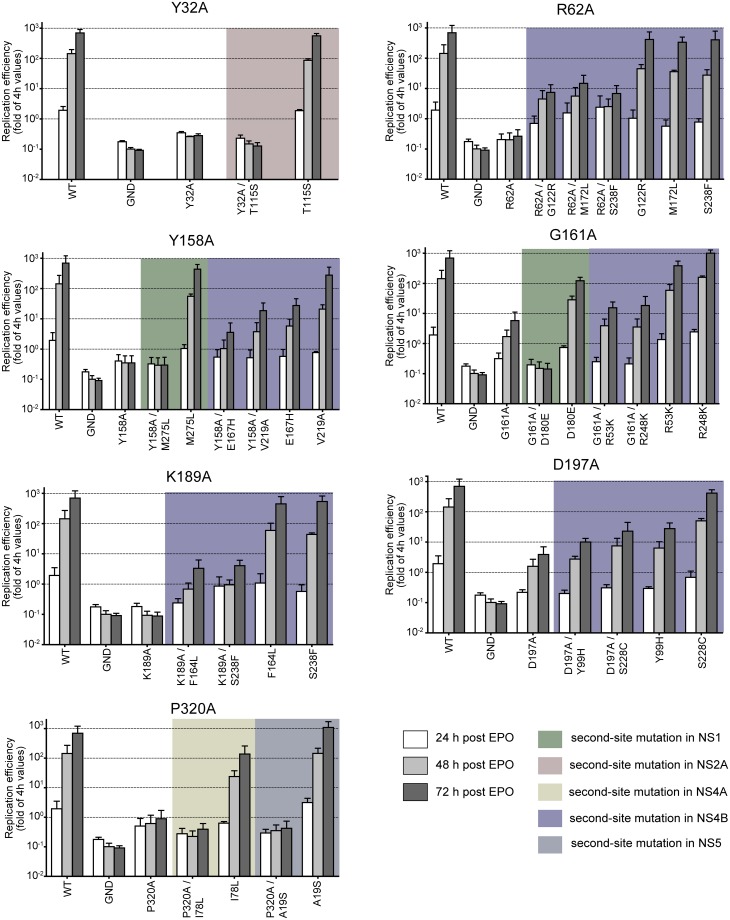
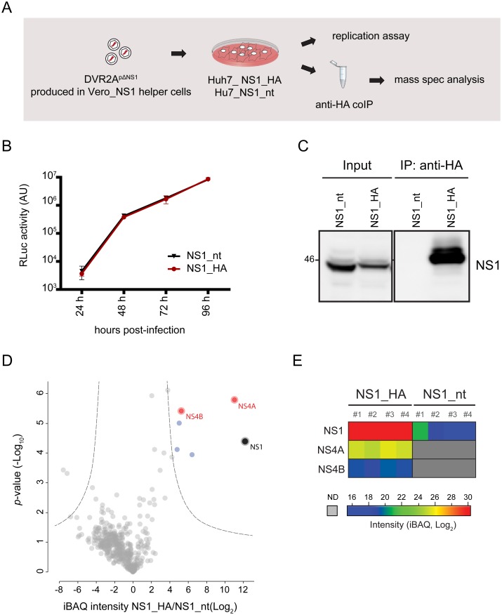
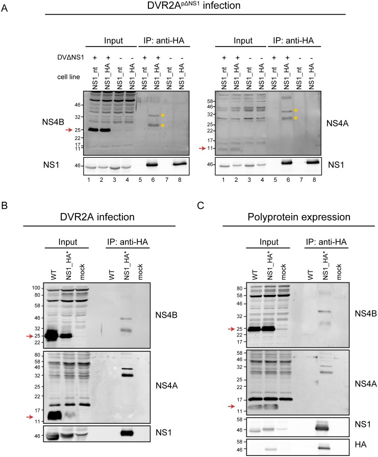
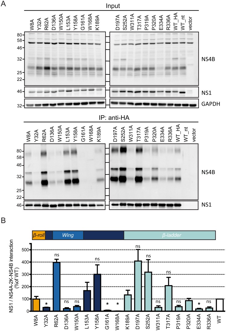
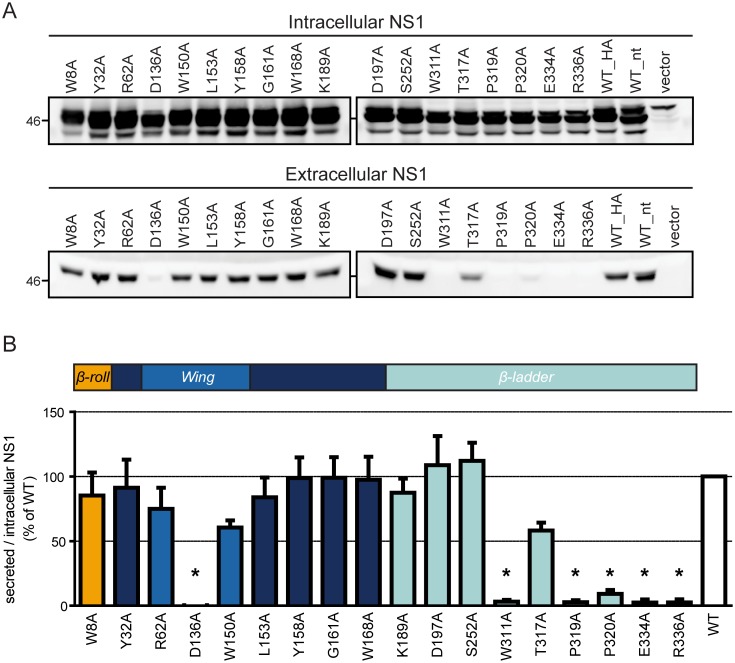
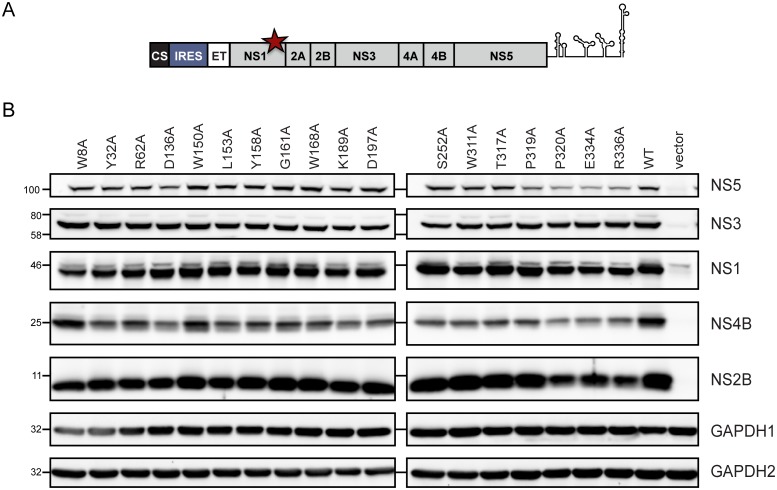
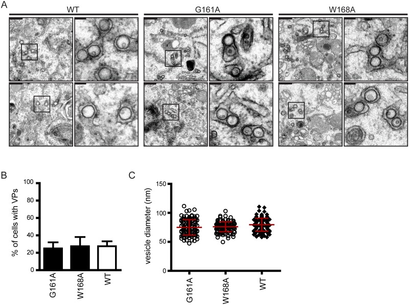
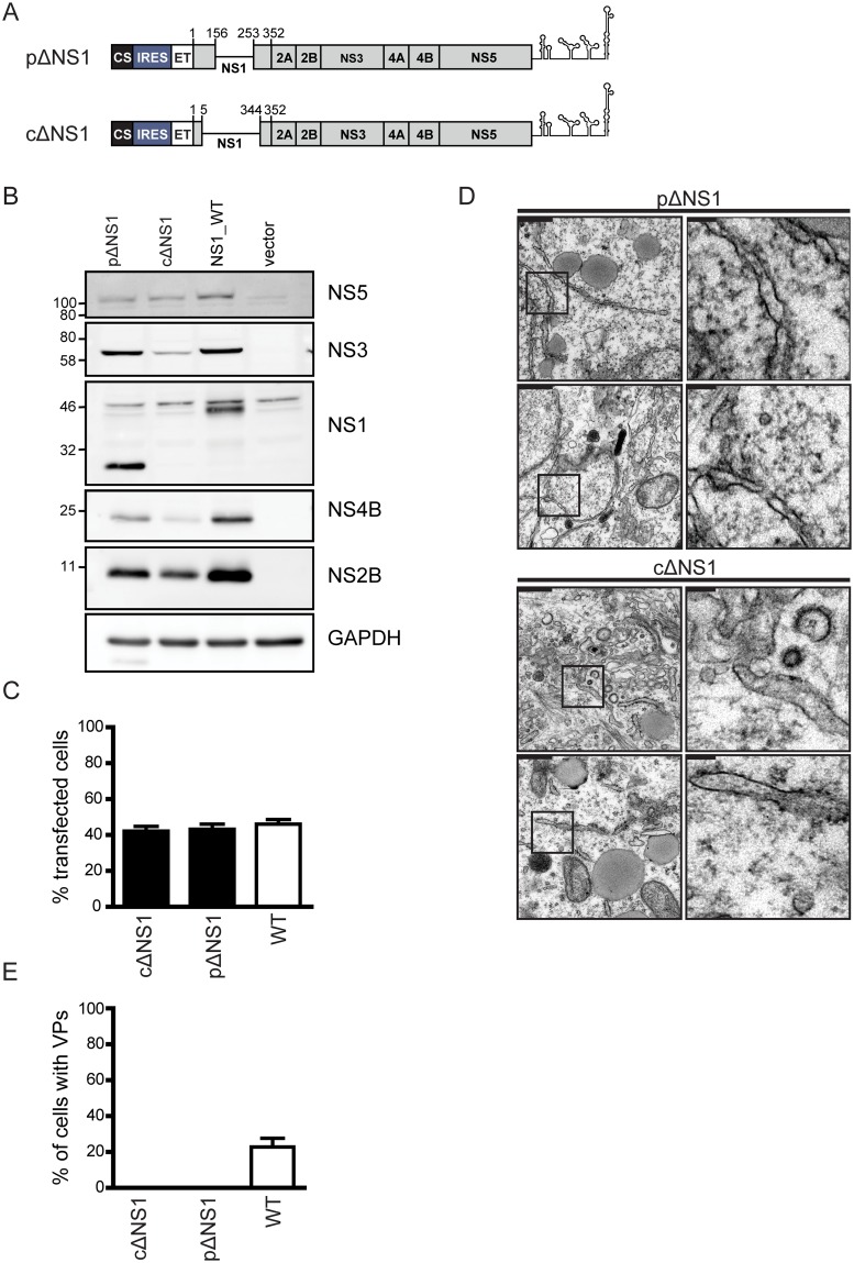
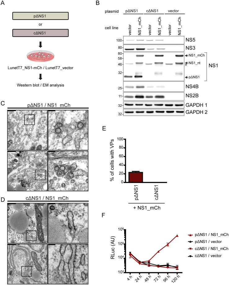
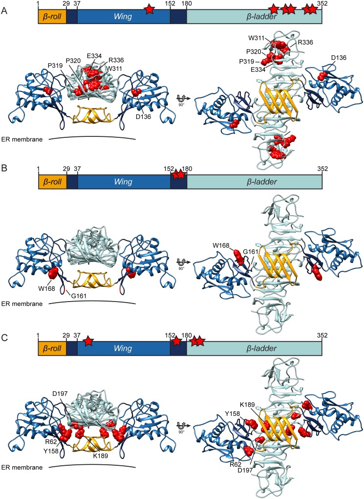
Dengue virus (DENV) has emerged as major human pathogen. Despite the serious socio-economic impact of DENV-associated diseases, antiviral therapy is missing. DENV replicates in the cytoplasm of infected cells and induces a membranous replication organelle, formed by invaginations of the endoplasmic reticulum membrane and designated vesicle packets (VPs). Nonstructural protein 1 (NS1) of DENV is a multifunctional protein. It is secreted from cells to counteract antiviral immune responses, but also critically contributes to the severe clinical manifestations of dengue. In addition, NS1 is indispensable for viral RNA replication, but the underlying molecular mechanism remains elusive. In this study, we employed a combination of genetic, biochemical and imaging approaches to dissect the determinants in NS1 contributing to its various functions in the viral replication cycle. Several important observations were made. First, we identified a cluster of amino acid residues in the exposed region of the β-ladder domain of NS1 that are essential for NS1 secretion. Second, we revealed a novel interaction of NS1 with the NS4A-2K-4B cleavage intermediate, but not with mature NS4A or NS4B. This interaction is required for RNA replication, with two residues within the connector region of the NS1 "Wing" domain being crucial for binding of the NS4A-2K-4B precursor. By using a polyprotein expression system allowing the formation of VPs in the absence of viral RNA replication, we show that the NS1 -NS4A-2K-4B interaction is not required for VP formation, arguing that the association between these two proteins plays a more direct role in the RNA amplification process. Third, through analysis of polyproteins containing deletions in NS1, and employing a trans-complementation assay, we show that both cis and trans acting elements within NS1 contribute to VP formation, with the capability of NS1 mutants to form VPs correlating with their capability to support RNA replication. In conclusion, these results reveal a direct role of NS1 in VP formation that is independent from RNA replication, and argue for a critical function of a previously unrecognized NS4A-2K-NS4B precursor specifically interacting with NS1 and promoting viral RNA replication.


Conflict of interest statement

The authors have declared that no competing interests exist.

Figures

Fig 1
Fig 1. A forward genetic screen identifies pseudoreversions in NS4A and NS4B compensating replication-inactivating mutations in NS1.
(A) Homology model of the NS1 dimer based on the DENV NS1 structure (PDB 4O6B) with missing residues modelled according to the ZIKV NS1 structure (PDB 5K6K). The model was built using the MOE 2015 software package and molecular graphics were prepared with UCSF Chimera [66]. Wing, β-ladder and β-roll domains are shown in blue, turquoise and orange, respectively. The connector subdomains in Wing domain are shown in dark blue. The membrane is indicated with a horizontal line. (B) Left panel: schematic of the experimental approach used to select for pseudoreversions rescuing replication of NS1 mutants. Point mutations in NS1 were inserted into a selectable subgenomic DENV replicon (sgDVH2A) encoding a hygromycin phosphotransferase gene (HygroR) downstream of by the cyclisation sequence residing in the capsid-coding region (CS) and upstream of the 2A protease of Thosea asigna virus (2AP) and the signal sequence of NS1 corresponding to the last 24 amino acid residues of E (ET). VeroE6 cells were electroporated with selectable replicon RNAs containing given mutations and cultured in the presence of hygromycin B. After three to four weeks, single cell clones were propagated; total RNA was extracted and amplified for sequence analysis. Right panel: number of hygromycin B-resistant cell colonies obtained after selection for each primary mutation in NS1 (indicated at the bottom) with the color of the bar corresponding to the color of the NS1 domain into which the respective mutation was introduced. (C) Schematic summarizing the localization of second site mutations in the NS4A-2K-NS4B polyprotein that is drawn according to its hypothetical membrane topology [7,36]. The circle color of primary mutations in NS1 (lower left) corresponds to pseudoreversions identified for each respective mutant.
Fig 2
Fig 2. Partial rescue of RNA replication of NS1 mutants by pseudoreversions residing in NS4B.
Point mutations in NS1 were inserted alone or together with mutations specified in the bottom of each panel into the full length genome of the DENV Renilla luciferase (RLuc) reporter virus DVR2A. VeroE6 cells were electroporated with in vitro transcripts derived from each construct. Cells were lysed at indicated time points after electroporation and RLuc activity was measured to quantify viral RNA replication. For each construct, values were normalized to the 4 h-value to account for differences in transfection efficiency. Results shown are mean values from three independent experiments performed in duplicates with two independent RNA preparations; error bars indicate SD.
Fig 3
Fig 3. Identification of viral proteins interacting with NS1.
(A) Schematic of the experimental approach. Cells stably expressing NS1 were infected with DVR2A containing an in-frame deletion in NS1 (DVR2ApΔNS1) and processed to measure viral RNA replication or for affinity purification of HA-tagged NS1 and subsequent analysis of protein complexes by mass spectrometry. (B) DVR2ApΔNS1 replication in Huh7 cells stably expressing C-terminally HA-tagged or non-tagged (nt) NS1. Cells were infected with DVR2ApΔNS1 (MOI = 1) and lysed at indicated times points post-infection. Viral replication was measured by luciferase assay. (C) Huh7 cells stably expressing NS1_nt or NS1_HA were infected with DVR2ApΔNS1 (MOI = 1). Seventy-two hours post infection cells were collected and subjected to HA-specific pull-down. Immune precipitated complexes were analyzed by western blot using a NS1-specific rabbit antiserum. (D) Immune purified complexes prepared as in (C) were subjected to mass spectrometry analysis. Four independent affinity purifications were performed for each bait. Shown is a volcano plot displaying the average degree of enrichment by NS1_HA over NS1_nt (ratio of Intensity-Based Absolute Quantification [iBAQ] protein intensities) and the P value (Student’s t-test) for each protein. Significantly enriched proteins are separated from background proteins by a hyperbolic curve (dotted grey line). Viral and host proteins specifically binding to NS1_HA are represented as red and blue dots, respectively. (E) Heat map showing non-imputed log2-transformed iBAQ intensities for each individual replicate (see color scale). Only the bait protein and the viral interaction partners are depicted. Gray color represents missing values (not determined [ND]).
Fig 4
Fig 4. The NS4A-2K-4B precursor polyprotein is the main interaction partner of NS1.
(A) Huh7 cells stably expressing carboxy-terminally HA tagged or non-tagged (nt) NS1 were infected with the DVR2ApΔNS1 virus (MOI = 1) or mock infected and cell lysates prepared 72 h after infection were subjected to immunoprecipitation (IP) using beads coated with HA-specific antibodies as shown in Fig 3A. Samples were analyzed by western blot using NS4B-, NS4A- or NS1-specific antisera. Mature forms of NS4B and NS4A migrate with an apparent molecular weight of ~25 kDa and ~11 kDa, respectively (indicated by red arrows on the left side of each panel). The NS4A-2K-4B precursor bands migrate at ~30 kDa and ~35 kDa and are highlighted with yellow dots. (B) Huh7 cells were infected with the DVR2A wildtype (WT) or the DVR2A variant encoding an internally HA-tagged NS1 (NS1_HA*) and cell lysates prepared 72 h post infection were subjected to anti-HA immunoprecipitation followed by western blot as described in (A). (C) Huh7-Lunet_T7 cells were transfected with an NS1 to 5 polyprotein expression construct encoding either wildtype or internally HA-tagged NS1 under control of the T7 RNA polymerase promoter. Cells were lysed 20 h post transfection and lysates were subjected to anti HA immunoprecipitation as described in (A).
Fig 5
Fig 5. Characterization of the interaction between NS1 and the NS2A-2K-4B cleavage intermediate.
(A) Schematic of the experimental approach. (B) Huh7 cells stably expressing the T7 RNA polymerase and DENV NS2B-3 (with protease) or only the T7 polymerase (w/o protease) were co-transfected with plasmids encoding carboxy-terminally HA-tagged or non-tagged NS1 and NS4A and/or NS4B constructs indicated above each lane. Sixteen hours post transfection cell lysates were prepared and subjected to anti-HA immunoprecipitation. Captured protein complexes were analyzed by immunoblotting using rabbit sera reacting with DENV proteins indicated on the right.
Fig 6
Fig 6. Two residues in the connector region of the Wing domain are essential for NS1 interaction with the NS4A-2K-4B precursor.
(A) Huh7 cells stably expressing the T7 RNA polymerase and proteolytically active DENV NS2B-3 were co-transfected with constructs encoding HA-tagged wildtype or mutated NS1 and the NS4A-2K-4B polyprotein construct. Cell lysates were processed as described in Fig 4B and immunoblots were probed with NS1- and NS4B-specific antisera. A representative result of four independent experiments is shown. (B) Quantification of NS1 and NS4B-specific signals from all four experiments. In the case of NS4B, the signals of the two bands at ~30 and ~35 kDa were added and used to calculate pull down efficiency. Cells expressing non-tagged NS1 were used to determine the background of the assay that was subtracted from the NS1_HA values. Bars represent the means of the NS4B/NS1 signal ratio, normalized to the wildtype, from four independent experiments. Error bars indicate SEM. *, p<0,002. In all other cases, the difference to the wildtype was not significant (ns).
Fig 7
Fig 7. A cluster of conserved amino acid residues in the carboxy-terminal β-ladder of NS1 is essential for NS1 secretion.
(A) Huh7 cells stably expressing the T7 RNA polymerase were transfected with plasmids encoding HA-tagged NS1 that contained point mutations specified above each lane. Sixteen hours later cell culture supernatants were collected and cleared by centrifugation. Equal volumes of supernatants and cell lysates were analyzed by immunoblotting using an NS1-specific antiserum. (B) Quantification of NS1 signals from (A). The bars are means of the ratio of secreted to intracellular NS1, normalized to the wildtype, from three independent experiments. Error bars indicate SEM. *, p<0,002. In all other cases, the difference to the wildtype was not significant.
Fig 8
Fig 8. Replication-blocking mutations in NS1 do not impact polyprotein processing.
(A) A schematic of the expression cassette used to produce the DENV-2 polyprotein. (B) Huh7-Lunet T7 cells were transfected with the polyprotein expression constructs harboring mutations in NS1 specified above each lane. Eighteen hours post transfection cells were lysed and processed for western blot by using antisera specific to DENV proteins given in the right of each panel. GAPDH was used as loading control. GAPDH 1 corresponds to membranes probed for NS1, NS2B and NS5; GAPDH 2 for membranes probed with NS3 and NS4B. A representative result of three independent experiments is shown.
Fig 9
Fig 9. Formation of DENV vesicle packets is independent from the interaction between NS1 and the NS4A-2K-4B precursor.
Huh7-Lunet T7 cells were transfected with the DENV polyprotein expression construct shown in Fig 8A and 18 h later, cells were fixed and processed for transmission electron microscopy. (A) Representative images of membrane invaginations observed in cells transfected with the wildtype (WT) polyprotein, the G161A or the W168A mutant, respectively. Scale bars (upper left of each panel) correspond to 500 nm in the overview and 100 nm in the cropped sections that are indicated with black rectangles in the overviews. (B) Quantification of the number of cells containing VPs. Results show the mean of two independent experiments, counting at least 20 cells per construct and experiment. The error bars indicate SD. Transfection efficiency as determined by immunofluorescence was ~45%. (C) Quantitation of the vesicle diameter in cells transfected with WT, G161A or W168A polyprotein construct. Scatter plots indicate the diameter of >100 vesicles from two independent experiments; horizontal lines indicate means and error bars indicate SD.
Fig 10
Fig 10. NS1 is essential for the formation of vesicle packets.
(A) Schematic representation of the expression constructs containing a partial deletion in NS1 (pΔNS1) or lacking NS1 completely (cΔNS1). Huh7-Lunet_T7 cells were transfected with pΔNS1, cΔNS1 or the wildtype (WT) construct, fixed 18 h post transfection and processed for electron microscopy, western blot or immunofluorescence. (B) Western blot of cell lysates prepared 18 h post transfection and analyzed by using antibodies indicated on the right. A representative experiment of three repetitions is shown. (C) Transfection efficiency of Huh7-Lunet_T7 cells as determined by immunofluorescence. Data are the mean from three independent experiments using two independent plasmid preparations and counting each time at least 200 cells per sample. The error bars indicate the SEM. (D) Representative transmission electron microscopy images of Huh7-Lunet_T7 cells transfected with expression constructs containing a partial or complete NS1 deletion. Scale bars (upper left of each panel) correspond to 500 nm in the overview and 100 nm in the cropped sections that are indicated with black rectangles in the overviews. (E) Quantification of the EM analysis. The percentage of cells with VPs is shown. Note the absence of regular VPs in cells expressing either NS1 deletion mutant. Data are based on 4 independent experiments, using two independent plasmid preparations and counting at least 20 cells per construct and per repetition.
Fig 11
Fig 11. Rescue of vesicle packet formation by NS1 provided in trans.
(A) Huh7-Lunet_T7 cells stably expressing NS1 with a carboxy-terminal mCherry tag (NS1_mCh) or the empty vector were transfected with pΔNS1, cΔNS1 or the wildtype (WT) polyprotein expression constructs. Cells were harvested 18 h post-transfection and processed for western blot and EM analysis. (B) Expression of DENV proteins in transfected Huh7-Lunet_T7 cells. Black arrows indicate NS1 variants; the star indicates an unspecific background signal. GAPDH was used as loading control. GAPDH 1 corresponds to membranes probed for NS1, NS2B and NS5; GAPDH 2 for membranes probed with NS3 and NS4B. (C) Representative electron micrographs of VPs in cells expressing NS1-mCh and transfected with the pΔNS1 polyprotein expression construct. (D) Absence of VPs in NS1_mCh expressing cells transfected with the cΔNS1 polyprotein expression construct. Scale bars (upper left of each panel) in (C) and (D) correspond to 500 nm in the overview and 100 nm in the cropped sections that are indicated with black rectangles in the overviews. (E) Quantification of the number of cells containing VPs. Data are mean from 2 independent experiments, counting at least 20 cells per condition; error bar indicates SD. (F) Replication of DVR2A containing a partial (pΔNS1) or complete (cΔNS1) deletion of NS1 in Huh-7-Lunet_T7 cells expressing NS1_mCh or control cells (stably transduced with the empty vector). Cells were transfected with in vitro transcribed RNA derived from the respective construct by electroporation, lysed at indicated time points post transfection and RLuc activity was measured to quantify viral RNA replication. Results shown are mean values from two independent experiments performed in triplicates; error bars indicate SEM.
Fig 12
Fig 12. Overview of amino acid residues in NS1 required for NS1 secretion, interaction with the NS4A-2K-4B precursor and RNA replication.
Replication-impairing mutations that (A) abolish NS1 secretion, (B) abrogate binding between NS1 and the NS4A-2K-4B precursor, and (C) can be complemented by second site mutations in NS4B. Upper panels show the linear map of NS1 with mutated residues indicated by red stars; bottom panels show the homology model of the 3D structure of NS1 based on PDB entries 4O6B and 5K6K as described in Fig 1(A) with mutated residues shown as van der Waal spheres in red. Wing, β-ladder and β-roll domains are shown in blue, turquoise and orange, respectively. Connector subdomains in Wing domain are shown in dark blue.

Similar articles

Cited by

References

    1. Bhatt S, Gething PW, Brady OJ, Messina JP, Farlow AW, Moyes CL, Drake JM, Brownstein JS, Hoen AG, Sankoh O, Myers MF, George DB, Jaenisch T, Wint GR, Simmons CP, Scott TW, Farrar JJ, Hay SI (2013) The global distribution and burden of dengue. Nature 496: 504–507. 10.1038/nature12060 - DOI - PMC - PubMed
    1. World Health Organization (2016) Dengue vaccine: WHO position paper—July 2016. Wkly Epidemiol Rec 91: 349–364. - PubMed
    1. Acosta EG, Kumar A, Bartenschlager R (2014) Revisiting dengue virus-host cell interaction: new insights into molecular and cellular virology. Adv Virus Res 88:1–109. 10.1016/B978-0-12-800098-4.00001-5: 1–109. - DOI - PubMed
    1. Welsch S, Miller S, Romero-Brey I, Merz A, Bleck CK, Walther P, Fuller SD, Antony C, Krijnse-Locker J, Bartenschlager R (2009) Composition and three-dimensional architecture of the dengue virus replication and assembly sites. Cell Host Microbe 5: 365–375. 10.1016/j.chom.2009.03.007 - DOI - PMC - PubMed
    1. Junjhon J, Pennington JG, Edwards TJ, Perera R, Lanman J, Kuhn RJ (2014) Ultrastructural characterization and three-dimensional architecture of replication sites in dengue virus-infected mosquito cells. J Virol 88: 4687–4697. 10.1128/JVI.00118-14 - DOI - PMC - PubMed

Publication types

MeSH terms

Substances

Grants and funding

This project was supported by the Deutsche Forschungsgemeinschaft (SFB1129, TP11 and BA1505/8-1, both to R.B. and TRR179 TP11 to A.Pi.). A.Pl. was funded via the European Union Horizon 2020 Marie Sklodowska-Curie ETN ‘ANTIVIRALS’, grant agreement number 642434. C.J.N was funded by a European Molecular Biology Organization (EMBO) Long-Term Fellowship (ALTF 466-2016). A.Pi. was funded by an ERC consolidator grant (ProDAP 817798). The funders had no role in study design, data collection and analysis, decision to publish, or preparation of the manuscript.