Review
. 2019 Jun 1;126(6):1782-1789.
doi: 10.1152/japplphysiol.00676.2018. Epub 2019 May 9.

Passive force enhancement in striated muscle

Affiliations
Review

Passive force enhancement in striated muscle

Walter Herzog. J Appl Physiol (1985). .

Abstract

Passive force enhancement is defined as the increase in passive, steady-state, isometric force of an actively stretched muscle compared with the same muscle stretched passively to that same length. Passive force enhancement is long lasting, increases with increasing muscle length and increasing stretch magnitudes, contributes to the residual force enhancement in skeletal and cardiac muscle, and is typically only observed at muscle lengths at which passive forces occur naturally. Passive force enhancement is typically equal to or smaller than the total residual force enhancement, it persists when a muscle is deactivated and reactivated, but can be abolished instantaneously when a muscle is shortened quickly from its stretched length. There is strong evidence that the passive force enhancement is caused by the filamentous sarcomeric protein titin, although the detailed molecular mechanisms underlying passive force enhancement remain unknown. Here I propose a tentative mechanism based on experimental evidence that associates passive force enhancement with the shortening of titin's free spring length in the I-band region of sarcomeres. I suggest that this shortening is accomplished by titin binding to actin and that the trigger for titin-actin interactions is associated with the formation of strongly bound cross bridges between actin and myosin that exposes actin attachment sites for titin through movement of the regulatory proteins troponin and tropomyosin.

Keywords: cross bridge theory; residual force enhancement; sarcomere nonuniformity; sliding filament theory; titin.


Conflict of interest statement

No conflicts of interest, financial or otherwise, are declared by the author.

Figures

Fig. 1.
Fig. 1.
Example of force as a function of time for 3 different contractile conditions for a cat soleus muscle (35°C). Bottom trace (at 4 s) is a passive stretch of the soleus, where stretching occurs between ~2.5 and 3.5 s. Middle trace (at 4 s) represents an isometric contraction at the stretched length. Finally, top trace (at 4 s) represents stretching of the activated muscle in the same manner as the passive stretch (same magnitude and same speed of stretching). FE indicates the residual force enhancement, which is the increase in steady-state isometric force after active muscle stretching compared with the corresponding (same length, same activation) isometric force for a purely isometric contraction. PFE indicates the passive force enhancement, which corresponds to the increase in steady-state passive force following stretching of an activated muscle compared with the same stretching of the passive muscle.
Fig. 2.
Fig. 2.
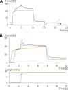
Force-time and muscle length change-time histories of cat soleus muscle for a variety of experimental conditions (at 35°C). A: passive force enhancement is long lasting but can be abolished instantaneously by a shortening-stretch cycle (at ~15 s) that brings the muscle to its prestretch length in the shortening phase and back to its original stretched length in the stretch phase. i refers to the isometric reference contraction; 9 refers to the muscle that was stretched by 9 mm. B: passive force enhancement (like the total residual force enhancement) increases with increasing stretch magnitudes. The 9-mm stretch of the activated soleus muscle produces more force enhancement and passive force enhancement (difference between yellow and red lines at 12 s) than the 3-mm stretch (difference between yellow and blue lines).
Fig. 3.
Fig. 3.
Schematic illustration of a sarcomere with Z band, actin, myosin, and titin filaments in their supposed arrangement. Titin runs from the middle of the sarcomere (M line) in either direction toward the Z band. Titin is thought to be firmly attached to myosin and then run freely across the I-band region of the sarcomere until ~50–100 nm away from the Z band, where it is thought to attach to actin and then enter the Z band bound to actin. Below the schematic sarcomere are illustrations of the structure of titin in cardiac muscle and in 2 selected skeletal muscles (psoas and soleus). Note that cardiac titin isoforms are much smaller than those of skeletal muscles and that skeletal muscles have different isoforms that are thought to reflect the passive requirements of the muscle. Specific segments (N2B, N2A, N2BA, and PEVK) are indicated. The brownish rectangles represent immunoglobulin domains. Adapted from Granzier and Labeit (16) with permission from Wolters Kluwer Health, Inc.
Fig. 4.
Fig. 4.
Peak force required to unfold immunoglobulin (Ig) domains in the absence (Control) and presence (Calcium) of calcium, simulating activation of a muscle. Note the substantially increased force required to unfold and stretch immunoglobulin domains in the presence of calcium. Only the first 5 (of 8) immunoglobulin domains are shown, and only results where at least 5 domains unfolded were included in this analysis. Unfolding of 6 or more immunoglobulin domains was rare, and thus the number of observations was deemed inappropriate for statistical testing. Unfolding of 5 of the 8 immunoglobulin domains was observed in >300 individual test specimens for each of the calcium and the control conditions. Adapted from DuVall et al. (9) with permission from Springer Nature.
Fig. 5.
Fig. 5.
Titin segmental elongations as a function of sarcomere lengths. Titin is divided into a proximal (close to the Z band) and a distal (close to myosin) segment by a fluorescently labeled antibody in the PEVK domain. For passive stretching (not shown), the distal and proximal segments elongate continuously when sarcomeres are stretched and sarcomere length increases. During active stretching shown here, the proximal segment of titin only elongates for part of the sarcomere stretching and then stops elongating. This occurs at a sarcomere length of ~3.0 µm for the particular myofibril shown here as an example from >120 experiments. In other myofibrils, titin proximal segment elongation stopped occurring at sarcomere lengths ranging from 2.2 to 3.4 µm, and this occurrence seemed to depend crucially on the sarcomere lengths at which myofibril activation occurred. When the proximal segment stops elongating, all sarcomere elongation is taken up by the distal segment of titin. I interpret this result as showing that titin binds to actin during active stretching. This binding of titin to actin does not occur through activation (calcium release) but seems to require cross-bridge binding to actin. Therefore, this phenomenon is independent of calcium and is thought to persist when the muscle is deactivated and calcium is sequestered back into the sarcoplasmic reticulum of muscle fibers.
Fig. 6.
Fig. 6.
Proposed mechanism for the passive force enhancement property of skeletal muscle. In the initial condition, the sarcomere is short and in the passive state (A). If stretched passively from that initial configuration, the sarcomere will become longer and all titin segments are stretched and produce a force in accordance with the titin properties (B). If the sarcomere is activated via calcium but cross-bridge binding to actin is inhibited (for example, by 2,3-butanedione monoxime or by troponin C deletion) and then stretched, calcium will bind to titin (indicated by the brownish color in C) but titin will not bind to actin. Titin is stiffer in this configuration because of the bound calcium. If the sarcomere is stretched while activated and cross-bridge forces are allowed to develop (normal activation and force production, eccentric muscle action), titin will bind calcium and will bind to actin (I propose somewhere in the PEVK region), and will produce more force than in C, because of the titin binding to actin that is made possible by the cross-bridge binding to actin (D). When the actively stretched muscle (in which cross bridges were allowed to bind to actin) is now deactivated, calcium is released from titin and titin will remain bound to actin, thereby producing the passive force enhancement observed in skeletal muscles after stretching of an activated muscle (E). If now, in the deactivated state, the muscle is quickly shortened to its initial length and immediately stretched back to its original length (indicated by double arrow in F), titin is released from actin and the passive force enhancement is immediately abolished, while without this quick shortening-stretch cycle, the passive force enhancement would persist for minutes.

Similar articles

Cited by

References

    1. Abbott BC, Aubert XM. The force exerted by active striated muscle during and after change of length. J Physiol 117: 77–86, 1952. - PMC - PubMed
    1. Astier C, Raynaud F, Lebart MC, Roustan C, Benyamin Y. Binding of a native titin fragment to actin is regulated by PIP2. FEBS Lett 429: 95–98, 1998. doi:10.1016/S0014-5793(98)00572-9. - DOI - PubMed
    1. Avrova SV, Shelud’ko NS, Borovikov YS, Galler S. Twitchin of mollusc smooth muscles can induce “catch”-like properties in human skeletal muscle: support for the assumption that the “catch” state involves twitchin linkages between myofilaments. J Comp Physiol B 179: 945–950, 2009. doi:10.1007/s00360-009-0375-z. - DOI - PubMed
    1. Bartoo ML, Linke WA, Pollack GH. Basis of passive tension and stiffness in isolated rabbit myofibrils. Am J Physiol Cell Physiol 273: C266–C276, 1997. doi:10.1152/ajpcell.1997.273.1.C266. - DOI - PubMed
    1. Bianco P, Nagy A, Kengyel A, Szatmári D, Mártonfalvi Z, Huber T, Kellermayer MS. Interaction forces between F-actin and titin PEVK domain measured with optical tweezers. Biophys J 93: 2102–2109, 2007. doi:10.1529/biophysj.107.106153. - DOI - PMC - PubMed

Publication types

Substances

LinkOut - more resources