Flexible-to-rigid transition is central for substrate transport in the ABC transporter BmrA from Bacillus subtilis
- PMID: 31044174
- PMCID: PMC6488656
- DOI: 10.1038/s42003-019-0390-x
Flexible-to-rigid transition is central for substrate transport in the ABC transporter BmrA from Bacillus subtilis
Abstract
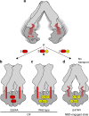
ATP-binding-cassette (ABC) transporters are molecular pumps that translocate molecules across the cell membrane by switching between inward-facing and outward-facing states. To obtain a detailed understanding of their mechanism remains a challenge to structural biology, as these proteins are notoriously difficult to study at the molecular level in their active, membrane-inserted form. Here we use solid-state NMR to investigate the multidrug ABC transporter BmrA reconstituted in lipids. We identify the chemical-shift differences between the inward-facing, and outward-facing state induced by ATP:Mg2+:Vi addition. Analysis of an X-loop mutant, for which we show that ATPase and transport activities are uncoupled, reveals an incomplete transition to the outward-facing state upon ATP:Mg2+:Vi addition, notably lacking the decrease in dynamics of a defined set of residues observed in wild-type BmrA. This suggests that this stiffening is required for an efficient transmission of the conformational changes to allow proper transport of substrate by the pump.
Keywords: Bacteria; Solid-state NMR.
Conflict of interest statement
The authors declare no competing interests.
Figures


Similar articles
-
ATP Analogues for Structural Investigations: Case Studies of a DnaB Helicase and an ABC Transporter.Molecules. 2020 Nov 12;25(22):5268. doi: 10.3390/molecules25225268. Molecules. 2020. PMID: 33198135 Free PMC article. Review.
-
The transport activity of the multidrug ABC transporter BmrA does not require a wide separation of the nucleotide-binding domains.J Biol Chem. 2024 Jan;300(1):105546. doi: 10.1016/j.jbc.2023.105546. Epub 2023 Dec 9. J Biol Chem. 2024. PMID: 38072053 Free PMC article.
-
Characterization of YvcC (BmrA), a multidrug ABC transporter constitutively expressed in Bacillus subtilis.Biochemistry. 2004 Jun 15;43(23):7491-502. doi: 10.1021/bi0362018. Biochemistry. 2004. PMID: 15182191
-
Myristic Acid Inhibits the Activity of the Bacterial ABC Transporter BmrA.Int J Mol Sci. 2021 Dec 17;22(24):13565. doi: 10.3390/ijms222413565. Int J Mol Sci. 2021. PMID: 34948362 Free PMC article.
-
Structural basis for the mechanism of ABC transporters.Biochem Soc Trans. 2015 Oct;43(5):889-93. doi: 10.1042/BST20150047. Biochem Soc Trans. 2015. PMID: 26517899 Review.
Cited by
-
Not Just Transporters: Alternative Functions of ABC Transporters in Bacillus subtilis and Listeria monocytogenes.Microorganisms. 2021 Jan 13;9(1):163. doi: 10.3390/microorganisms9010163. Microorganisms. 2021. PMID: 33450852 Free PMC article. Review.
-
ATP Analogues for Structural Investigations: Case Studies of a DnaB Helicase and an ABC Transporter.Molecules. 2020 Nov 12;25(22):5268. doi: 10.3390/molecules25225268. Molecules. 2020. PMID: 33198135 Free PMC article. Review.
-
The ABC transporter MsbA adopts the wide inward-open conformation in E. coli cells.Sci Adv. 2022 Oct 14;8(41):eabn6845. doi: 10.1126/sciadv.abn6845. Epub 2022 Oct 12. Sci Adv. 2022. PMID: 36223470 Free PMC article.
-
The transport activity of the multidrug ABC transporter BmrA does not require a wide separation of the nucleotide-binding domains.J Biol Chem. 2024 Jan;300(1):105546. doi: 10.1016/j.jbc.2023.105546. Epub 2023 Dec 9. J Biol Chem. 2024. PMID: 38072053 Free PMC article.
-
Backbone NMR assignment of the nucleotide binding domain of the Bacillus subtilis ABC multidrug transporter BmrA in the post-hydrolysis state.Biomol NMR Assign. 2022 Apr;16(1):81-86. doi: 10.1007/s12104-021-10063-2. Epub 2022 Jan 5. Biomol NMR Assign. 2022. PMID: 34988902 Free PMC article.
References
Publication types
MeSH terms
Substances
LinkOut - more resources
Full Text Sources
Molecular Biology Databases

