Review
. 2019 Jun;1862(6):634-642.
doi: 10.1016/j.bbagrm.2019.04.004. Epub 2019 Apr 28.

A novel role of U1 snRNP: Splice site selection from a distance

Affiliations
Review

A novel role of U1 snRNP: Splice site selection from a distance

Ravindra N Singh et al. Biochim Biophys Acta Gene Regul Mech. 2019 Jun.

Abstract

Removal of introns by pre-mRNA splicing is fundamental to gene function in eukaryotes. However, understanding the mechanism by which exon-intron boundaries are defined remains a challenging endeavor. Published reports support that the recruitment of U1 snRNP at the 5'ss marked by GU dinucleotides defines the 5'ss as well as facilitates 3'ss recognition through cross-exon interactions. However, exceptions to this rule exist as U1 snRNP recruited away from the 5'ss retains the capability to define the splice site, where the cleavage takes place. Independent reports employing exon 7 of Survival Motor Neuron (SMN) genes suggest a long-distance effect of U1 snRNP on splice site selection upon U1 snRNP recruitment at target sequences with or without GU dinucleotides. These findings underscore that sequences distinct from the 5'ss may also impact exon definition if U1 snRNP is recruited to them through partial complementarity with the U1 snRNA. In this review we discuss the expanded role of U1 snRNP in splice-site selection due to U1 ability to be recruited at more sites than predicted solely based on GU dinucleotides.

Keywords: Cryptic splice site; ISS-N1; SMA; SMN; Splicing; U1 snRNP.


Conflict of interest statement

Conflict of Interest Statement:

Authors declare no conflict of interest.

Figures

Fig. 1.
Fig. 1.
Diagrammatic representation of human U1 snRNP. U1 snRNP is composed of one U1 snRNA, seven common Sm proteins and three U1 snRNP-specific proteins (U1-70K, U1A and U1C) [49]. The 5′ end of U1 snRNA is tri-methyl-guanosine-capped. ψ indicates post transcriptional modification, pseudo-Uridine. Secondary structure of U1 snRNA consists of four stem-loops (SL) and an H helix highlighted in purple. Nucleotides forming H helix are shown. In addition, U1 snRNA sequences relevant to RNA:protein or RNA:5′ss interactions are given as well [42]. The loop portion of SL1 is drawn according to the crystal structure described in [42]. It is closed by a trans WC/Hoogsteen base pair formed between A29 and A36 [42]. Protein components of U1 snRNP, their sizes and their approximate locations are shown as well [49]. The Sm ring formed by the Sm proteins shown as green circles bind to the Sm site, which is boxed. U1-70K shown in red recognizes SL1. U1A shown in yellow binds SL2. U1C shown in blue is recruited to U1 snRNP through protein:protein interactions with U1-70 K and Sm proteins. The broken arrow signifies interactions between U1C and Sm ring.
Fig. 2.
Fig. 2.
Diagrammatic representation of the splice site selection from a distance in an SMN2 exon 7 model system. Three eU1s indicated in different colors were designed to form maximum possible number of base pairs with their intended targets. Sizes of exons and introns and the relative positioning of the splice sites are not to the scale. Cr1 refers to the cryptic 5′ss located 23 nts downstream of the authentic 5′ss of exon 7 [36]. Splice sites and Cr1 are indicated by green arrows. Black arrows represent the major splicing events. Red arrows represent splicing events with the major splice products shown. C>U refers to C-to-U transition at the 6th exonic position that distinguishes SMN2 from SMN1. (i) Overexpression of wild type U1 snRNA shown in blue is unable to rescue exon 7 inclusion. eU1s annealing to the authentic 5′ss of exon 7 (ii), to the Cr1 site (iii), as well as upstream (iv) and downstream (v) of Cr1, promote SMN2 exon 7 inclusion through activation of the authentic 5′ss. Models are based on results reported in [36].
Fig. 3.
Fig. 3.
Diagrammatic representation of SMN intron 7 secondary structure. It is based on experimental structure probing results [28]. Last twenty-two nucleotides of exon 7 are given as well. Exonic sequences are shown in black, intronic, in blue. Negative and neutral numbering of nucleotides starts from the end of exon 7 and the beginning of intron 7, respectively. Regulatory cis-element that affect exon 7 splicing, including ISS-N1 with its hnRNP A1/A2 binding sites, GC-rich sequence and TIA1 binding sites, are highlighted with colors [85]. Structural elements that contribute to exon 7 skipping, including terminal stem loops (TSL) 2 and 3 as well as internal stem formed by long-distance interactions (ISTL) 1, are shown [85]. The authentic and the cryptic (Cr1 and Cr2) 5′ splice sites are indicated by red arrows. Cr1 and Cr2 are described in [36].
Fig. 4.
Fig. 4.
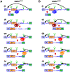
Diagrammatic representation of splice site selection from a distance in the context of SMA-associated site-specific mutations within the 5′ss of SMN1 exon 7. Three eU1s indicated in different colors were designed to form maximum possible number of base pairs with their intended targets. Sizes of exons and introns and the relative positioning of the splice sites are not to the scale. Mutated nucleotides are highlighted in red. Abbreviations and markings are the same as in Fig. 2. (A) Effect of eU1s on activation of the 5′ss (authentic or cryptic) from a distance in the context of SMN1 carrying a pathogenic G-to-C mutation at the 1st position of intron 7. (i) Overexpression of wild type U1 snRNA (in blue) is unable to rescue exon 7 inclusion. (ii) eU1 (in brown) annealing to the mutated authentic 5′ss of exon 7 promotes intron 7 retention. (iii) eU1 (in red) annealing to the Cr1 site promotes exon 7 inclusion through activation of Cr1. (iv) eU1 (in orange) annealing downstream of Cr1 promotes exon 7 inclusion through activation of Cr1. (v) eU1 (in green) annealing upstream of Cr1 promotes exon 7 inclusion through activation of Cr1. (B) Effect of eU1s on activation of the 5′ss (authentic or cryptic) from a distance in the context of SMN1 carrying a pathogenic U-to-G mutation at the 6th position of intron 7. (i) Overexpression of wild type U1 snRNA (in blue) is unable to rescue exon 7 inclusion. (ii) eU1 (in brown) designed to anneal to the wild type authentic 5′ss of exon 7 promotes exon 7 inclusion through activation of the mutated 5′ss. (iii) eU1 (in red) annealing to the Cr1 site promotes exon 7 inclusion through activation of Cr1. (iv) eU1 (in orange) annealing downstream of Cr1 promotes exon 7 inclusion through activation of Cr1. (v) eU1 (in green) anneals upstream of Cr1 and promotes exon 7 inclusion through activation of Cr1. Models are based on the results reported in [36].
Fig. 5.
Fig. 5.
Diagrammatic representation of splice site selection from a distance in the context of SMA-associated deletion mutations at the 5′ and 3′ss of SMN1 exon 7. Three eU1s indicated in different colors were designed to form maximum possible number of base pairs with their intended targets. Sizes of exons and introns and the relative positioning of the splice sites are not to the scale. Abbreviations and markings are the same as in Fig. 2. (A) Effect of eU1s on activation of the cryptic 5′ss (Cr1) from a distance in SMN1 carrying a pathogenic deletion at the 5′ss of exon 7. The 4-nt deletion is indicated by red dashes. (i) Overexpression of wild type U1 snRNA (in blue) is unable to rescue exon 7 inclusion. (ii) eU1 (in light blue) annealing to the mutated authentic 5′ss of exon 7 promotes intron 7 retention. (iii) eU1 (in red) annealing to the Cr1 site promotes exon 7 inclusion through activation of Cr1. (iv) eU1 (in orange) annealing downstream of Cr1 promotes exon 7 inclusion through activation of Cr1. (B) Effect of eU1s on activation of the 5′ss from a distance in the context of SMN1 carrying a pathogenic deletion at the 3′ss of exon 7. Δ refers to a 7-nt deletion within the polypyrimidine tract of the 3′ss of exon 7. (i) Overexpression of wild type U1 snRNA (in blue) is unable to rescue exon 7 inclusion. (ii) eU1 (in brown) annealing to the authentic 5′ss of exon 7 promotes its inclusion through activation of this site. (iii) eU1 (in red) annealing to the Cr1 site promotes exon 7 inclusion through activation of Cr1. (iv) eU1 (in orange) annealing downstream of Cr1 promotes exon 7 inclusion through activation of the authentic 5′ss. Models are based on the results reported in [36].

Similar articles

Cited by

References

    1. Sharp PA, Split genes and RNA splicing, Cell 77 (1994) 805–815. - PubMed
    1. Ebbesen KK, Kjems J, Hansen TB, Circular RNAs: Identification, biogenesis and function, Biochim. Biophys. Acta 1859 (2016) 163–168. - PubMed
    1. Wilusz JE, A 360° view of circular RNAs: From biogenesis to functions, Wiley Interdiscip Rev RNA 9 (2018), e1478. - PMC - PubMed
    1. Yang L, Splicing noncoding RNAs from the inside out, Wiley Interdiscip Rev RNA. 6 (2015), 651–660. - PMC - PubMed
    1. Kelemen O, Convertini P, Zhang Z, Wen Y, Szhen M, Falaleeva M, Stamm S, Function of alternative splicing, Gene 514 (2013), 1–30. - PMC - PubMed

Publication types

LinkOut - more resources