. 2019 Jun 14;294(24):9416-9429.
doi: 10.1074/jbc.RA119.007485. Epub 2019 Apr 29.

Partial ligand-receptor engagement yields functional bias at the human complement receptor, C5aR1

Affiliations

Partial ligand-receptor engagement yields functional bias at the human complement receptor, C5aR1

Shubhi Pandey et al. J Biol Chem. .

Abstract

The human complement component, C5a, binds two different seven-transmembrane receptors termed C5aR1 and C5aR2. C5aR1 is a prototypical G-protein-coupled receptor that couples to the Gαi subfamily of heterotrimeric G-proteins and β-arrestins (βarrs) following C5a stimulation. Peptide fragments derived from the C terminus of C5a can still interact with the receptor, albeit with lower affinity, and can act as agonists or antagonists. However, whether such fragments might display ligand bias at C5aR1 remains unexplored. Here, we compare C5a and a modified C-terminal fragment of C5a, C5apep, in terms of G-protein coupling, βarr recruitment, endocytosis, and extracellular signal-regulated kinase 1/2 mitogen-activated protein kinase activation at the human C5aR1. We discover that C5apep acts as a full agonist for Gαi coupling as measured by cAMP response and extracellular signal-regulated kinase 1/2 phosphorylation, but it displays partial agonism for βarr recruitment and receptor endocytosis. Interestingly, C5apep exhibits full-agonist efficacy with respect to inhibiting lipopolysaccharide-induced interleukin-6 secretion in human macrophages, but its ability to induce human neutrophil migration is substantially lower compared with C5a, although both these responses are sensitive to pertussis toxin treatment. Taken together, our data reveal that compared with C5a, C5apep exerts partial efficacy for βarr recruitment, receptor trafficking, and neutrophil migration. Our findings therefore uncover functional bias at C5aR1 and also provide a framework that can potentially be extended to chemokine receptors, which also typically interact with chemokines through a biphasic mechanism.

Keywords: G-protein–coupled receptor (GPCR); arrestin; cell signaling; complement; signal transduction.


Conflict of interest statement

The authors declare that they have no conflicts of interest with the contents of this article

Figures

Figure 1.
Figure 1.
A modified C terminus C5a peptide, C5apep, is a full agonist for Gαi coupling. A, schematic representation of C5a binding to C5aR1. There are two sites of interaction, one involving the N terminus of C5aR1 and the other involving the extracellular loops. B, the primary sequence and modification of C5apep, which is derived based on the C terminus of C5a. C, C5apep behaves as a full-agonist in GloSensor-based cAMP assay. HEK-293 cells expressing C5aR1 were transfected with F22 plasmids. 24 h post-transfection, the cells were stimulated with indicated concentrations of C5a and C5apep followed by recording of bioluminescence readout. D, Gαi coupling induced by C5a and C5apep measured in CHO cells using LANCE cAMP assay. CHO cells stably expressing C5aR1 were stimulated with the indicated concentrations of respective ligands. The data presented in C and D represent the means ± S.E. of three to five independent experiments, and the EC50 values of C5a and C5apep were analyzed using unpaired t test. ***, p < 0.001.
Figure 2.
Figure 2.
C5apep is a partial agonist for βarr recruitment. A, HEK-293 cells expressing FLAG-C5aR1 and either βarr1 or 2 were stimulated with the indicated concentrations of different ligands (W54011, 0.1 μm; C5a, 1 μm; C5apep, 10 μm) followed by cross-linking using DSP. Subsequently, FLAG-C5aR1 was immunoprecipitated using anti-FLAG antibody agarose, and co-elution of βarrs was visualized using Western blotting. B, densitometry-based quantification of data presented in A (averages ± S.E.; n = 2) normalized with respect to signal for C5a-βarr1 condition (treated as 100%) and analyzed using two-way ANOVA. ***, p < 0.001. C, a time-course co-immunoprecipitation experiment to measure the interaction of C5aR1 with βarr2. The experiment was performed following the protocol as indicated except that cells were stimulated for different time points. D, densitometry-based quantification of the data presented in C (average ± S.E.; n = 2) normalized with respect to signal for C5a at 30 min condition (treated as 100%) and analyzed using two-way ANOVA. *, p < 0.05.
Figure 3.
Figure 3.
C5apep induces slower endosomal trafficking of βarrs. HEK-293 cells expressing C5aR1 and βarr1/2 were stimulated with C5a (1 μm) and C5apep (10 μm), and the trafficking patterns of βarrs were visualized using confocal microscopy for indicated time points. Both C5a and C5apep induced surface localization of βarrs at early time points; however, somewhat slower endosomal trafficking of βarrs were observed for C5apep as assessed by localization of βarrs in endosomal vesicles. The figure shows representative images from three independent experiments.
Figure 4.
Figure 4.
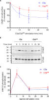
C5apep exhibits a bias between receptor endocytosis and ERK1/2 MAP kinase phosphorylation. A, HEK-293 cells expressing C5aR1 were stimulated with C5a (1 μm) and C5apep (10 μm) for indicated time points followed by the assessment of surface receptor levels using a whole-cell ELISA assay. C5apep displays a weaker efficacy in promoting C5aR1 endocytosis compared with C5a. The data represent averages ± S.E. from five independent experiments. B, C5apep induces robust phosphorylation of ERK1/2 MAP kinase at levels similar to C5a. HEK-293 cells expressing C5aR1 were stimulated with respective ligands for indicated time points followed by measurement of ERK1/2 phosphorylation using Western blotting. C, densitometry-based quantification of the ERK1/2 phosphorylation data presented in B (averages ± S.E.) of five independent experiments.
Figure 5.
Figure 5.
C5a/C5apep-induced ERK1/2 phosphorylation is sensitive to PTX treatment. A, HEK-293 cells stably expressing C5aR1 were incubated with 100 ng/ml PTX for 12–16 h followed by serum starvation and ligand stimulation. Subsequently, ERK1/2 phosphorylation was measured by Western blotting. B, densitometry-based quantification of data presented in A from three independent experiments normalized with respect to maximal response (treated as 100%) and analyzed using one-way ANOVA. ***, p < 0.001. C, effect of PTX treatment on C5apep-induced ERK1/2 phosphorylation measured as in A above. D, densitometry-based quantification of data presented in C, normalized, and analyzed as in B.
Figure 6.
Figure 6.
C5apep exhibits full-agonist efficacy for cAMP response and ERK1/2 phosphorylation for a chimeric C5aR1, C5a-V2R. A, schematic representation of the C terminus of C5aR1 and a chimeric construct harboring the C terminus of AVPR2 (V2R), referred to as C5a-V2R. V2R tail in the chimeric construct is highlighted in red. B, C5apep behaves as a full agonist in GloSensor-based cAMP assay. HEK-293 cells expressing C5a-V2R were transfected with F22 plasmids. 24 h post-transfection, the cells were stimulated with the indicated concentrations of C5a and C5apep followed by recording of bioluminescence readout. The data represent averages ± S.E. of three independent experiments, each carried out in duplicate, and the EC50 values of C5a and C5apep were analyzed using unpaired t test. ***, p < 0.001. C, C5apep induces robust phosphorylation of ERK1/2 MAP kinase at levels similar to C5a. HEK-293 cells expressing C5a-V2R were stimulated with respective ligands (C5a, 1 μm; C5apep, 10 μm) for the indicated time points followed by measurement of ERK1/2 phosphorylation using Western blotting. D, densitometry-based quantification of ERK1/2 phosphorylation data presented in C (average ± S.E.) of five independent experiments.
Figure 7.
Figure 7.
C5apep induces differential recruitment of βarr1 and 2 at C5aV2R. A, HEK-293 cells expressing FLAG-C5a-V2R and βarr2 were stimulated with saturating concentrations of different ligands followed by cross-linking using DSP. Subsequently, FLAG-C5a-V2R was immunoprecipitated using anti-FLAG antibody agarose, and co-elution of βarr2 was visualized using Western blotting. B, densitometry-based quantification of data presented in A (average±S.E.; n = 2) normalized with respect to signal for C5a-βarr2 30 min condition (treated as 100%) and analyzed using two-way ANOVA. **, p < 0.01; *, p < 0.05. C, interaction of C5aR1 or C5a-V22R with βarr1 measured essentially in similar fashion as described in A above. D, densitometry-based quantification of data presented in C (averages ± S.E.; n = 3) normalized with respect to signal for C5a-C5aR1 condition (treated as 100%) and analyzed using two-way ANOVA. **, p < 0.01.
Figure 8.
Figure 8.
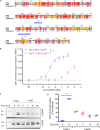
Sequence alignment of C5aR2 with C5aR1, βarr2 recruitment, and ERK1/2 phosphorylation. A, the sequences of human C5aR1 and C5aR2 were retrieved from Uniprot and aligned on M-coffee server with default parameters. Alignment reliability was assessed by core/TCS and generated alignment was visualized using Espript 3. Specific mutations in the DRY and NPXXY motif are highlighted. B, HEK-293 cells expressing C5aR2-Venus and βarr2-Rluc8 constructs were first incubated with luciferase-substrate for 2 h. Subsequently, the cells were stimulated with respective ligands, and BRET signals were monitored using a dose-response curve. The data represent averages ± S.E. of three independent experiments, and the EC50 values are compared using unpaired t test. ***, p < 0.001. C, HEK-293 cells expressing C5aR1 or C5aR2 were stimulated with C5a (100 nm) for indicated time points followed by detection of ERK1/2 phosphorylation using Western blotting. D, densitometry-based quantification of data presented in C normalized with C5a response for C5aR1 (treated as 100%) and analyzed using two-way ANOVA. ***, p < 0.001.
Figure 9.
Figure 9.
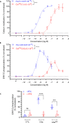
C5apep exhibits bias at the level of Ca2+ mobilization and ERK1/2 phosphorylation in HMDMs. A, C5apep is a full agonist in Ca2+ mobilization assay in HMDM cells. HMDMs were first loaded with Fluo-4 calcium indicator followed by the addition of respective ligands and subsequent measurement of fluorescence intensity. The data were normalized with maximal response obtained for C5a and represent means ± S.E. of triplicate experiments performed from three independent donors, and the EC50 values of C5a and C5apep were analyzed using unpaired t test. ***, p < 0.001. B, HMDMs were stimulated with respective ligands for 10 min at room temperature before being lysed. The phospho-ERK1/2 content in the lysate was detected using AlphaLISA Surefire Ultra p-ERK1/2 kit. The data were normalized with maximal response obtained for C5a and represent the means ± S.E. of triplicate experiments performed from five independent donors, and the EC50 values of C5a and C5apep were analyzed using unpaired t test. ****, p < 0.0001. C, C5a/C5apep-induced ERK1/2 phosphorylation in HMDMs is sensitive to PTX. HMDMs seeded in tissue culture-treated 96-well plates were preincubated with 200 ng/ml PTX or vehicle in serum-free IMDM overnight. Subsequently, the cells were stimulated with C5a/C5apep, and ERK1/2 phosphorylation was measured using AlphaLISA Surefire Ultra p-ERK1/2 kit. The data are normalized with maximal response obtained for C5a and represent the means ± S.E. of triplicate experiments performed from three independent donors and analyzed using paired two-way ANOVA. ***, p < 0.001; ****, p < 0.0001. CTL, control.
Figure 10.
Figure 10.
C5apep exhibits bias between LPS-induced IL-6 release and neutrophil migration. A, C5apep behaves as a full-agonist for lowering of LPS-induced IL-6 release in HMDM cells. HMDMs were stimulated using 10 ng/ml LPS in the absence or presence of C5a or C5apep. Subsequently, the levels of IL-6 present in the supernatant after 24 h of stimulation were quantified using ELISA, background-corrected with the values obtained for serum-free medium/BSA, and normalized with maximal response for LPS (i.e. treated as 100%). The data represent normalized means ± S.E. of triplicate measurements conducted in cells from four independent donors, analyzed using two-way ANOVA. *, p < 0.05; ****, p < 0.0001. B, LPS-induced IL-6 response is sensitive to PTX. HMDMs seeded in 96-well plates were incubated overnight with 200 ng/ml PTX or vehicle followed by stimulation with respective ligands. Subsequently, the IL-6 content in the supernatant was quantified using ELISA kits. The data are presented, normalized, and analyzed as in A above. C, C5apep elicits only partial-agonist response in migration of human PMNs. Freshly isolated hPMNs seeded into Transwell inserts were stimulated with respective ligands added to the receiver wells and then allowed to migrate for 1 h. The number of migrated cells was recorded and normalized to the maximal C5a-induced migration. The data represent the means ± S.E. of triplicate measurements conducted in cells from three independent donors. D, C5apep-induced neutrophil migration is sensitive to PTX. Freshly isolated hPMNs were preincubated with 3 μg/ml PTX or vehicle for 4 h, and cell migration was initiated by adding respective ligands. Ligand-induced migration was measured as in C, and the data are normalized with control (i.e. no stimulation) and analyzed suing two-way ANOVA. *, p < 0.05. CTL, control.

Similar articles

Cited by

References

    1. Manthey H. D., Woodruff T. M., Taylor S. M., and Monk P. N. (2009) Complement component 5a (C5a). Int. J. Biochem. Cell Biol. 41, 2114–2117 10.1016/j.biocel.2009.04.005 - DOI - PubMed
    1. Ward P. A. (2004) The dark side of C5a in sepsis. Nat. Rev. Immunol. 4, 133–142 10.1038/nri1269 - DOI - PubMed
    1. Klos A., Wende E., Wareham K. J., and Monk P. N. (2013) International Union of Basic and Clinical Pharmacology [corrected]: LXXXVII. Complement peptide C5a, C4a, and C3a receptors. Pharmacol. Rev. 65, 500–543 10.1124/pr.111.005223 - DOI - PubMed
    1. Schatz-Jakobsen J. A., Yatime L., Larsen C., Petersen S. V., Klos A., and Andersen G. R. (2014) Structural and functional characterization of human and murine C5a anaphylatoxins. Acta Crystallogr. D Biol. Crystallogr. 70, 1704–1717 10.1107/S139900471400844X - DOI - PMC - PubMed
    1. Siciliano S. J., Rollins T. E., DeMartino J., Konteatis Z., Malkowitz L., Van Riper G., Bondy S., Rosen H., and Springer M. S. (1994) Two-site binding of C5a by its receptor: an alternative binding paradigm for G protein-coupled receptors. Proc. Natl. Acad. Sci. U.S.A. 91, 1214–1218 10.1073/pnas.91.4.1214 - DOI - PMC - PubMed

Publication types

MeSH terms

LinkOut - more resources