. 2019 Aug 7;70(15):4049-4062.
doi: 10.1093/jxb/erz195.

A cytorhabdovirus phosphoprotein forms mobile inclusions trafficked on the actin/ER network for viral RNA synthesis

Affiliations

A cytorhabdovirus phosphoprotein forms mobile inclusions trafficked on the actin/ER network for viral RNA synthesis

Xiao-Dong Fang et al. J Exp Bot. .

Abstract

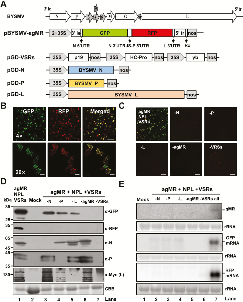
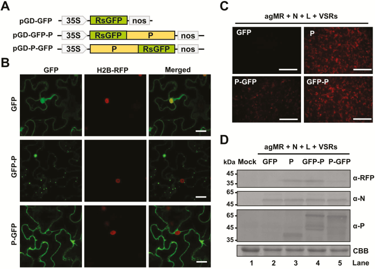
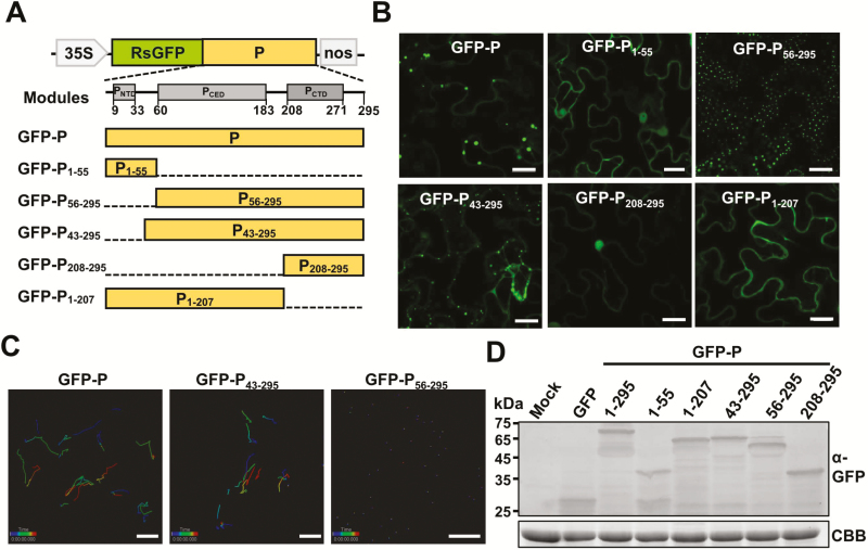
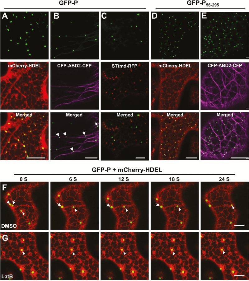
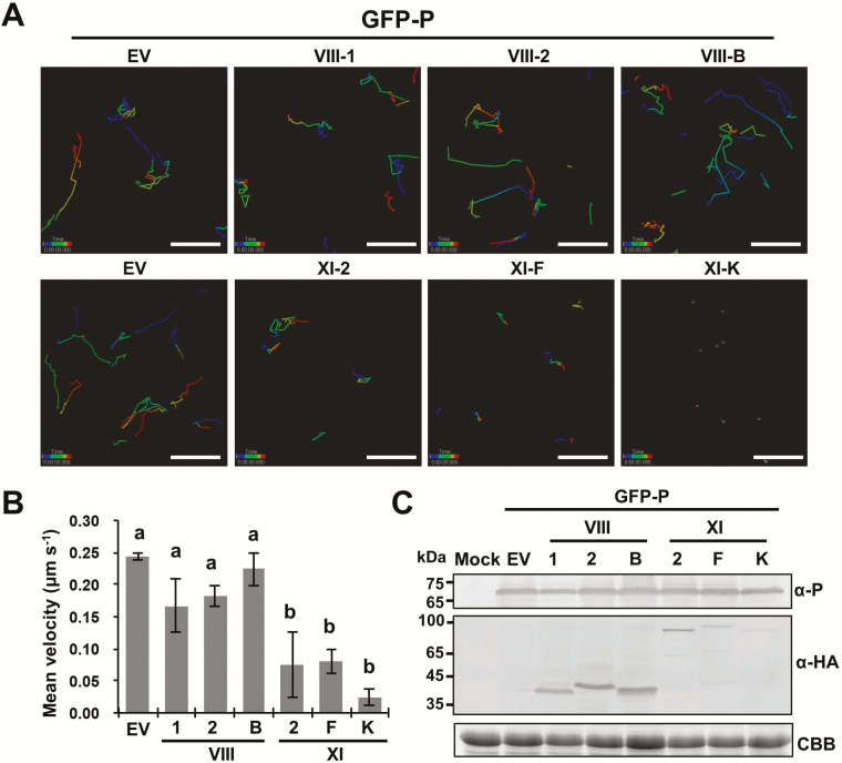
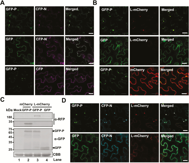
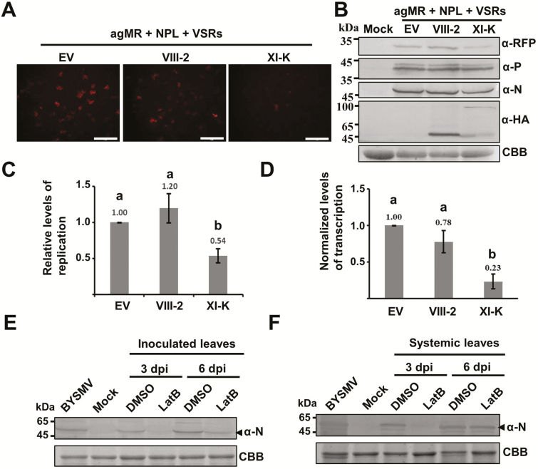
As obligate parasites, plant viruses usually hijack host cytoskeletons for replication and movement. Rhabdoviruses are enveloped, negative-stranded RNA viruses that infect vertebrates, invertebrates, and plants, but the mechanisms of intracellular trafficking of plant rhabdovirus proteins are largely unknown. Here, we used Barley yellow striate mosaic virus (BYSMV), a plant cytorhabdovirus, as a model to investigate the effects of the actin cytoskeleton on viral intracellular movement and viral RNA synthesis in a mini-replicon (MR) system. The BYSMV P protein forms mobile inclusion bodies that are trafficked along the actin/endoplasmic reticulum network, and recruit the N and L proteins into viroplasm-like structures. Deletion analysis showed that the N terminal region (aa 43-55) and the remaining region (aa 56-295) of BYSMV P are essential for the mobility and formation of inclusions, respectively. Overexpression of myosin XI-K tails completely abolishes the trafficking activity of P bodies, and is accompanied by a significant reduction of viral MR RNA synthesis. These results suggest that BYSMV P contributes to the formation and trafficking of viroplasm-like structures along the ER/actin network driven by myosin XI-K. Thus, rhabdovirus P appears to be a dynamic hub protein for efficient recruitment of viral proteins, thereby promoting viral RNA synthesis.

Keywords: Barley yellow striate mosaic virus; Actin cytoskeleton; P bodies; intracellular movement; myosin motor; rhabdovirus.


Figures

Fig. 1.
Fig. 1.
Construction of a BYSMV antigenomic-sense mini-replicon. (A) Illustration of binary Agrobacterium vectors designed to generate antigenomic-sense mini-replicon (agMR) RNA and to express the BYSMV N, P, and L proteins in vivo. In the pBYSMV-agMR plasmid, a reporter cassette of antigenomic-sense BYSMV derivative was inserted between the truncated CaMV double 35S promoter (2×35S) and the hepatitis delta (HDV) ribozyme sequence (RZ). Le, leader; tr, trailer; UTR, untranslated region; N 3′UTR-IS-P 5′UTR, intergenic sequences including N 3´UTR, intergenic sequence (IS), and P 5´UTR; nos, nopaline synthase terminator. (B) GFP and RFP foci in N. benthamiana leaves agro-infiltrated with BYSMV-agMR combinations at 5 d post inoculation (dpi). Scale bars in the top panels are 500 μm and the bars in the bottom panels are 100 μm. (C) Requirement of N, P, L, and viral suppressors of RNA silencing (VSRs) for expression of MR reporter genes. GFP and RFP fluorescence were observed at 5 dpi. Scale bars are 500 μm. (D) Western blot analysis showing accumulation of the GFP, RFP, N, P, and L proteins in the leaves shown in (C) as determined using rabbit antibodies against GFP, RFP, N, P, and a mouse Myc antibody for BYSMV L. The mock sample was infiltrated with Agrobacterium harboring the pGD vector. Coomassie brilliant blue (CBB) staining was used for protein loading controls. (E) Northern blot analysis of BYSMV agMR transcription and replication. Total RNA extracted from leaves shown in panel (C) was hybridized with an in vitro transcript of a positive-sense GFP probe to detect the genomic-sense MR (gMR) RNA. Randomly labeled GFP and RFP cDNA probes were used for detection of GFP and RFP mRNA, respectively. rRNAs stained with methylene blue were used as loading controls.
Fig. 2.
Fig. 2.
Subcellular localization and mobility of the BYSMV P protein. (A) Schematic diagram of pGD vectors for expression of free GFP, GFP-P, and P-GFP. (B) Confocal micrographs showing the subcellular distribution of free GFP, GFP-P, and P-GFP in epidermal cells of agro-infiltrated leaves of H2B-RFP transgenic N. benthamiana at 2 d post inoculation (dpi). Scale bars are 20 μm. (C) RFP foci at 6 dpi in leaves infiltrated with Agrobacterium engineered for expression of free GFP, P, GFP-P, or P-GFP in antigenomic-sense mini-replicon (agMR) combinations. Scale bars are 1 mm. (D) Western blotting analysis showing accumulation of RFP, and N and P proteins in infiltrated leaves with anti-RFP, anti-N, or anti-P polyclonal antibodies, respectively. Buffer-infiltrated leaves were used as a negative control (mock). Coomassie brilliant blue (CBB) staining was used for protein loading controls.
Fig. 3.
Fig. 3.
Identification of BYSMV P protein domains responsible for trafficking and formation of inclusion bodies. (A) Modular organization of the BYSMV P protein and schematic presentation of the designs of deletion mutants. Normalized disorder scores (D-scores) were calculated from 15 different predictors, and structured regions (grey boxes) were defined as locations within a D-score above a threshold of 0.5 and disordered regions (lines) were defined as having a D-score below 0.5. Numbers below the boxes indicate the limits of the structured regions. Deleted regions of BYSMV P are indicated by dashed lines. (B) Fluorescence micrographs showing localizations of GFP-fused P and deletion mutants. Images were taken at 2 d post inoculation. Scale bars are 20 μm. (C) Tracking of individual GFP-P, GFP-P43–295, and GFP-P56–295 bodies using Imaris software. Individual particles in epidermal cells of agro-infiltrated N. benthamiana leaves were tracked in 1-s bursts separated by 4-s dark intervals, and 50 images were collected. Different time intervals are indicated by the different colors. Scale bars are 20 μm. (D) Detection of GFP-tagged P protein and P mutant proteins by western blotting with anti-GFP polyclonal antibodies. Coomassie brilliant blue (CBB) staining was used as the protein loading control. Healthy N. benthamiana leaves served as mock samples.
Fig. 4.
Fig. 4.
Confocal micrographs showing the localization of BYSMV P protein bodies in relation to the ER/actin network. (A–E) Co-expression of GFP-P with mCherry-HDEL (A), CFP-ABD2-CFP (B), and STtmd-RFP (C), and co-expression of GFP-P56–295 with mCherry-HDEL (D) and CFP-ABD2-CFP (E) were monitored at 2 d post inoculation (dpi). Scale bars are 20 μm. (F, G) Time-lapse confocal images of GFP-P and mCherry-HDEL at 2 dpi in co-infiltrated N.benthamiana leaves treated with DMSO (F) or latrunculin B (LatB) to disrupt actin assembly (G) at 2 dpi. Images were taken 3 h after DMSO or 10 μM LatB treatments. Scale bars are 10 μm. In the DMSO-treated leaves (F), arrows indicate fusion of two GFP-P inclusion bodies, and arrowheads indicate movement of a GFP-P body along the ER network. In the LatB-treated leaves (G), arrowheads indicate an immobile GFP-P body.
Fig. 5.
Fig. 5.
Effects of myosin tail overexpression on trafficking of BYSMV-P protein bodies in co-infiltrated N. benthamiana. (A) Representative confocal images of GFP-P bodies in epidermal cells overexpressing myosin tails VIII-1, VIII-2, VIII-B, XI-2, XI-F, and XI-K at 2 d post inoculation. Individual bodies were recorded in a time-series and are indicated by connecting lines. Different time intervals are represented by the different colors. (B) Mean velocities of the BYSMV-P bodies in the infiltrated leaves shown in (A). The mean velocities were calculated from the velocities of at least 90 bodies over 4 min. Data are means (±SE) of three independent experiments. Different letters indicate significant differences as determined by ANOVA followed by Turkey’s multiple comparison test (P<0.05). (C) Accumulation of GFP-P and myosin tails in those N. benthamiana leaves shown in (A). Anti-P, and anti-HA antibodies were used to detect the accumulation of GFP-P and HA-tagged myosin tails, respectively. Uninfiltrated N. benthamiana leaves were used as mock controls. Coomassie brilliant blue (CBB) staining was used for loading controls. EV, empty vector.
Fig. 6.
Fig. 6.
Recruitment of the BYSMV N and L proteins into BYSMV-P viroplasm-like inclusion bodies. (A) Co-localization of CFP-N, but not CFP, with GFP-P bodies. GFP-P, GFP, CFP-N, and CFP proteins were expressed in different combinations via agro-infiltration in N. benthamiana leaves, and fluorescence images were observed at 2 d post inoculation (dpi). Scale bars are 20 μm. (B) Co-localization of L-mCherry, but not mCherry, with GFP-P bodies. Co-expression of GFP-P/L-mCherry, GFP-P/mCherry, and GFP/L-mCherry in N. benthamiana epidermal cells in fluorescence images taken at 2 dpi. Scale bars are 20 μm. (C) Accumulation of GFP-P and L-mCherry in the N. benthamiana leaves shown in (B). Anti-GFP and anti-RFP antibodies were used to detect accumulation of GFP-P and L-mCherry, respectively. Coomassie brilliant blue (CBB) staining was used as loading protein control. Healthy N. benthamiana leaves served as mock samples. (D) Accumulation of CFP-N and L-mCherry in GFP-P bodies, and lack of association with GFP. CFP-N and L-mCherry were expressed with GFP-P or GFP in agro-infiltrated N.benthamiana leaves. Fluorescence images were taken at 2 dpi. Scale bars are 20 μm.
Fig. 7.
Fig. 7.
Effects of BYSMV-P protein body trafficking on mini-genome RNA synthesis. (A) Representative confocal images of RFP fluorescence expressed from antigenomic-sense mini-replicon (agMR) combinations in epidermal cells of N. benthamiana treated with either the empty vector (EV) or with overexpressed myosin tails VIII-2 and XI-K at 6 d post inoculation (dpi). Scale bars are 1 mm. (B) Accumulation of RFP, P, N, and myosin tails shown in (A), as detected by western blotting with antibodies against RFP, P, N, and HA, respectively. Coomassie brilliant blue (CBB) staining was used as a loading control. Healthy N. benthamiana leaves served as mock samples. (C) Quantitative real-time PCR analysis of mini-genome RNA replication levels and (D) real-time quantitative PCR analysis of the relative levels of RFP mRNA versus mini-genome RNA in the samples shown in in (A). The EF1A housekeeping gene served as an internal control. The values of viral replication and relative transcription in the samples treated with the empty vector (EV) were set to 1. Data are means (±SE) of three independent experiments, and the mean values are indicated. Different letters indicate significant differences as determined by ANOVA followed by Turkey’s multiple comparison test (P<0.05). (E) Accumulation of BYSMV N protein in the inoculated leaves after treatment with DMSO or latrunculin B (LatB, 5 μM) to disrupt actin assembly, as analysed by western blotting. (F) Accumulation of BYSMV N protein in systemically infected leaves that emerged after viruliferous planthoppers had been given access to leaves that had been treated as described in (E). BYSMV-infected barley plants and healthy plants (mock) provided positive and negative controls, respectively. CBB) staining was used as loading controls.

Similar articles

Cited by

References

    1. Agbeci M, Grangeon R, Nelson RS, Zheng H, Laliberté JF. 2013. Contribution of host intracellular transport machineries to intercellular movement of turnip mosaic virus. PLoS Pathogens 9, e1003683. - PMC - PubMed
    1. Amari K, Di Donato M, Dolja VV, Heinlein M. 2014. Myosins VIII and XI play distinct roles in reproduction and transport of tobacco mosaic virus. PLoS Pathogens 10, e1004448. - PMC - PubMed
    1. Amari K, Lerich A, Schmitt-Keichinger C, Dolja VV, Ritzenthaler C. 2011. Tubule-guided cell-to-cell movement of a plant virus requires class XI myosin motors. PLoS Pathogens 7, e1002327. - PMC - PubMed
    1. Avisar D, Prokhnevsky AI, Dolja VV. 2008a. Class VIII myosins are required for plasmodesmatal localization of a closterovirus Hsp70 homolog. Journal of Virology 82, 2836–2843. - PMC - PubMed
    1. Avisar D, Prokhnevsky AI, Makarova KS, Koonin EV, Dolja VV. 2008b. Myosin XI-K Is required for rapid trafficking of Golgi stacks, peroxisomes, and mitochondria in leaf cells of Nicotiana benthamiana. Plant Physiology 146, 1098–1108. - PMC - PubMed

Publication types