. 2019 Jun 14;93(13):e00196-19.
doi: 10.1128/JVI.00196-19. Print 2019 Jul 1.

Hepatitis B Virus Precore Protein p22 Inhibits Alpha Interferon Signaling by Blocking STAT Nuclear Translocation

Affiliations

Hepatitis B Virus Precore Protein p22 Inhibits Alpha Interferon Signaling by Blocking STAT Nuclear Translocation

Bidisha Mitra et al. J Virol. .

Abstract

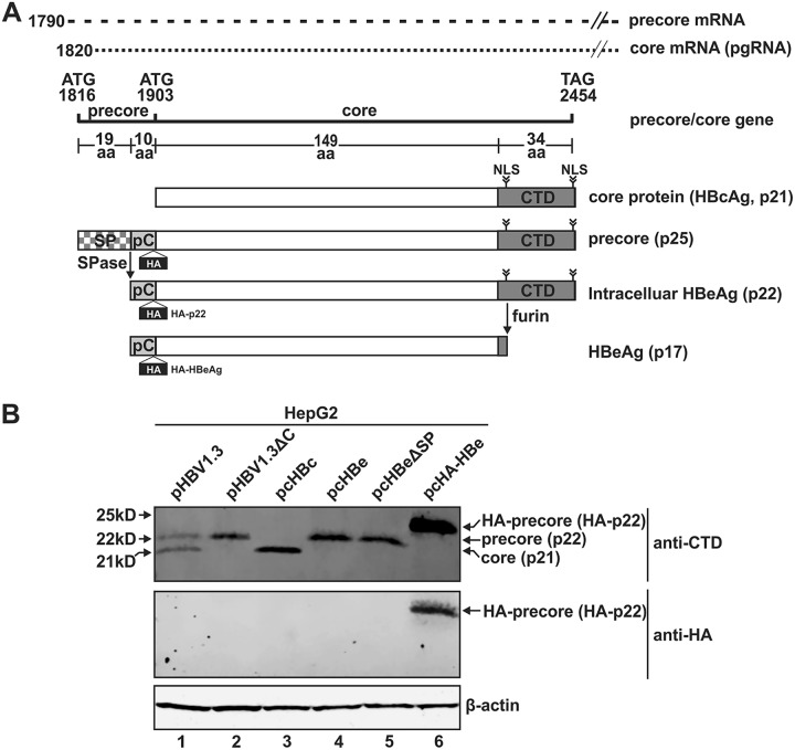
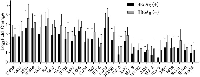
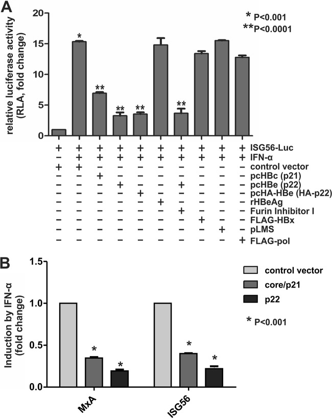
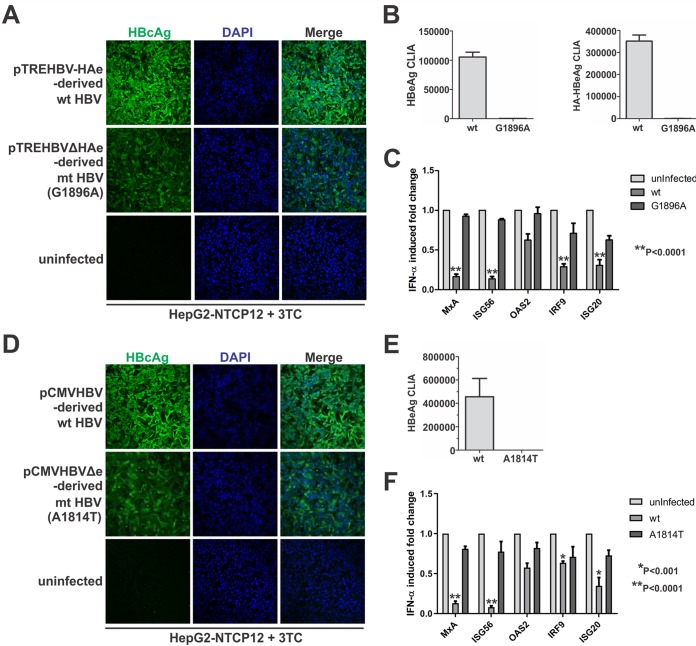
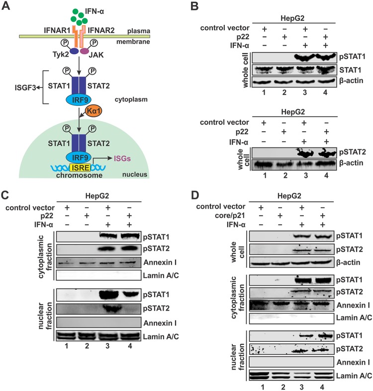
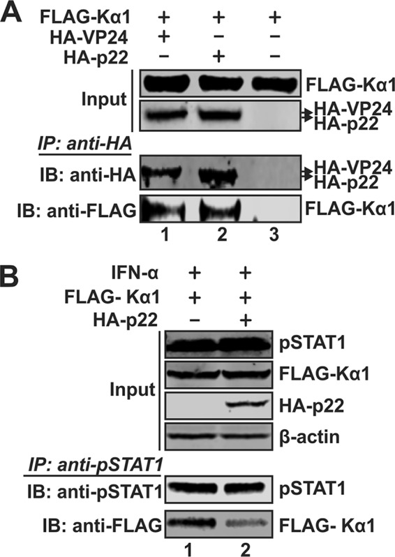
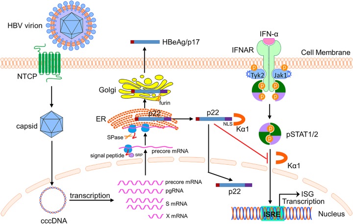
Antagonism of host immune defenses against hepatitis B virus (HBV) infection by the viral proteins is speculated to cause HBV persistence and the development of chronic hepatitis. The circulating hepatitis B e antigen (HBeAg, p17) is known to manipulate host immune responses to assist in the establishment of persistent viral infection, and HBeAg-positive (HBeAg+) patients respond less effectively to IFN-α therapy than do HBeAg-negative (HBeAg-) patients in clinical practice. However, the function(s) of the intracellular form of HBeAg, previously reported as the precore protein intermediate (p22) without the N-terminal signal peptide, remains elusive. Here, we report that the cytosolic p22 protein, but not the secreted HBeAg, significantly reduces interferon-stimulated response element (ISRE) activity and the expression of interferon-stimulated genes (ISGs) upon alpha interferon (IFN-α) stimulation in cell cultures. In line with this, HBeAg+ patients exhibit weaker induction of ISGs in their livers than do HBeAg- patients upon IFN-α therapy. Mechanistically, while p22 does not alter the total STAT1 or pSTAT1 levels in cells treated with IFN-α, it blocks the nuclear translocation of pSTAT1 by interacting with the nuclear transport factor karyopherin α1 through its C-terminal arginine-rich domain. In summary, our study suggests that HBV precore protein, specifically the p22 form, impedes JAK-STAT signaling to help the virus evade the host innate immune response and, thus, causes resistance to IFN therapy.IMPORTANCE Chronic hepatitis B virus (HBV) infection continues to be a major global health concern, and patients who fail to mount an efficient immune response to clear the virus will develop a life-long chronic infection that can progress to chronic active hepatitis, cirrhosis, and primary hepatocellular carcinoma. There is no definite cure for chronic hepatitis B, and alpha interferon (IFN-α) is the only available immunomodulatory drug, to which only a minority of chronic patients are responsive, with hepatitis B e antigen (HBeAg)-negative patients responding better than HBeAg-positive patients. We herein report that the intracellular HBeAg, also known as precore or p22, inhibits the antiviral signaling of IFN-α, which sheds light on the enigmatic function of precore protein in shaping HBV chronicity and provides a perspective toward areas that need to be further studied to make the current therapy better until a cure is achieved.

Keywords: hepatitis B virus; interferons.


Figures

FIG 1
FIG 1
HBV precore protein exists as a p22 form intracellularly. (A) Schematic illustration of the translation of HBV precore and core proteins. While HBV core protein is formed from the pregenomic RNA (pgRNA), the ORF of precore (p25) is translated from the precore mRNA and processed into p22 by removal of the N-terminal signal peptide (SP) by signal peptidase (SPase); p22 then gets further processed at the C-terminal domain (CTD) to form the secreted HBeAg (p17). The figure shows a comparison of the differences in the various domains among the above-mentioned proteins. The position of the HA tag insertion in the precore domain of HA-tagged precore is indicated. (B) HepG2 cells were seeded in 12-well plates and mock transfected or transfected with pHBV1.3, pHBV1.3ΔC, pcHBc, pcHBe, pcHBeΔSP, and pcHA-HBe, as indicated. Cells were harvested at day 5 posttransfection, and the proteins were analyzed by Western blotting using antibodies against CTD or HA tag. β-Actin served as the loading control.
FIG 2
FIG 2
Inability of precore/p22 to support HBV capsid formation and DNA replication. (A) HepG2 cells in 12-well plates were cotransfected with pHBV1.3 and control vector, pHBV1.3ΔC and control vector, or pcHBc, pcHBe, or pcHA-HBe, as indicated. Cells were harvested at day 5 posttransfection, and viral total RNA and encapsidated pgRNA were analyzed by Northern blotting, core DNA was detected by Southern blotting, and cytoplasmic capsid was analyzed by native capsid gel EIA. The expression of core and precore protein was detected by Western blotting using core antibody against the CTD domain, and the HA-tagged p22 was detected by Western blotting using anti-HA antibody. β-Actin served as the loading control. For RNA analysis, each lane was loaded with 10 μg of total RNA and probed with a genome-length, plus-strand-specific HBV riboprobe. The levels of ribosomal RNAs (28S and 18S) are presented as loading controls. The positions of HBV pgRNA (3.5 kb) and subgenomic surface RNAs (2.4 kb and 2.1 kb) are indicated. HBV core DNA was probed with genome-length, minus-strand-specific HBV riboprobe. The positions of relaxed circular (RC) and single-stranded (SS) DNAs are indicated. (B) HepG2 cells in 12-well plates were cotransfected with pHBV1.3 and control vector, pHBV1.3ΔC and control vector, or pcHBeC−7Q, as indicated, for 5 days. The analyses of viral RNA, DNA, and proteins were performed in the same way as described for panel A.
FIG 3
FIG 3
Subcellular localization of p22. (A) HepHA-HBe4 cells and noninduced (Tet+) and induced (Tet−; day 14) HepBHAe82 cells were subjected to immunofluorescence microscopy for HA-p22 (red) staining. The cell nuclei were stained by DAPI (blue). (B) HepG2 cells in 12-well plates were transfected with either control vector or pcHA-HBe (HA-p22). Cells were harvested at day 5 posttransfection, and subcellular fractionation was carried out to detect HA-p22 in cytoplasmic and nuclear lysates by Western blotting using anti-HA antibody. Annexin I and lamin A/C served as a marker and loading control for the cytoplasmic and the nuclear fractions, respectively, and β-actin served as a loading control for whole-cell lysate. (C) 293T cells in 12-well plates were transfected with HA-p22 and left untreated or treated with furin inhibitor I (1 μM). Cells were harvested at day 5 posttransfection and subjected to microsomal fractionation analysis. Membrane-associated HA-p22 and cytosolic HA-p22 were detected by Western blotting using anti-HA antibody. Annexin I and PDI served as a marker and loading control for the cytosolic and the membrane fractions, respectively, and β-actin served as a loading control for whole-cell lysate.
FIG 4
FIG 4
Induction of intrahepatic ISGs in HBeAg+ and HBeAg chronic hepatitis B patients under IFN-α treatment. Liver biopsies were performed for 11 HBeAg+ and 10 HBeAg patients at baseline and after 24 weeks of IFN-α monotherapy. Total cellular mRNA samples were subjected to RNA sequencing, and the expression levels of genes were determined. The fold changes of the expression abundance for 28 ISGs (filter criteria, P ≤ 0.05 and fold change of ≥2) in HBeAg+ and HBeAg groups were plotted. The histogram represents the ratio of FPKM for each gene between baseline and after therapy, whose values were then logarithmically transformed.
FIG 5
FIG 5
Suppression of IFN-α signaling by intracellular p22. (A) HepG2 cells were seeded in 96-well plates and cotransfected with ISG56-Luc and control vector or HBV protein-expressing plasmids, as indicated, together with pRL-CMV. Five days later, the transfected cells were left untreated or treated with human IFN-α (1,000 IU/ml) for 18 h. Where indicated, exogenous treatment with recombinant HBeAg (rHBeAg; 50 ng/ml) or treatment with furin inhibitor I (1 μM) was carried out. Firefly and Renilla luciferase activities were measured where the latter served as the internal control to normalize transfection efficiency. The relative luciferase activities were plotted as fold changes versus the results for the control group without IFN-α treatment (mean ± SD, n = 4). (B) HepG2 cells in 12-well plates were transfected with control vector, pcHBc (core/p21), or pcHBe (p22) as indicated. At day 5 posttransfection, the transfected cells were treated with human IFN-α (1,000 IU/ml) for 30 min and total cellular RNA was extracted and subjected to MxA and ISG56 mRNA qPCR analysis. The relative levels of individual gene induction were plotted as fold changes versus the results for the control group (mean ± SD, n = 4).
FIG 6
FIG 6
Precore downregulates IFN-α-mediated ISG induction in HBV infection system in vitro. HepG2-NTCP12 cells in 96-well plates were left uninfected or were infected with the wild-type HBV and G1896A and A1814T precore-null viruses, respectively, at 500 VGE/cell for 24 h. The infected cells were then cultured in the presence of 10 μM 3TC for 7 days. (A and D) HBV infectivity was assessed by HBcAg immunofluorescence. (B and E) The precore-null nature of the mutant viruses was confirmed with supernatant HBeAg and HA-HBeAg CLIA. (C and F) For another set of cells uninfected or infected with wild-type or mutant viruses in the same way as described above, the cells were treated with IFN-α (1,000 IU/ml) for 30 min prior to cell harvest, and the induced expression of MxA, ISG56, OAS2, IRF9, and ISG20 was measured by qPCR analysis. The relative levels of individual gene induction by IFN-α were plotted as fold changes versus the results for the uninfected control group (mean ± SD, n = 3).
FIG 7
FIG 7
p22 blocks nuclear translocation of STAT1/2. (A) Schematic illustration of IFN-α-elicited JAK-STAT signaling and the activation of ISG expression. IFN-α binds to its cognate receptor IFNAR1 and IFNAR2 on the cell membrane and activates the JAK-STAT pathway. The phosphorylated STAT1 and STAT2 form a heterodimer, followed by interaction with IRF9 to form interferon-stimulated gene factor 3 (ISGF3). Karyopherin α1 (Kα1), a nuclear transporter protein binding ISGF3, is essential to mediate the nuclear import of ISGF3. The ISGF3 binds to interferon-stimulated response element (ISRE) in DNA to activate transcription of ISGs. (B) HepG2 cells in 12-well plates were transfected with either control vector or pcHBe (p22). At day 5 posttransfection, the transfected cells were left untreated or treated with IFN-α (1,000 IU/ml) for 30 min. Total STAT1, pSTAT1, and pSTAT2 were detected by Western blotting, with β-actin serving as a loading control. (C) HepG2 cells in 6-well plates were transfected with control vector or p22 for 5 days, followed by mock treatment or IFN-α treatment (1,000 IU/ml) for 30 min. The harvested cells were subjected to cell fractionation, and the levels of the pSTATs were detected in cytoplasmic (top) and nuclear (bottom) fractions. Annexin I and lamin A/C served as a marker and loading control for the cytoplasmic and nuclear fraction, respectively. (D) HepG2 cells in 6-well-plate were transfected with control vector or core/p21. At day 5 posttransfection, the cells were left untreated or treated with IFN-α (1,000 IU/ml) for 30 min. Cell fractionation and pSTAT1/2 detection in cytoplasmic and nuclear fractions were conducted as described for panel C.
FIG 8
FIG 8
p22 competes with the binding of Kα1 with pSTAT1. (A) 293T cells in 6-well plates were cotransfected with FLAG-Kα1 and HA-VP24, HA-p22, or control vector as indicated. Five days later, the transfected cells were lysed and subjected to coimmunoprecipitation with beads coated by anti-HA antibody, and the presence of coprecipitated proteins was analyzed by Western blotting using anti-HA and anti-FLAG antibodies. (B) 293T cells in 6-well plates were cotransfected with FLAG-Kα1 plus control vector or HA-p22 for 5 days, followed by mock treatment or IFN-α treatment (1,000 IU/ml) for 30 min. The cells were then subjected to coimmunoprecipitation with anti-pSTAT1 antibody-conjugated beads, and the presence of pulled-down FLAG-Kα1 was analyzed by Western blotting using anti-FLAG antibody.
FIG 9
FIG 9
The CTD domain is required for the p22-mediated inhibition of IFN signaling. (A) HepG2 cells in 6-well plates were transfected with plasmid expressing HA-p22 or HA-p22ΔCTD for 5 days and subjected to cell fractionation assay. The presence of full-length and CTD-truncated HA-p22 in the whole-cell and subcellular fractions was assessed by Western blotting using anti-HA antibody. β-Actin served as the loading control for whole-cell lysate. Annexin I and lamin A/C served as a marker and loading control for cytoplasmic and nuclear lysate, respectively. (B) 293T cells in 6-well plates were cotransfected with FLAG-Kα1-expressing plasmid plus HA-p22- or HA-p22ΔCTD-expressing plasmid or control vector as indicated for 5 days. The transfected cells were then lysed and subjected to coimmunoprecipitation with anti-HA antibody-coated beads, and the coimmunoprecipitated FLAG-Kα1 was detected by Western Blotting using anti-FLAG antibody. (C) HepG2 cells in 96-well plates were cotransfected with ISG56-Luc and control vector or p22, HA-p22, or HA-p22ΔCTD as indicated, plus pRL-CMV. Five days later, the transfected cells were left untreated or treated with IFN-α (1,000 IU/ml) for 18 h. The cells were then harvested for dual-luciferase assay. Renilla luciferase activity served as the internal control to normalize transfection efficiency. The relative firefly luciferase activities in each sample were plotted as fold changes versus the results for the control group without IFN-α treatment (mean ± SD, n = 3).
FIG 10
FIG 10
Model of action of p22-mediated inhibition of IFN-α signaling. HBV virion infects hepatocyte via sodium-taurocholate cotransporting polypeptide (NTCP)-mediated entry, followed by nuclear import of nucleocapsid and the establishment of cccDNA episome. cccDNA serves as the transcription template to produce viral mRNAs. During the translation of precore protein from the 3.5-kb precore mRNA, the initially translated N-terminal signal peptide is recognized by the cellular signal recognition particle (SRP) receptor and directed to the rough ER. After the cleavage of signal peptide by ER-associated signal peptidase (SPase), the translation resumes and p22 is synthesized and enters the ER lumen. Subsequently, p22 is sorted into the Golgi apparatus, where the CTD is cleaved off by Golgi apparatus-resident endopeptidase furin to generate the secreted HBeAg/p17. In the meantime, a portion of p22 is retrotranslocated into the cytosol from the ER and can be further imported into the nuclear compartment due to the presence of the NLS motif on the CTD domain, which is recognized by cellular nuclear transportation receptor karyopherin α1 (Kα1). When the cell receives IFN-α treatment, the cytoplasmic p22 can blunt the IFN-α-elicited JAK-STAT pathway through competing with the binding of Kα1 with activated pSTAT, leading to downregulated IFN signaling and ISG expression. This mode of action represents a viral strategy to evade the innate antiviral response. This scheme was illustrated by using the Biology Bundle of Motifolio Drawing Toolkits.

Similar articles

Cited by

References

    1. Seeger C, Mason WS. 2000. Hepatitis B virus biologies. Microbiol Mol Biol Rev 64:51–68. doi:10.1128/MMBR.64.1.51-68.2000. - DOI - PMC - PubMed
    1. Block TM, Guo H, Guo JT. 2007. Molecular virology of hepatitis B virus for clinicians. Clin Liver Dis 11:685–706. doi:10.1016/j.cld.2007.08.002. - DOI - PMC - PubMed
    1. Alter H, Block T, Brown N, Brownstein A, Brosgart C, Chang KM, Chen PJ, Chisari FV, Cohen C, El-Serag H, Feld J, Gish R, Glenn J, Greten T, Guo H, Guo JT, Hoshida Y, Hu J, Kowdley KV, Li W, Liang J, Locarnini S, Lok AS, Mason W, McMahon B, Mehta A, Perrillo R, Revill P, Rice CM, Rinaudo J, Schinazi R, Seeger C, Shetty K, Tavis J, Zoulim F. 2018. A research agenda for curing chronic hepatitis B virus infection. Hepatology 67:1127–1131. doi:10.1002/hep.29509. - DOI - PMC - PubMed
    1. Nassal M. 2015. HBV cccDNA: viral persistence reservoir and key obstacle for a cure of chronic hepatitis B. Gut 64:1972–1984. doi:10.1136/gutjnl-2015-309809. - DOI - PubMed
    1. Guo JT, Guo H. 2015. Metabolism and function of hepatitis B virus cccDNA: implications for the development of cccDNA-targeting antiviral therapeutics. Antiviral Res 122:91–100. doi:10.1016/j.antiviral.2015.08.005. - DOI - PMC - PubMed

Publication types

MeSH terms