Review
. 2019 Jul 1;317(1):R1-R13.
doi: 10.1152/ajpregu.00008.2019. Epub 2019 Apr 24.

Current paradigms and new perspectives on fetal hypoxia: implications for fetal brain development in late gestation

Affiliations
Review

Current paradigms and new perspectives on fetal hypoxia: implications for fetal brain development in late gestation

Charles E Wood et al. Am J Physiol Regul Integr Comp Physiol. .

Abstract

The availability of oxygen to the fetus is limited by the route taken by oxygen from the atmosphere to fetal tissues, aided or diminished by pregnancy-associated changes in maternal physiology and, ultimately, a function of atmospheric pressure and composition of the mother's inspired gas. Much of our understanding of the fetal physiological response to hypoxia comes from experiments designed to elucidate the cardiovascular and endocrine responses to transient hypoxia. Complementing this work is equally impactful research into the origins of intrauterine growth restriction in which animal models designed to restrict the transfer of oxygen from the maternal to the fetal circulation were used. A common assumption has been that outcomes measured after a period of hypoxia are related to cellular deprivation of oxygen and reoxygenation: an assumption based on a focus on what we can see "under the streetlights." Recent studies demonstrate that availability of oxygen may not tell the whole story. Transient hypoxia in the fetal sheep stimulates transcriptomics responses that mirror inflammation. This response is accompanied by the appearance of bacteria in the fetal brain and other tissues, likely resulting from a hypoxia-stimulated release of bacteria from the placenta. The appearance of bacteria in the fetus after transient hypoxia complements the recent discovery of bacterial DNA in the normal human placenta and in the tissues of fetal sheep. An understanding of the mechanism of the physiological, cellular, and molecular responses to hypoxia requires an appreciation of stimuli other than cellular oxygen deprivation: stimuli that we would have never known about without looking "between the streetlights," illuminating direct responses to the manipulated variables.

Keywords: bacteria; chronic; fetus; hypoxia; transcriptome.


Conflict of interest statement

No conflicts of interest, financial or otherwise, are declared by the authors.

Figures
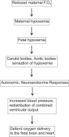
Fig. 1.
Fig. 1.
The generally accepted mechanism of the cardiovascular response to transient hypoxic hypoxia involves stimulation of fetal arterial chemoreceptors, increase in sympathetic nervous system activity, and secretion of neurohormones (e.g., arginine vasopressin and epinephrine) that redistribute fetal combined ventricular output to support oxygen delivery to the fetal heart and brain. FiO2, fraction of inspired oxygen.
Fig. 2.
Fig. 2.
Influence of altitude on partial pressure of oxygen in arterial blood of men exposed to transient hypobaric conditions that mimic exposure to high altitude (100), men breathing ambient air during ascent of Mount Everest (127), pregnant women at 400 and 3,600 m (97), pregnant sheep at 3,600 m (54), nonpregnant sheep at 1,600 and 4,340 m (6), and pregnant sheep at 3,820 m (66). As ambient pressure decreases, arterial oxygen partial pressure decreases, as predicted by ambient pressures and the alveolar gas equation, regardless of species or pregnant state.
Fig. 3.
Fig. 3.
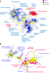
A: network of gene ontology (GO) significantly overrepresented by hypothalamic genes upregulated 1 h (red) and 24 h (blue) or both 1 and 24 h (yellow) after 30 min of transient hypoxic hypoxia in late-gestation fetal sheep. Statistical significance of the individual GO terms, represented as nodes in the merged network, is indicated by intensity of color (higher intensity represents greater statistical significance). GO terms were inferred using BiNGO (80) and networks were inferred using GeneMania (126), plug-in programs for the Cytoscape (113) framework. B: network analysis of significantly overrepresented GO terms were performed as described for upregulated genes in the cerebral cortex after hypoxic hypoxia, comparing upregulated processes 1 h (red color) and 24 h (blue color) or both 1 and 24 h (yellow color) after 30 min of partial umbilical cord occlusion. TLR, Toll-like receptor.
Fig. 4.
Fig. 4.
Venn diagram illustrating overlap of upregulated gene expression 24 h after transient hypoxia in the hypothalamus (blue), hippocampus (yellow), and cerebral cortex (green). The 91 upregulated genes that were common to the 3 brain regions were significantly associated with gene ontology terms related to inflammation and bacterial invasion.
Fig. 5.
Fig. 5.
Proposed mechanism of bacterial invasion of the fetus after transient hypoxia. We propose that bacteria (shown disproportionate to size as green round shapes) in the placenta (1) transit into the fetal circulation and transit to the fetal brain, as well as other organs, perhaps including the kidney and spleen. Bacteria released from the placenta during or after transient hypoxia may have originated from other commensurate bacterial populations in the mother, including the mouth, gastrointestinal tract, or vagina. We and others have demonstrated that hypoxia increases permeability (leakiness of the fetal blood-brain barrier to plasma protein), which may be important for transit of bacteria from fetal blood into fetal brain tissue. We propose that hypoxia may similarly increase vascular permeability to proteins (and perhaps microorganisms) in the placenta.
Fig. 6.
Fig. 6.
Result of a consensus-weighted gene coexpression analysis of ontogenetic trajectories of gene expression for immune-related genes in 4 fetal/neonatal brain regions (cerebral cortex, hypothalamus, hippocampus, and medullary brain stem). Expression of genes related to the hematopoietic lineage increases with fetal age and on day 1 of extrauterine life (blue line and symbols). Expression of genes related to immune system activation is strongly induced after birth (red line and symbols). Values represent average gene expression (log2 of signal intensity). Graph is redrawn from previously reported data (99).
Fig. 7.
Fig. 7.
Transient hypoxia stimulates short- and long-term responses. Short-term responses (changes in autonomic nervous tone, redistribution of fetal combined ventricular output toward heart and brain, and secretion of vasoregulatory and glucose regulatory hormones) aid in short-term survival of the fetus. Longer-term responses (hormonal effects on development, tissue inflammation, and exposure of the fetus to bacteria liberated from the placenta) are either less well understood (e.g., effects of fetal and maternal hormones on fetal programming) or only recently discovered (exposure of the fetus to bacteria after transient hypoxia).

References

    1. Aagaard K, Ma J, Antony KM, Ganu R, Petrosino J, Versalovic J. The placenta harbors a unique microbiome. Sci Transl Med 6: 237ra265, 2014. doi:10.1126/scitranslmed.3008599. - DOI - PMC - PubMed
    1. Alonso JG, Okai T, Longo LD, Gilbert RD. Cardiac function during long-term hypoxemia in fetal sheep. Am J Physiol Heart Circ Physiol 257: H581–H589, 1989. doi:10.1152/ajpheart.1989.257.2.H581. - DOI - PubMed
    1. Baburamani AA, Ek CJ, Walker DW, Castillo-Melendez M. Vulnerability of the developing brain to hypoxic-ischemic damage: contribution of the cerebral vasculature to injury and repair? Front Physiol 3: 424, 2012. doi:10.3389/fphys.2012.00424. - DOI - PMC - PubMed
    1. Barcroft J, Barron DH, Cowie AT, Forsham PH. The oxygen supply of the foetal brain of the sheep and the effect of asphyxia on foetal respiratory movement. J Physiol 97: 338–346, 1940. doi:10.1113/jphysiol.1940.sp003812. - DOI - PMC - PubMed
    1. Barron DH, Battaglia FC. The oxygen concentration gradient between the plasmas in the maternal and fetal capillaries of the placenta of the rabbit. Yale J Biol Med 28: 197–207, 1955. - PMC - PubMed