. 2019 May 31;294(22):8991-9006.
doi: 10.1074/jbc.RA118.005804. Epub 2019 Apr 18.

The redox regulator sulfiredoxin forms a complex with thioredoxin domain-containing 5 protein in response to ER stress in lung cancer cells

Affiliations

The redox regulator sulfiredoxin forms a complex with thioredoxin domain-containing 5 protein in response to ER stress in lung cancer cells

Hedy A Chawsheen et al. J Biol Chem. .

Abstract

Sulfiredoxin (Srx) reduces hyperoxidized 2-cysteine-containing peroxiredoxins (Prxs) and protects cells against oxidative stress. Previous studies have shown that Srx is highly expressed in primary specimens of lung cancer patients and plays a pivotal role in lung tumorigenesis and cancer progression. However, the oncogenic mechanisms of Srx in cancer are incompletely understood. In this study, we found that Srx knockdown sensitizes lung cancer cells to endoplasmic reticulum (ER) stress-induced cell death. Through MS analysis, we determined that Srx forms a complex with the ER-resident protein thioredoxin domain-containing protein 5 (TXNDC5). Using reciprocal co-immunoprecipitation, immunofluorescence imaging, subcellular fractionation, and domain-mapping assays with site-specific mutagenesis and purified recombinant proteins, we further characterized the Srx-TXNDC5 interaction. In response to ER stress but not to oxidative stress, Srx exhibits an increased association with TXNDC5, facilitating the retention of Srx in the ER. Of note, TXNDC5 knockdown in lung cancer cells inhibited cell proliferation and repressed anchorage-independent colony formation and migration, but increased cell invasion and activation of mitogen-activated protein kinases. Using immunohistochemical staining, we demonstrate that TXNDC5 is highly expressed in patient-derived lung cancer specimens. Bioinformatics analysis of publicly available data sets revealed that those with high Srx levels have significantly shorter survival and that those with high TXNDC5 levels have longer survival. We conclude that the cellular levels of Srx and TXNDC5 may be useful as biomarkers to predict the survival of individuals with lung cancer.

Keywords: antioxidant; enzyme mechanism; oxidative stress; protein-disulfide isomerase; protein-protein interaction; proteomics; pulmonary carcinoma; sulfiredoxin; thioredoxin-domain containing 5 (TXNDC5); tumorigenesis.


Conflict of interest statement

The authors declare that they have no conflicts of interest with the contents of this article

Figures

Figure 1.
Figure 1.
Knockdown of Srx sensitizes human lung cancer A549 cells to ER stress–induced cell death. A, representative results of Srx knockdown using two different shSrx constructs in human lung cancer cells. B, knockdown of Srx sensitizes cells to tunicamycin-induced death. Data from six replicates are presented as mean ± S.D. (error bars), and the IC50 values were calculated through log transformation and linear regression analysis. Calculated IC50 values of control cells (ShNT) and Srx knockdown cells (ShSrx) are 1.69 and 0.36 μg/ml, respectively. C, knockdown of Srx induces a rapid response of UPR, as indicated by the accelerated splicing of XBP-1 mRNA. D, expression of spliced XBP protein and activation of ATF6α in the presence of tunicamycin in A549 control and Srx knockdown cells. NS, nonspecific band. The bar graph with a dot plot on the right indicates the quantitative results (*, p < 0.05, t test).
Figure 2.
Figure 2.
Identification of TXNDC5 as an interacting protein of Srx. A, silver staining of protein pulldown by anti-FLAG-Srx IP in human HEK293T cells. The identity of the differential band was determined by RPLC-MS analysis. B, Srx and TXNDC5 were pulled down by reciprocal co-IP in HEK293T and A549 cells. The numbers indicate independent, experimental repeats.
Figure 3.
Figure 3.
Colocalization of Srx and TXNDC5 in the ER. A, subcellular fractionation and Western blotting indicate the presence of Srx and TXNDC5 in the ER. The numbers above the bands indicate independent, experimental repeats. The bar graph with a dot plot on the right indicates the quantitative results. B, immunofluorescence staining of overexpressed FLAG-Srx (red) in HEK293T cells and its colocalization with endogenous TXNDC5 (green) in the ER. The appearance of yellow in the merged image indicates possible colocalization. C, immunofluorescence staining of endogenous Srx (red) in A549 cells and its colocalization with endogenous TXNDC5 (green) in the ER. In these results, endogenously expressed calnexin, an ER-resident protein, is used as a specific marker that is only present in the ER. Error bars, S.D.
Figure 4.
Figure 4.
Characterization and mapping of domains in TXNDC5 that directly interact with Srx. A, recombinant human Srx and TXNDC5 were purified from E. coli and visualized by Coomassie Blue staining (left). The purified proteins were mixed in the binding solution. Reciprocal IP was performed and examined by Western blotting (right). B, predicted interaction of Srx and TXNDC5 based on their structures using I-TASSER and ZDOCK software. The thioredoxin domains (with the key CGHC motif) in TXNDC5 are highlighted in red. The prediction indicates that Srx (blue) interacts with TXNDC5 (green) through the first and the third thioredoxin domain of TXNDC5. C, plasmids that express c-Myc–tagged TXNDC5 or its deletion mutants were expressed in HEK293T-FLAGSrx cells, and cell lysates were used in anti-FLAG IP and examined by Western blotting. Deletion of the first or the third CGHC motif in TXNDC5 leads to significant loss of binding to Srx. D, plasmids that express c-Myc–tagged TXNDC5 or its cysteine-specific mutants were expressed in HEK293T-FLAGSrx cells, and cell lysates were used for anti-FLAG IP and examined by Western blotting. Mutation of two cysteines in the first or the third CGHC motif in TXNDC5 leads to significant loss of binding to Srx. The bar graph with a dot plot on the right indicates the quantitative results (compared with WT; *, p < 0.05, t test). Error bars, S.D.
Figure 5.
Figure 5.
The Srx–TXNDC5 interaction is not affected by the treatment of cells with exogenous H2O2. A, HEK293-FLAGSrx or A549 cells were treated with vehicle or increasing concentrations of H2O2 for 10 min. Cell lysates were collected, and IPs were performed using anti-FLAG or anti-Srx antibodies. IP eluates were separated by SDS-PAGE under reducing conditions. Western blotting results indicate that treatment of cells with H2O2 does not affect the amount of endogenous TXNDC5 pulldown by FLAG-Srx in HEK293T or Srx in A549 cells. B, IP eluates from A were separated by SDS-PAGE under nonreducing conditions, and Western blotting indicates the position of monomer as well as possible disulfide bond formation between Srx and TXNDC5 as bands indicated by an asterisk. C, FLAG-Srx or its cysteine mutant (C99A) was expressed in HEK293T cells. Cell lysates were collected and IPs were performed using anti-FLAG antibody. IP eluates were separated by SDS-PAGE under reducing conditions. Western blotting results indicate that mutation of cysteine 99 in Srx does not affect its ability to interact with TXNDC5.
Figure 6.
Figure 6.
Knockdown of TXNDC5 in lung cancer cells leads to more localization of Srx in the cytosol. A, two shRNAs targeting different coding regions of TXNDC5 were used to establish stable knockdown in A549 cells. Knockdown of TXNDC5 does not affect the endogenous expression of Srx or TXNDC7, a close member of TXNDC5 in the PDI family. B, subcellular fractionation of A549 control (ShNT) or TXNDC5 knockdown cells for the distribution of Srx in ER and cytosol. Results from three independent replicates were shown. The bar graph with a dot plot on the right indicates the quantitative results (*, p < 0.05, ANOVA). Error bars, S.D.
Figure 7.
Figure 7.
Knockdown of TXNDC5 sensitizes human lung cancer cells to ER stress–induced cell death. A, knockdown of TXNDC5 (ShTX) sensitizes cells to tunicamycin-induced death. Data from six replicates are shown. B, knockdown of TXNDC5 induces a rapid response to UPR, as indicated by the accelerated splicing of XBP-1 and activation of ATF6α in the presence of tunicamycin. NS, nonspecific band. The bar graph with a dot plot indicates the quantitative results (*, p < 0.05, one-way ANOVA). Error bars, S.D.
Figure 8.
Figure 8.
Knockdown of TXNDC5 in A549 cells inhibits cell proliferation but promotes cell invasion. A, representative results of cell proliferation evaluated by the modified XTT assay. B, anchorage-independent colony formation in soft agar. C and D, wound-healing assay as shown in culture (C) and quantitation (D). E and F, Matrigel invasion assay showing invaded cells (C) and quantitation (D). The bar graph with a dot plot indicates the quantitative results (*, p < 0.05; NS, no significance; one-way ANOVA). Error bars, S.D.
Figure 9.
Figure 9.
Knockdown of TXNDC5 enhances EGF-induced MAPK activation. A549 control and TXNDC5 knockdown cells were serum-starved overnight and treated with fresh medium containing EGF for the indicated period of time. Cell lysates were harvested for Western blotting. Quantitative results of ERK1/2 activation were shown on the right. *, significant difference by comparing the area under the curve (*, p < 0.05, t test). Error bars, S.D.
Figure 10.
Figure 10.
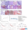
Expression profile of TXNDC5 in human normal tissue and lung cancer. A and B, anti-TXNDC5 staining (brown) with counterstaining by hematoxylin (blue) was performed on tissue microarray slides of human normal organs (A) and lung normal and lung tumor specimens from patients (B). C, quantitative data from tissue microarray were shown. * p < 0.05 (t test), compared with normal lung. D, an example data set from Oncomine indicates amplified DNA copy numbers in human lung cancer. E and F, Kaplan–Meier Plotter analysis of the TCGA data set in human lung cancer indicates the negative association of Srx with patient survival (E) and the positive association of TXNDC5 with patient survival (F). G, a proposed model of Srx and TXNDC5 in lung cancer and their value as potential biomarkers to predict patient survival. Error bars, S.D.

Similar articles

Cited by

References

    1. Kinnula V. L., Pääkkö P., and Soini Y. (2004) Antioxidant enzymes and redox regulating thiol proteins in malignancies of human lung. FEBS Lett. 569, 1–6 10.1016/j.febslet.2004.05.045 - DOI - PubMed
    1. Hampton M. B., Vick K. A., Skoko J. J., and Neumann C. A. (2018) Peroxiredoxin Involvement in the Initiation and Progression of Human Cancer. Antioxid. Redox Signal. 28, 591–608 10.1089/ars.2017.7422 - DOI - PMC - PubMed
    1. Jiang H., Wu L., Mishra M., Chawsheen H. A., and Wei Q. (2014) Expression of peroxiredoxin 1 and 4 promotes human lung cancer malignancy. Am. J. Cancer Res. 4, 445–460 - PMC - PubMed
    1. Kim J. H., Bogner P. N., Baek S. H., Ramnath N., Liang P., Kim H. R., Andrews C., and Park Y. M. (2008) Up-regulation of peroxiredoxin 1 in lung cancer and its implication as a prognostic and therapeutic target. Clin. Cancer Res. 14, 2326–2333 10.1158/1078-0432.CCR-07-4457 - DOI - PubMed
    1. Hwang J. A., Song J. S., Yu D. Y., Kim H. R., Park H. J., Park Y. S., Kim W. S., and Choi C. M. (2015) Peroxiredoxin 4 as an independent prognostic marker for survival in patients with early-stage lung squamous cell carcinoma. Int. J. Clin. Exp. Pathol. 8, 6627–6635 - PMC - PubMed

Publication types

MeSH terms

LinkOut - more resources