. 2019 Jun 14;93(13):e00388-19.
doi: 10.1128/JVI.00388-19. Print 2019 Jul 1.

Complex Membrane Remodeling during Virion Assembly of the 30,000-Year-Old Mollivirus Sibericum

Affiliations

Complex Membrane Remodeling during Virion Assembly of the 30,000-Year-Old Mollivirus Sibericum

E R Quemin et al. J Virol. .

Abstract

Cellular membranes ensure functional compartmentalization by dynamic fusion-fission remodeling and are often targeted by viruses during entry, replication, assembly, and egress. Nucleocytoplasmic large DNA viruses (NCLDVs) can recruit host-derived open membrane precursors to form their inner viral membrane. Using complementary three-dimensional (3D)-electron microscopy techniques, including focused-ion beam scanning electron microscopy and electron tomography, we show that the giant Mollivirus sibericum utilizes the same strategy but also displays unique features. Indeed, assembly is specifically triggered by an open cisterna with a flat pole in its center and open curling ends that grow by recruitment of vesicles never reported for NCLDVs. These vesicles, abundant in the viral factory (VF), are initially closed but open once in close proximity to the open curling ends of the growing viral membrane. The flat pole appears to play a central role during the entire virus assembly process. While additional capsid layers are assembled from it, it also shapes the growing cisterna into immature crescent-like virions and is located opposite to the membrane elongation and closure sites, thereby providing virions with a polarity. In the VF, DNA-associated filaments are abundant, and DNA is packed within virions prior to particle closure. Altogether, our results highlight the complexity of the interaction between giant viruses and their host. Mollivirus assembly relies on the general strategy of vesicle recruitment, opening, and shaping by capsid layers similar to all NCLDVs studied until now. However, the specific features of its assembly suggest that the molecular mechanisms for cellular membrane remodeling and persistence are unique.IMPORTANCE Since the first giant virus Mimivirus was identified, other giant representatives are isolated regularly around the world and appear to be unique in several aspects. They belong to at least four viral families, and the ways they interact with their hosts remain poorly understood. We focused on Mollivirus sibericum, the sole representative of "Molliviridae," which was isolated from a 30,000-year-old permafrost sample and exhibits spherical virions of complex composition. In particular, we show that (i) assembly is initiated by a unique structure containing a flat pole positioned at the center of an open cisterna, (ii) core packing involves another cisterna-like element seemingly pushing core proteins into particles being assembled, and (iii) specific filamentous structures contain the viral genome before packaging. Altogether, our findings increase our understanding of how complex giant viruses interact with their host and provide the foundation for future studies to elucidate the molecular mechanisms of Mollivirus assembly.

Keywords: Mollivirus sibericum; electron tomography; focused-ion beam scanning electron microscopy; giant viruses; membrane remodeling; nucleocytoplasmic large DNA viruses; viral factory; virus assembly.


Figures

FIG 1
FIG 1
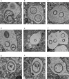
Transmission electron micrographs on the entry of Mollivirus sibericum. At 3 h postinfection, cells of Acanthamoeba castellanii were fixed and processed for transmission electron microscopy after cryo-fixation and freeze substitution. (a to i) Entry of Mollivirus sibericum leads to substitution of the viral inner content (a) and occurs via endocytosis with multiple particles present in one vacuole with different shape and content (b to f) and in individual particles with internal structures (g to i).
FIG 2
FIG 2
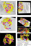
Analysis of whole infected cells by focused-ion beam scanning electron microscopy. At 8 h postinfection, cells of Acanthamoeba castellanii were fixed and processed for focused-ion beam scanning electron microscopy. (a) Slice 535 through the reconstructed volume (acquired at 10 by 10 by 10 nm) with 3D surface rendering of infected cells. Scale bar = 2 μm. (b) Slice 242 of the acquired volume and 3D surface rendering. (c) Projection with 3D surface rendering. (d) Slice 592 through the reconstructed volume (acquired at 10 by 10 by 10 nm) with 3D surface rendering of infected cells. Scale bar = 1 μm. (e) Slice 592 of the acquired volume and 3D surface rendering at different angles. (a to e) The video of volume reconstruction with segmentation is shown in Movie S2 (a to c) and Movie S3 (d to e). As indicated in the key, mitochondria are in yellow, endoplasmic reticulum is in black, vesicles are in green, empty virions are in magenta, mature virions are in dark blue, closure of virions is in red, viral crescents are in orange, and membrane precursors are in cyan.
FIG 3
FIG 3
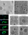
Observations of the different assembly stages of Mollivirus sibericum. At 8 h postinfection, cells of Acanthamoeba castellanii were fixed and processed for scanning transmission electron tomography and transmission electron microscopy after cryo-fixation and freeze substitution. (a) Slice 40 through a tomogram acquired by scanning transmission electron tomography; bottom, projections of 3D surface rendering of particles being assembled. A detailed overview of slices through the tomogram including 3D surface rendering is shown in Fig. 8. The video of the reconstructed volume with segmentation is shown in Movie S4. The tegument is in blue, additional capsid layers are in dark green, and the inner viral membrane is in light green. Scale bar = 300 nm. (b) Different stages of viral assembly can be observed by transmission electron microscopy, as follows: 1, curved membrane intermediates with open curling ends associated with a flat pole or covered with the tegument and additional capsid layers; 2, typical open crescents associated with cisternae with segregated ribosomes; 3, closure of particles beginning with the inner viral membrane on the opposite side of the flat pole; and 4, mature particles with thick tegument covered with fibers and encasing additional capsid layers, the inner viral membrane, and dense material. Scale bar = 500 nm. (c) Enlargements of the particles at different stages of assembly as depicted by black squares in panel b. Membrane precursors are indicated by white stars in squares 1 and 3, the cisternae associated with viral crescents and segregated ribosomes are indicated by white arrowheads in square 2, the flat pole is indicated by a black bracket in square 3, and the different capsid layers are numbered in square 4. Note the top views and tangential planes of particles in squares 3 and 4 which are indicated by a white pound sign and show the striations of the capsid outer surface.
FIG 4
FIG 4
DNA-associated filaments involved in packaging during assembly of Mollivirus sibericum. At 8 h postinfection, cells of Acanthamoeba castellanii were fixed and processed for immunolabeling of thawed cryo-sections. DNA labeling is associated with viral filamentous structures and present in particles at the end of the assembly. Scale bar = 500 nm.
FIG 5
FIG 5
3D analysis of membrane precursor during assembly of Mollivirus sibericum particles. At 8 h postinfection, cells of Acanthamoeba castellanii were fixed and processed for transmission electron tomography. (a) Slices 12 (left) and 94 (right) through the tomogram showing curved membrane intermediates, with open curling ends associated with a flat pole and typical open crescents associated with cisternae. Scale bars = 200 nm. The flat pole is indicated by a black bracket. (b) Projection images of 3D surface rendering of the particle shown in panel a with different angles. The video of volume reconstruction with segmentation is shown in Movie S5. The tegument is in blue, the additional capsid layers are in dark green, the inner viral membrane is in light green, and the flat pole is in orange.
FIG 6
FIG 6
Transmission electron micrographs showing the flat pole on viral crescents. At 8 h postinfection, cells of Acanthamoeba castellanii were fixed and processed for transmission electron microscopy after substitution at room temperature. Different stages of virus assembly are shown. The flat pole, indicated by a black bracket, is always visible at the opposite of the site where assembly proceeds. Scale bars = 200 nm (a and b) or 100 nm (c).
FIG 7
FIG 7
3D analysis of viral crescent during assembly of Mollivirus sibericum particles. At 8 h postinfection, cells of Acanthamoeba castellanii were fixed and processed for transmission electron tomography. (a) Slices 16, 24, and 56 (from left to right) through the tomogram showing a typical viral crescent associated with a cisterna and displaying open curling ends. On the open curling end highlighted with a black square, a vesicle comes into contact with the inner membrane and subsequently opens up. Scale bars = 200 nm. The flat pole is indicated by black bracket, and one coated vesicle is labeled CV. (b) Projection images of 3D surface rendering of the particle shown in panel a with different angles. The video of volume reconstruction with segmentation is shown in Movie S5. The tegument is in blue, the additional capsid layers are in dark green, the inner viral membrane is in light green, the flat pole is in orange, and the cisternae are in red.
FIG 8
FIG 8
Elongation of the viral inner membrane is triggered by binding to and opening of vesicles. At 8 h postinfection, cells of Acanthamoeba castellanii were fixed and processed for transmission electron microscopy after cryo-fixation and freeze substitution. Slices 20, 40, 60, and 80 (from top to bottom) through the tomogram are shown. The video of the reconstructed volume with segmentation is shown in Movie S4. On the open curling end highlighted with a black square, a vesicle comes into contact with the inner membrane. Scale bars = 200 nm. The tegument is in blue, additional capsid layers are in dark green, and the inner viral membrane is in light green. On the open curling end of a viral crescent highlighted with a black square, a vesicle comes into contact with the inner membrane.
FIG 9
FIG 9
Coassembly of multiple particles of Mollivirus sibericum. At 8 h postinfection, cells of Acanthamoeba castellanii were fixed and processed for transmission electron tomography. (a) Slices 17 (left) and 80 (right) through the tomogram showing three particles being coassembled, including two connected through the inner viral membrane. Scale bars = 200 nm. (b) Projection images of 3D surface rendering of the particle shown in panel a with different angles. The video of volume reconstruction with segmentation is shown in Movie S7. The tegument is in blue, the additional capsid layers are in dark green, the inner viral membrane is in light green, and the cisternae are in red.
FIG 10
FIG 10
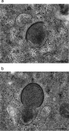
Viral crescents are associated with cisterna-like structures. At 8 h postinfection, cells of Acanthamoeba castellanii were fixed and processed for transmission electron microscopy after cryo-fixation and freeze substitution. The assembly of viral crescents early (a) or late (b) is associated with cisterna-like structures which are seemingly pushing the viroplasm inside the particle being assembled. Scale bars = 250 nm. The cisternae associated with viral crescents with segregated ribosomes are indicated by white arrowheads.
FIG 11
FIG 11
3D analysis of closure of particles during assembly of Mollivirus sibericum particles. At 8 h postinfection, cells of Acanthamoeba castellanii were fixed and processed for transmission electron tomography. (a) Slices 53 (left) and 85 (right) through the tomogram showing closure of the inner membrane of particles. Scale bars = 200 nm. (b) Projection images of 3D surface rendering of the particle shown in panel a with different angles. The video of volume reconstruction with segmentation is shown in Movie S6. The tegument is in blue, the additional capsid layers are in dark green, and the inner viral membrane is in light green. Due to the orientation of the particle in the tomogram, the flat-shaped pole is not visible and therefore not represented.
FIG 12
FIG 12
DNA packaging occurs at the end of the assembly of Mollivirus sibericum. At 8 h postinfection, cells of Acanthamoeba castellanii were fixed and processed for immunolabeling of thawed cryo-sections. DNA labeling is absent from half-assembled particles (a) and observed inside mature and associated with viral filamentous structures (b and c). Scale bars = 200 nm (a) and 500 nm (b and c). The flat pole is indicated by a black bracket.
FIG 13
FIG 13
Comparison of virions of Mollivirus sibericum prepared by classical embedding or the Tokuyasu method. At 8 h postinfection, cells of Acanthamoeba castellanii were processed for transmission electron microscopy after either cryo-fixation and freeze-substitution (left) or fixation and immunolabeling of thawed cryo-sections (right). The white arrows indicate the appearance of the two capsid layers by the classical embedding method and in the Tokuyasu-prepared sample, which resemble membranes. Scale bars = 200 nm (A) and 250 nm (B).
FIG 14
FIG 14
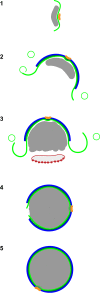
Schematic model of the assembly of Mollivirus sibericum. (1) Membrane assembly is initiated by an open cisternal element (light green) containing a flat pole (orange) at its center, and core proteins (gray) are recruited at the opposite side of the membrane. (2) From this pole, additional protein layers, the coat (light blue) and inner capsid layer (dark green), are recruited and shape the growing membrane into an open sphere or crescent. (3) The membrane grows by recruitment of vesicles that open upon contacting the open ends and final packaging of viral core proteins in crescents by a cisterna (red) located at the exact opposite of the flat pole; note that ribosomes are segregated on the side facing the cytoplasm. (4) Elongation of the membrane, packing of core proteins, and recruitment of the tegument layer result in an almost completed particle with a remaining opening located at the opposite side of the flat pole, likely required to take up the viral genome (not shown). (5) Closure of the internal membrane with removal of material in excess.

Similar articles

Cited by

References

    1. Schmick M, Bastiaens P. 2014. The interdependence of membrane shape and cellular signal processing. Cell 156:1132–1138. doi:10.1016/j.cell.2014.02.007. - DOI - PubMed
    1. Harayama T, Riezman H. 2018. Understanding the diversity of membrane lipid composition. Nat Rev Mol Cell Biol 19:281–296. doi:10.1038/nrm.2017.138. - DOI - PubMed
    1. Yamauchi Y, Helenius A. 2013. Virus entry at a glance. J Cell Sci 126:1289–1295. doi:10.1242/jcs.119685. - DOI - PubMed
    1. Reid CR, Airo AM, Hobman TC. 2015. The virus-host interplay: biogenesis of +RNA replication complexes. Viruses 7:4385–4413. doi:10.3390/v7082825. - DOI - PMC - PubMed
    1. Rossman JS, Lamb RA. 2011. Influenza virus assembly and budding. Virology 411:229–236. doi:10.1016/j.virol.2010.12.003. - DOI - PMC - PubMed

Publication types

MeSH terms

LinkOut - more resources