

Microcomputed tomography of the femur of diabetic rats: alterations of trabecular and cortical bone microarchitecture and vasculature-a feasibility study
- PMID: 30972589
- PMCID: PMC6458201
- DOI: 10.1186/s41747-019-0094-5
Microcomputed tomography of the femur of diabetic rats: alterations of trabecular and cortical bone microarchitecture and vasculature-a feasibility study
Abstract
Background: To better understand bone fragility in type 2 diabetes mellitus and define the contribution of microcomputed tomography (micro-CT) to the evaluation of bone microarchitecture and vascularisation, we conducted an in vitro preliminary study on the femur of Zucker diabetic fatty (ZDF) rats and Zucker lean (ZL) rats. We first analysed bone microarchitecture, then determined whether micro-CT allowed to explore bone vascularisation, and finally looked for a link between these parameters.
Methods: Eight ZDF and six ZL rats were examined for bone microarchitecture (group 1), and six ZDF and six ZL rats were studied for bone vascularisation after Microfil® perfusion which is a radiopaque casting agent (group 2). In group 1, we used micro-CT to examine the trabecular and cortical bone microarchitecture of the femoral head, neck, shaft, and distal metaphysis. In group 2, micro-CT was used to study the blood vessels in the head, neck, and distal metaphysis.
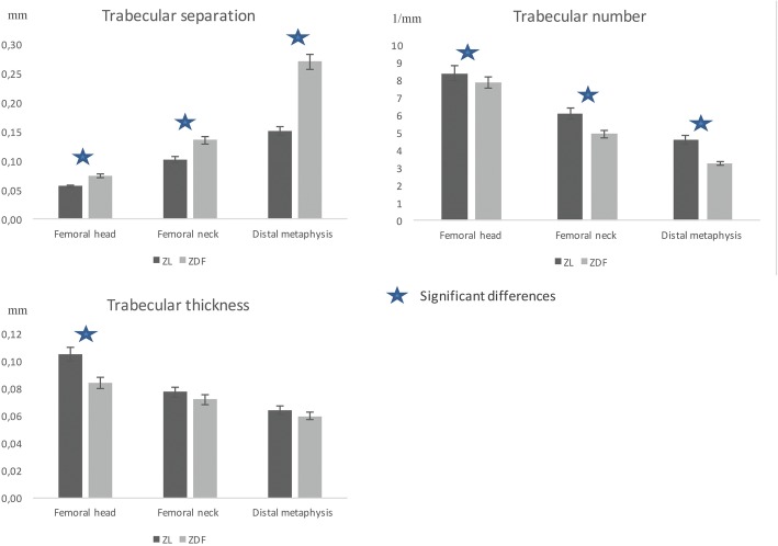
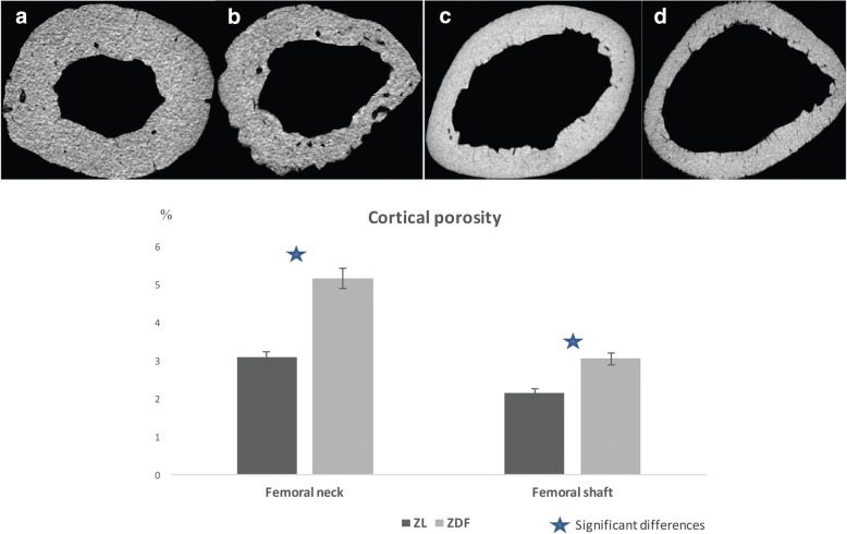
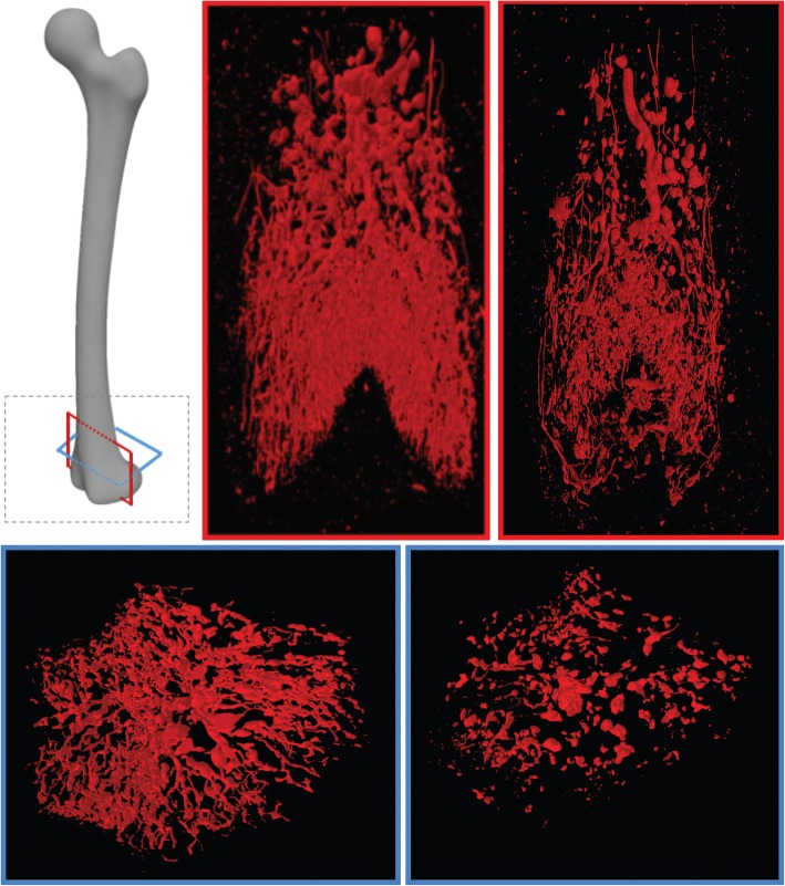
Results: Compared to ZL rats, the ZDF rats exhibited significantly lower trabecular bone volume and number and higher trabecular separation in the three locations (p = 0.02, p = 0.02, p = 0.003). Cortical porosity was significantly higher in the ZDF rats at the neck and shaft (p = 0.001 and p = 0.005). We observed a dramatically poorer bone vascularisation in the femur of ZDF rats, especially in distal metaphysis (p < 0.047).
Conclusions: Micro-CT demonstrated not only significant alterations in the bone microarchitecture of the femurs of ZDF rats, but also significant alterations in bone vascularisation. Further studies are required to demonstrate the causal link between poor vascularisation and impaired bone architecture.
Keywords: Bone and bones; Diabetes mellitus (type 2); Rats (Zucker); Vascular diseases; X-ray microtomography.
Conflict of interest statement
Ethics approval and consent to participate
Ethics Committee of the University Paris Diderot (N°01610.01/S69), Paris, France.
Consent for publication
Not applicable.
Competing interests
The authors declare that they have no competing interests.
Publisher’s Note
Springer Nature remains neutral with regard to jurisdictional claims in published maps and institutional affiliations.
Figures

Similar articles
-
Cortical Bone Morphological and Trabecular Bone Microarchitectural Changes in the Mandible and Femoral Neck of Ovariectomized Rats.PLoS One. 2016 Apr 29;11(4):e0154367. doi: 10.1371/journal.pone.0154367. eCollection 2016. PLoS One. 2016. PMID: 27127909 Free PMC article.
-
Sclerostin does not play a major role in the pathogenesis of skeletal complications in type 2 diabetes mellitus.Osteoporos Int. 2017 Jan;28(1):309-320. doi: 10.1007/s00198-016-3718-0. Epub 2016 Jul 28. Osteoporos Int. 2017. PMID: 27468901 Free PMC article.
-
Abaloparatide improves cortical geometry and trabecular microarchitecture and increases vertebral and femoral neck strength in a rat model of male osteoporosis.Bone. 2019 Jul;124:148-157. doi: 10.1016/j.bone.2019.04.025. Epub 2019 Apr 30. Bone. 2019. PMID: 31051317
-
A comparison of micro-CT and dental CT in assessing cortical bone morphology and trabecular bone microarchitecture.PLoS One. 2014 Sep 16;9(9):e107545. doi: 10.1371/journal.pone.0107545. eCollection 2014. PLoS One. 2014. PMID: 25226587 Free PMC article.
-
Type 2 diabetes impairs angiogenesis and osteogenesis in calvarial defects: MicroCT study in ZDF rats.Bone. 2018 Jul;112:161-172. doi: 10.1016/j.bone.2018.04.009. Epub 2018 Apr 25. Bone. 2018. PMID: 29702250
Cited by
-
Diabetes mellitus impairs bone regeneration and biomechanics.J Orthop Surg Res. 2023 Mar 6;18(1):169. doi: 10.1186/s13018-023-03644-5. J Orthop Surg Res. 2023. PMID: 36872328 Free PMC article.
-
The Hyperglycemia and Hyperketonemia Impaired Bone Microstructures: A Pilot Study in Rats.Front Endocrinol (Lausanne). 2020 Oct 22;11:590575. doi: 10.3389/fendo.2020.590575. eCollection 2020. Front Endocrinol (Lausanne). 2020. PMID: 33193101 Free PMC article.
-
Cornelian Cherry Pulp Has Beneficial Impact on Dyslipidemia and Reduced Bone Quality in Zucker Diabetic Fatty Rats.Animals (Basel). 2020 Dec 19;10(12):2435. doi: 10.3390/ani10122435. Animals (Basel). 2020. PMID: 33352633 Free PMC article.
-
Comparison of Quantitative Computed Tomography and Dual X-Ray Absorptiometry: Osteoporosis Detection Rates in Diabetic Patients.Cureus. 2022 Mar 13;14(3):e23131. doi: 10.7759/cureus.23131. eCollection 2022 Mar. Cureus. 2022. PMID: 35433140 Free PMC article.
-
The Effect of Type 2 Diabetes on Bone Biomechanics.Curr Osteoporos Rep. 2019 Oct;17(5):291-300. doi: 10.1007/s11914-019-00526-w. Curr Osteoporos Rep. 2019. PMID: 31392668 Free PMC article. Review.
References
Publication types
MeSH terms
LinkOut - more resources
Full Text Sources