

Inhibition of Ubiquitin-Specific Protease 14 Suppresses Cell Proliferation and Synergizes with Chemotherapeutic Agents in Neuroblastoma
- PMID: 30962318
- PMCID: PMC6565366
- DOI: 10.1158/1535-7163.MCT-18-0146
Inhibition of Ubiquitin-Specific Protease 14 Suppresses Cell Proliferation and Synergizes with Chemotherapeutic Agents in Neuroblastoma
Abstract
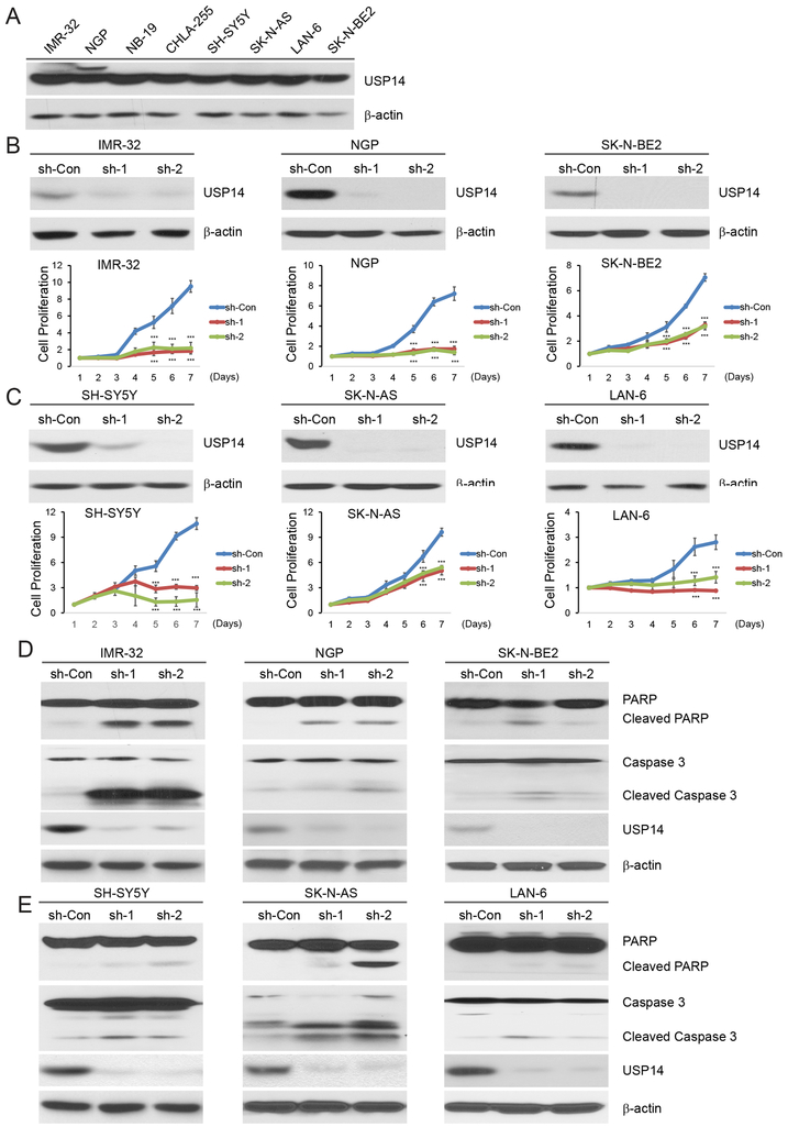
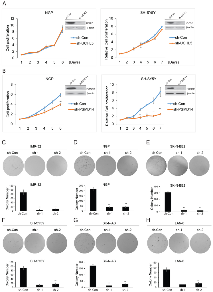
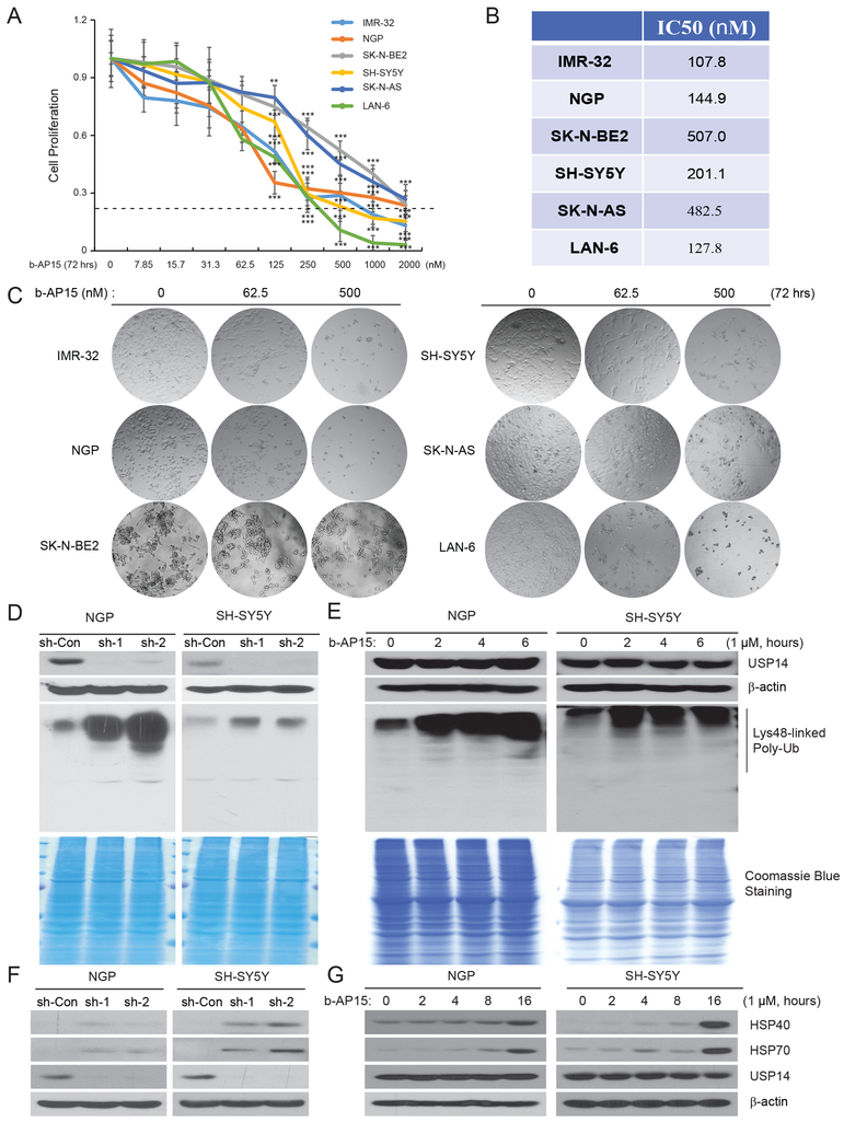
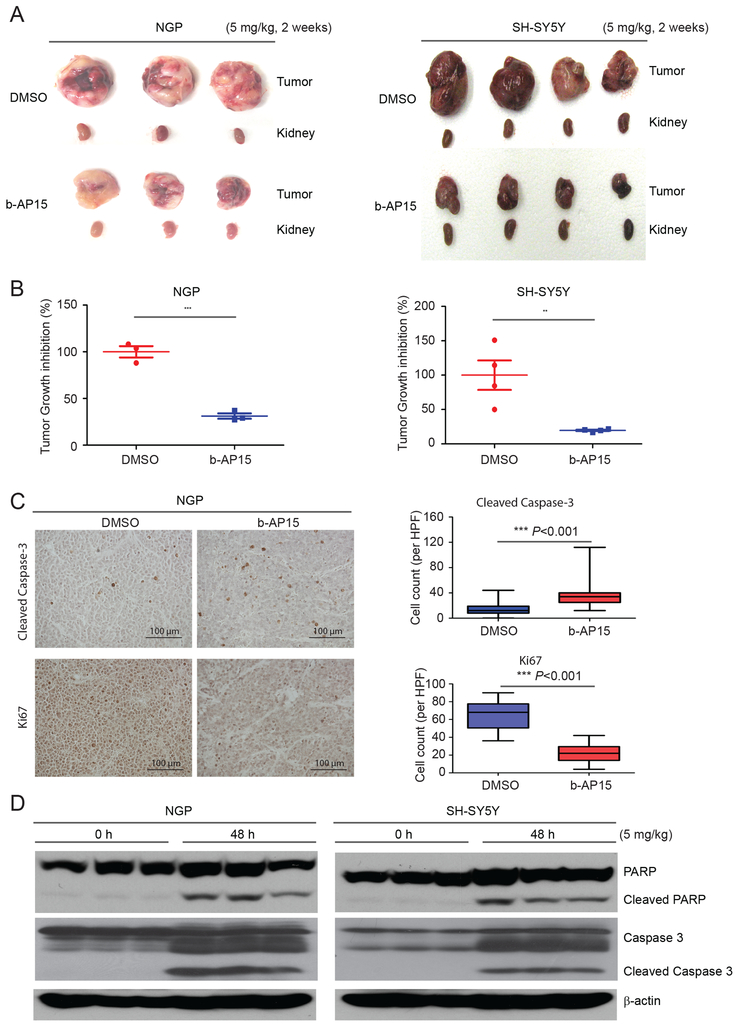
Neuroblastoma is the most common extracranial malignant solid tumor in children, and drug resistance is a major reason for poor outcomes. Elevated proteasome activity plays an important role in neuroblastoma tumor development and resistance to conventional chemotherapy. Ubiquitin-specific protease 14 (USP14), one of three deubiquitinases associated with the regulatory subunit of the proteasome, is emerging as a potential therapeutic target in multiple tumor types. However, the role of USP14 in neuroblastoma is yet to be elucidated. We found that USP14 inhibition in neuroblastoma via knockdown or a specific inhibitor such as b-AP15 suppressed cell proliferation by inducing cell apoptosis. Furthermore, b-AP15 significantly inhibited neuroblastoma tumor growth in NGP and SH-SY5Y xenograft mouse models. For combination treatment, b-AP15 plus conventional chemotherapeutic agents such as doxorubicin or VP-16 resulted in synergistic antitumor effects on neuroblastoma. Our study demonstrates that USP14 is required for cell viability and is a novel therapeutic target in neuroblastoma. Moreover, USP14 inhibition may add value in combination therapy due to its powerful synergistic effects in treating neuroblastoma.
©2019 American Association for Cancer Research.
Conflict of interest statement
The authors declare no conflicts of interest.
Figures
Similar articles
-
Targeting the ubiquitin-proteasome system: a novel therapeutic strategy for neuroblastoma.Front Oncol. 2024 Sep 26;14:1443256. doi: 10.3389/fonc.2024.1443256. eCollection 2024. Front Oncol. 2024. PMID: 39391247 Free PMC article. Review.
-
A novel small molecule inhibitor of deubiquitylating enzyme USP14 and UCHL5 induces apoptosis in multiple myeloma and overcomes bortezomib resistance.Blood. 2014 Jan 30;123(5):706-16. doi: 10.1182/blood-2013-05-500033. Epub 2013 Dec 6. Blood. 2014. PMID: 24319254 Free PMC article.
-
USP7 inhibitor P22077 inhibits neuroblastoma growth via inducing p53-mediated apoptosis.Cell Death Dis. 2013 Oct 17;4(10):e867. doi: 10.1038/cddis.2013.400. Cell Death Dis. 2013. PMID: 24136231 Free PMC article.
-
Ubiquitin-specific protease 14 regulates cell proliferation and apoptosis in oral squamous cell carcinoma.Int J Biochem Cell Biol. 2016 Oct;79:350-359. doi: 10.1016/j.biocel.2016.08.038. Epub 2016 Aug 31. Int J Biochem Cell Biol. 2016. PMID: 27592452
-
Targeted inhibition of the deubiquitinating enzymes, USP14 and UCHL5, induces proteotoxic stress and apoptosis in Waldenström macroglobulinaemia tumour cells.Br J Haematol. 2015 May;169(3):377-90. doi: 10.1111/bjh.13304. Epub 2015 Feb 17. Br J Haematol. 2015. PMID: 25691154 Free PMC article.
Cited by
-
Targeting the ubiquitin-proteasome system: a novel therapeutic strategy for neuroblastoma.Front Oncol. 2024 Sep 26;14:1443256. doi: 10.3389/fonc.2024.1443256. eCollection 2024. Front Oncol. 2024. PMID: 39391247 Free PMC article. Review.
-
CRISPR/Cas9-based genome-wide screening of the deubiquitinase subfamily identifies USP3 as a protein stabilizer of REST blocking neuronal differentiation and promotes neuroblastoma tumorigenesis.J Exp Clin Cancer Res. 2023 May 12;42(1):121. doi: 10.1186/s13046-023-02694-1. J Exp Clin Cancer Res. 2023. PMID: 37170124 Free PMC article.
-
Deubiquitinating Enzymes Orchestrate the Cancer Stem Cell-Immunosuppressive Niche Dialogue: New Perspectives and Therapeutic Potential.Front Cell Dev Biol. 2021 Jun 9;9:680100. doi: 10.3389/fcell.2021.680100. eCollection 2021. Front Cell Dev Biol. 2021. PMID: 34179009 Free PMC article. Review.
-
Analysis of determinants for in vitro resistance to the small molecule deubiquitinase inhibitor b-AP15.PLoS One. 2019 Oct 22;14(10):e0223807. doi: 10.1371/journal.pone.0223807. eCollection 2019. PLoS One. 2019. PMID: 31639138 Free PMC article.
-
Drug resistance mechanisms and treatment strategies mediated by Ubiquitin-Specific Proteases (USPs) in cancers: new directions and therapeutic options.Mol Cancer. 2024 May 3;23(1):88. doi: 10.1186/s12943-024-02005-y. Mol Cancer. 2024. PMID: 38702734 Free PMC article. Review.
References
-
- Castleberry RP. Neuroblastoma. European journal of cancer. 1997;33:1430–7; discussion 7–8. - PubMed
-
- Brodeur GM, Seeger RC, Schwab M, Varmus HE, Bishop JM. Amplification of N-myc in untreated human neuroblastomas correlates with advanced disease stage. Science. 1984;224:1121–4. - PubMed
-
- Look AT, Hayes FA, Nitschke R, McWilliams NB, Green AA. Cellular DNA content as a predictor of response to chemotherapy in infants with unresectable neuroblastoma. The New England journal of medicine. 1984;311:231–5. - PubMed
-
- Fong CT, Dracopoli NC, White PS, Merrill PT, Griffith RC, Housman DE, et al. Loss of heterozygosity for the short arm of chromosome 1 in human neuroblastomas: correlation with N-myc amplification. Proceedings of the National Academy of Sciences of the United States of America. 1989;86:3753–7. - PMC - PubMed
Publication types
MeSH terms
Substances
Grants and funding
LinkOut - more resources
Full Text Sources
Other Literature Sources
Medical
Research Materials
