

Major glucuronide metabolites of testosterone are primarily transported by MRP2 and MRP3 in human liver, intestine and kidney
- PMID: 30959153
- PMCID: PMC7075494
- DOI: 10.1016/j.jsbmb.2019.03.027
Major glucuronide metabolites of testosterone are primarily transported by MRP2 and MRP3 in human liver, intestine and kidney
Abstract
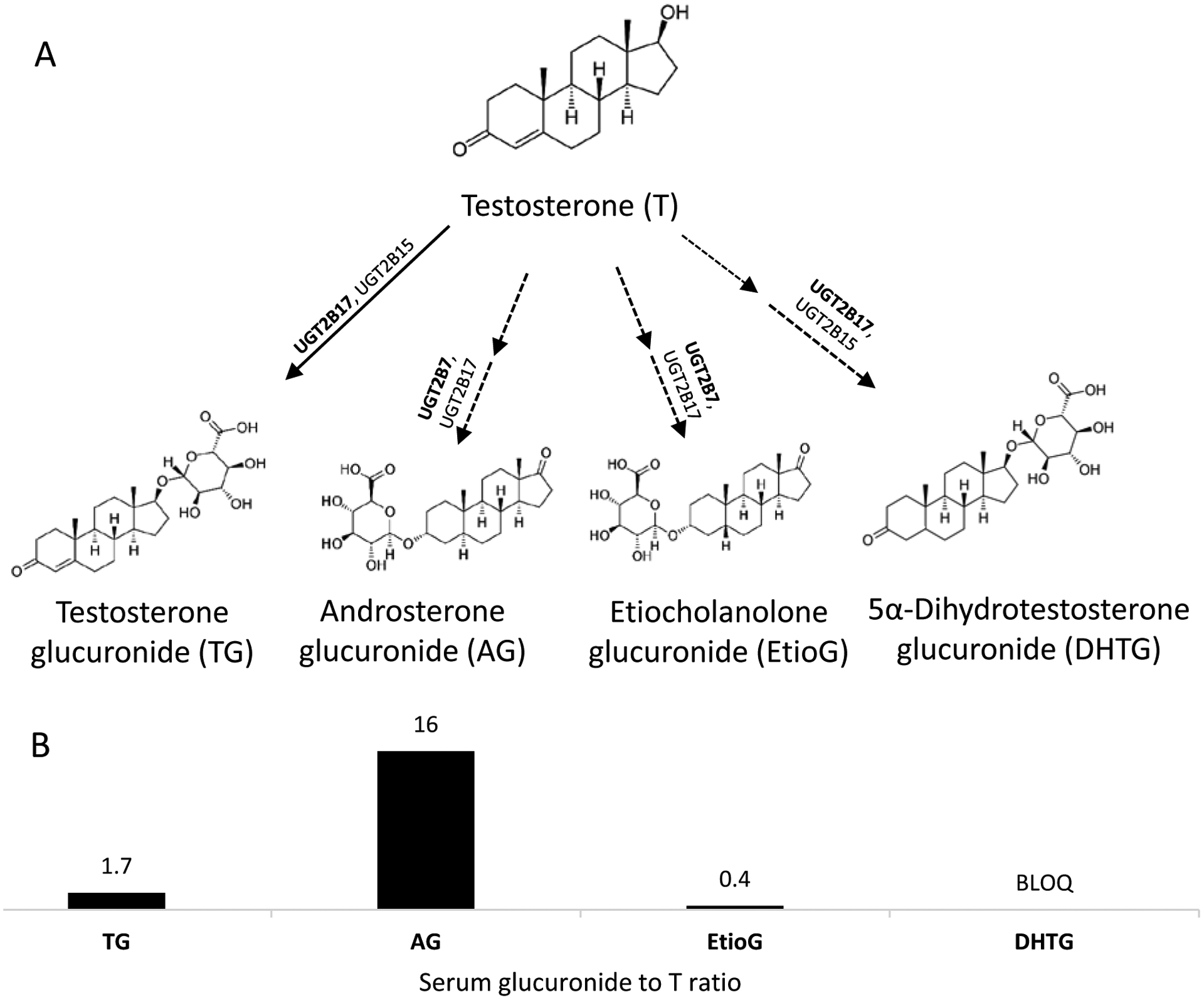
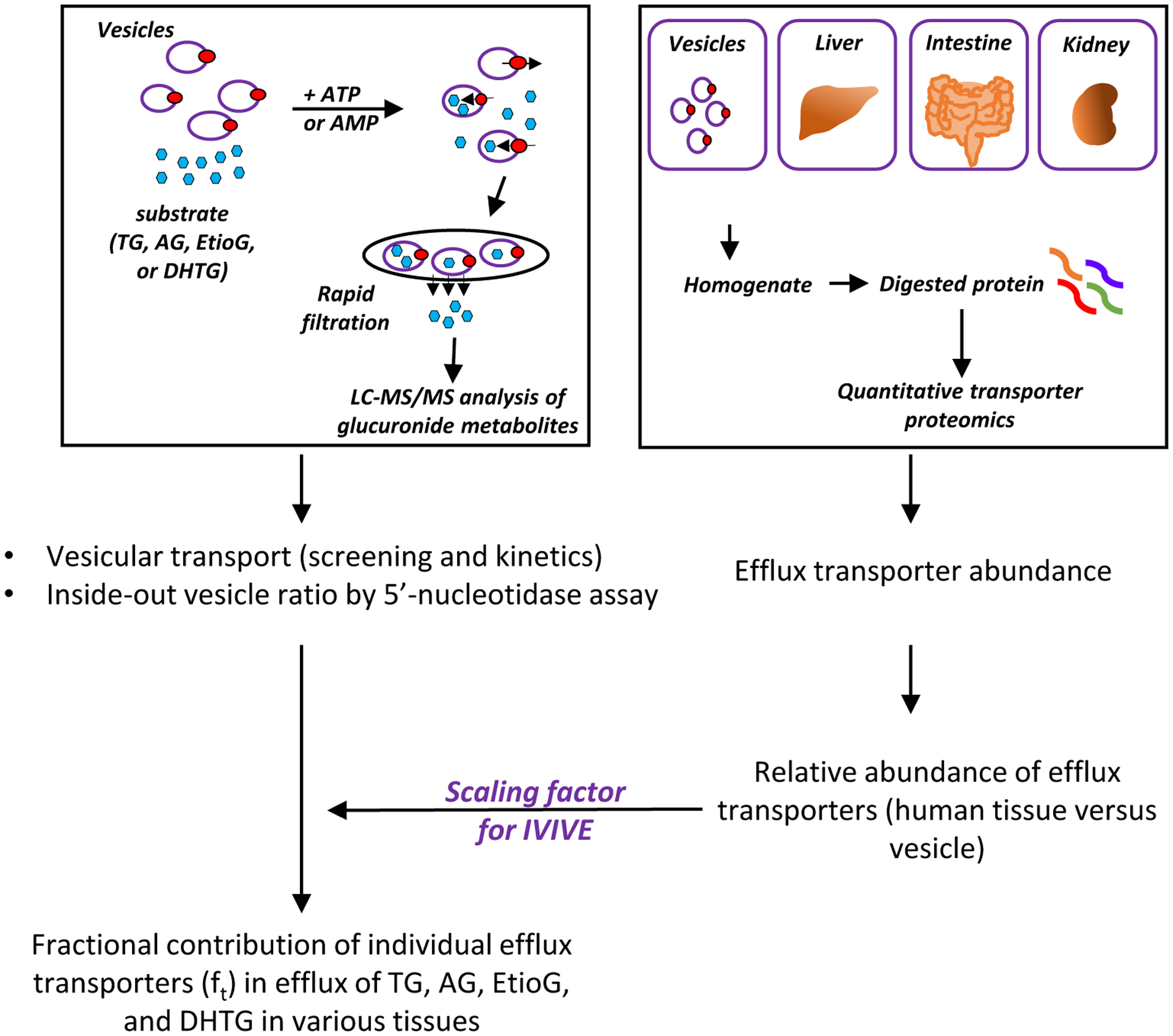
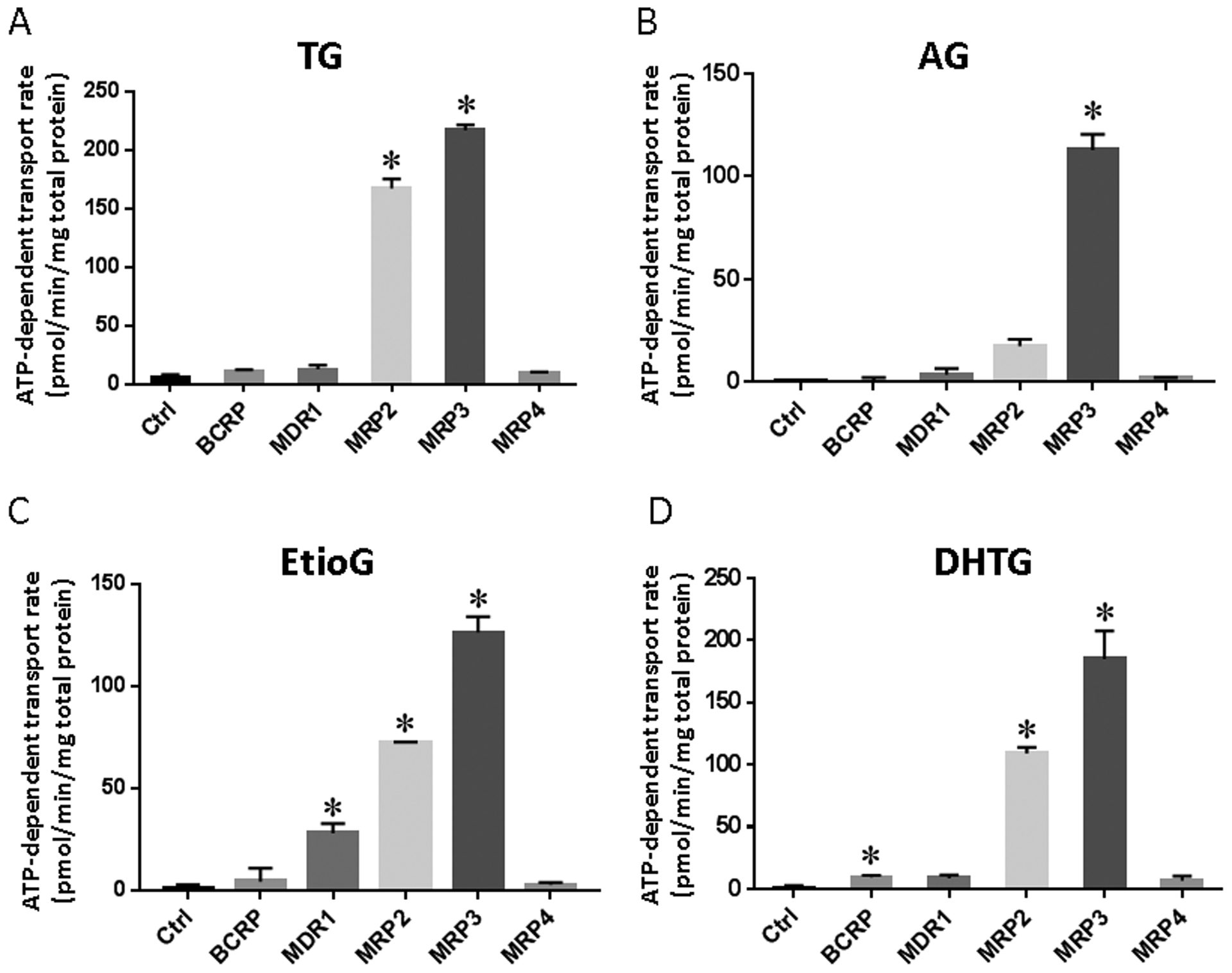
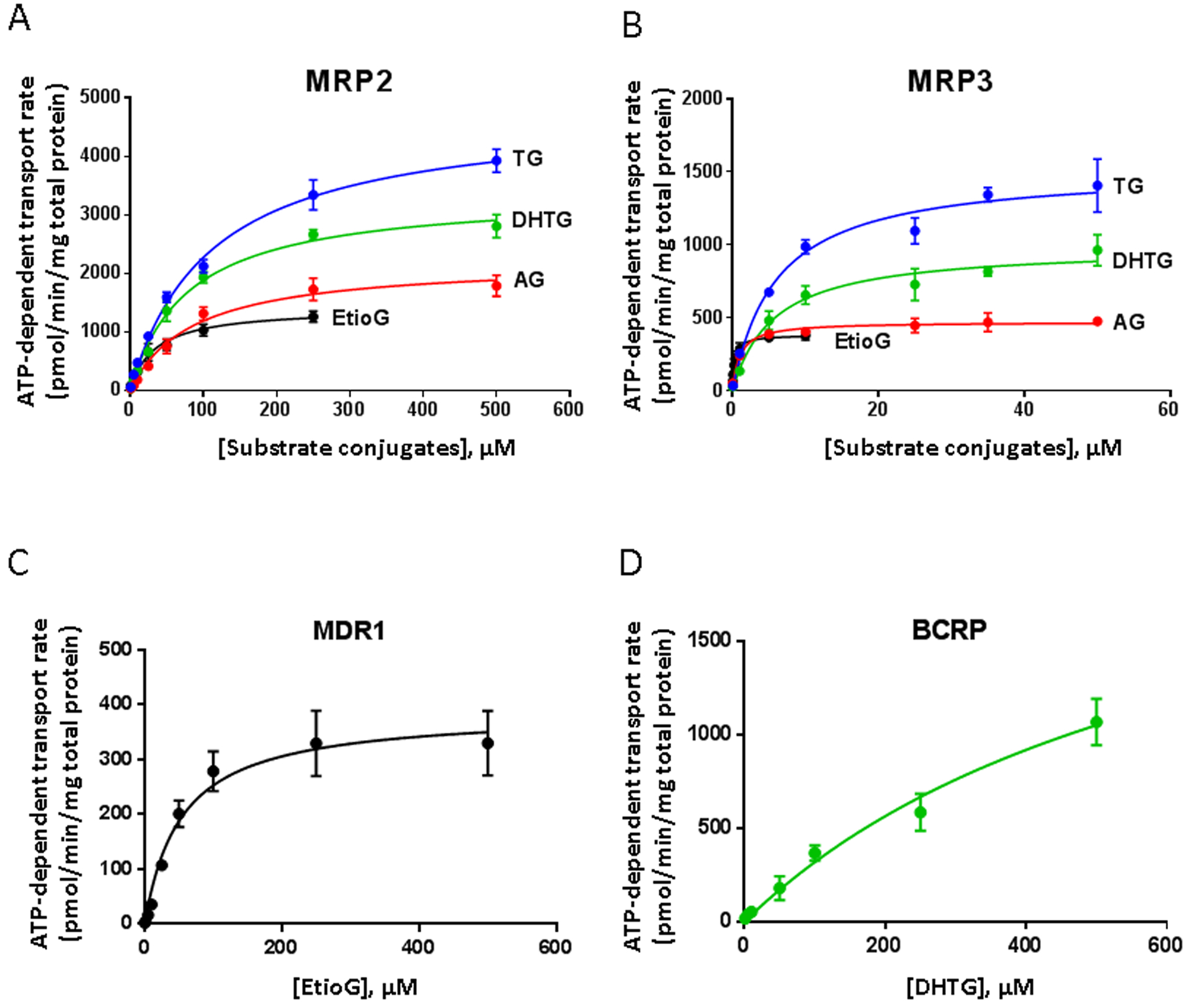
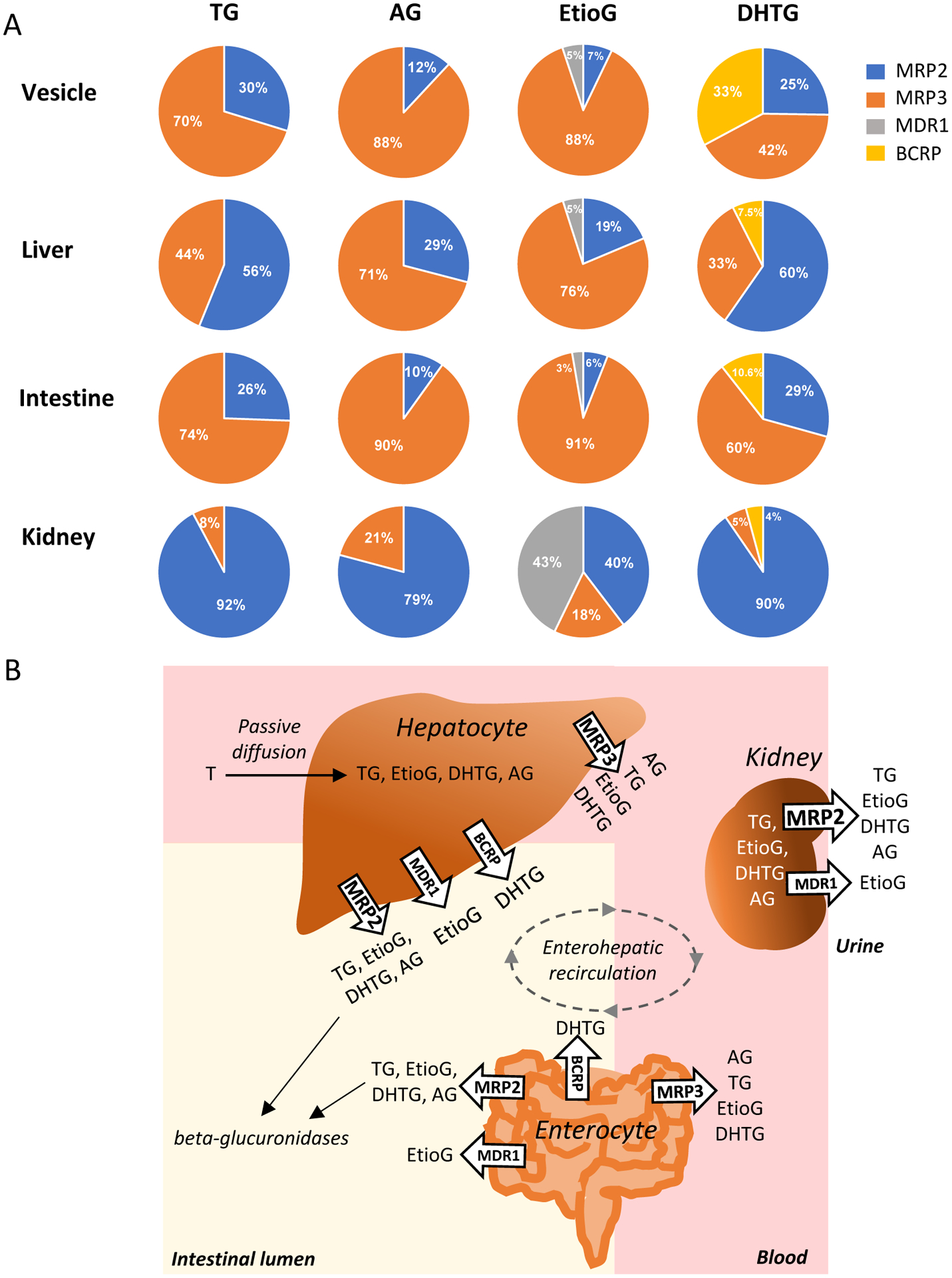
Testosterone glucuronide (TG), androsterone glucuronide (AG), etiocholanolone glucuronide (EtioG) and dihydrotestosterone glucuronide (DHTG) are the major metabolites of testosterone (T), which are excreted in urine and bile. Glucuronides can be deconjugated to active androgen in gut lumen after biliary excretion, which in turn can affect physiological levels of androgens. The goal of this study was to quantitatively characterize the mechanisms by which TG, AG, EtioG and DHTG are eliminated from liver, intestine, and kidney utilizing relative expression factor (REF) approach. Using vesicular transport assay with recombinant human MRP2, MRP3, MRP4, MDR1 and BCRP, we first identified that TG, AG, EtioG, and DHTG were primarily substrates of MRP2 and MRP3, although lower levels of transport were also observed with MDR1 and BCRP vesicles. The transport kinetic analyses revealed higher intrinsic clearances of TG by MRP2 and MRP3 as compared to that of DHTG, AG, and EtioG. MRP3 exhibited higher affinity for the transport of the studied glucuronides than MRP2. We next quantified the protein abundances of these efflux transporters in vesicles and compared the same with pooled total membrane fractions isolated from human tissues by quantitative LC-MS/MS proteomics. The fractional contribution of individual transporters (ft) was estimated by proteomics-based physiological scaling factors, i.e., transporter abundance in whole tissue versus vesicles, and corrected for inside-out vesicles (determined by 5'-nucleotidase assay). The glucuronides of inactive androgens, AG and EtioG were preferentially transported by MRP3, whereas the glucuronides of active androgens, TG and DHTG were mainly transported by MRP2 in liver. Efflux by bile canalicular transport may indicate the potential role of enterohepatic recirculation in regulating the circulating active androgens after deconjugation in the gut. In intestine, MRP3 possibly contributes most to the efflux of these glucuronides. In kidney, all studied glucuronides seemed to be preferentially effluxed by MRP2 and MDR1 (for EtioG). These REF based analysis need to be confirmed with in vivo findings. Overall, characterization of the efflux mechanisms of T glucuronide metabolites is important for predicting the androgen disposition and interindividual variability, including drug-androgen interaction in humans. The mechanistic data can be extrapolated to other androgen relevant organs (e.g. prostate, testis and placenta) by integrating these data with quantitative tissue proteomics data.
Keywords: Efflux transporters; Glucuronides; Quantitative proteomics; Testosterone; Vesicular transport.
Copyright © 2019 Elsevier Ltd. All rights reserved.
Figures
Similar articles
-
Human efflux transport of testosterone, epitestosterone and other androgen glucuronides.J Steroid Biochem Mol Biol. 2020 Mar;197:105518. doi: 10.1016/j.jsbmb.2019.105518. Epub 2019 Nov 6. J Steroid Biochem Mol Biol. 2020. PMID: 31704245
-
Organic Anion Transporting Polypeptide-Mediated Hepatic Uptake of Glucuronide Metabolites of Androgens.Mol Pharmacol. 2020 Sep;98(3):234-242. doi: 10.1124/mol.120.119891. Epub 2020 Jun 25. Mol Pharmacol. 2020. PMID: 32587096
-
Selectivity in the Efflux of Glucuronides by Human Transporters: MRP4 Is Highly Active toward 4-Methylumbelliferone and 1-Naphthol Glucuronides, while MRP3 Exhibits Stereoselective Propranolol Glucuronide Transport.Mol Pharm. 2017 Oct 2;14(10):3299-3311. doi: 10.1021/acs.molpharmaceut.7b00366. Epub 2017 Sep 13. Mol Pharm. 2017. PMID: 28850245
-
The roles of MRP2, MRP3, OATP1B1, and OATP1B3 in conjugated hyperbilirubinemia.Drug Metab Dispos. 2014 Apr;42(4):561-5. doi: 10.1124/dmd.113.055772. Epub 2014 Jan 23. Drug Metab Dispos. 2014. PMID: 24459177 Review.
-
[Analysis of xenobiotic detoxification system mediated by efflux transporters].Yakugaku Zasshi. 1999 Nov;119(11):822-34. doi: 10.1248/yakushi1947.119.11_822. Yakugaku Zasshi. 1999. PMID: 10590710 Review. Japanese.
Cited by
-
The implications of ABCC3 in cancer drug resistance: can we use it as a therapeutic target?Am J Cancer Res. 2021 Sep 15;11(9):4127-4140. eCollection 2021. Am J Cancer Res. 2021. PMID: 34659880 Free PMC article. Review.
-
Emerging Role of ABC Transporters in Glia Cells in Health and Diseases of the Central Nervous System.Cells. 2024 Apr 24;13(9):740. doi: 10.3390/cells13090740. Cells. 2024. PMID: 38727275 Free PMC article. Review.
-
Quantitative Proteomics in Translational Absorption, Distribution, Metabolism, and Excretion and Precision Medicine.Pharmacol Rev. 2022 Jul;74(3):769-796. doi: 10.1124/pharmrev.121.000449. Pharmacol Rev. 2022. PMID: 35738681 Free PMC article. Review.
-
In Vitro Glucuronidation of Caribbean Ciguatoxins in Fish: First Report of Conjugative Ciguatoxin Metabolites.Chem Res Toxicol. 2021 Aug 16;34(8):1910-1925. doi: 10.1021/acs.chemrestox.1c00181. Epub 2021 Jul 28. Chem Res Toxicol. 2021. PMID: 34319092 Free PMC article.
-
Causal role of blood metabolites in HER-positive and HER-negative breast cancer: a Mendelian randomization (MR) study.Aging (Albany NY). 2024 Aug 2;16(15):11626-11655. doi: 10.18632/aging.206042. Epub 2024 Aug 2. Aging (Albany NY). 2024. PMID: 39103210 Free PMC article.
References
-
- Allan CA, McLachlan RI, Age-related changes in testosterone and the role of replacement therapy in older men, Clin Endocrinol (Oxf), 60 (2004) 653–670. - PubMed
-
- Gray A, Feldman HA, McKinlay JB, Longcope C, Age, disease, and changing sex hormone levels in middle-aged men: results of the Massachusetts Male Aging Study, J Clin Endocrinol Metab, 73 (1991) 1016–1025. - PubMed
-
- Harman SM, Metter EJ, Tobin JD, Pearson J, Blackman MR, Baltimore A Longitudinal Study of, Longitudinal effects of aging on serum total and free testosterone levels in healthy men. Baltimore Longitudinal Study of Aging, J Clin Endocrinol Metab, 86 (2001) 724–731. - PubMed
Publication types
MeSH terms
Substances
Grants and funding
LinkOut - more resources
Full Text Sources
Research Materials
Miscellaneous
