. 2019 Jun 12;39(24):4797-4813.
doi: 10.1523/JNEUROSCI.0839-18.2019. Epub 2019 Apr 1.

Modulators of Kv3 Potassium Channels Rescue the Auditory Function of Fragile X Mice

Affiliations

Modulators of Kv3 Potassium Channels Rescue the Auditory Function of Fragile X Mice

Lynda El-Hassar et al. J Neurosci. .

Abstract

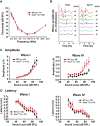
Fragile X syndrome (FXS) is characterized by hypersensitivity to sensory stimuli, including environmental sounds. We compared the auditory brainstem response (ABR) recorded in vivo in mice lacking the gene (Fmr1-/y ) for fragile X mental retardation protein (FMRP) with that in wild-type animals. We found that ABR wave I, which represents input from the auditory nerve, is reduced in Fmr1-/y animals, but only at high sound levels. In contrast, wave IV, which represents the activity of auditory brainstem nuclei is enhanced at all sound levels, suggesting that loss of FMRP alters the central processing of auditory signals. Current-clamp recordings of neurons in the medial nucleus of the trapezoid body in the auditory brainstem revealed that, in contrast to neurons from wild-type animals, sustained depolarization triggers repetitive firing rather than a single action potential. In voltage-clamp recordings, K+ currents that activate at positive potentials ("high-threshold" K+ currents), which are required for high-frequency firing and are carried primarily by Kv3.1 channels, are elevated in Fmr1-/y mice, while K+ currents that activate near the resting potential and inhibit repetitive firing are reduced. We therefore tested the effects of AUT2 [((4-({5-[(4R)-4-ethyl-2,5-dioxo-1-imidazolidinyl]-2-pyridinyl}oxy)-2-(1-methylethyl) benzonitrile], a compound that modulates Kv3.1 channels. AUT2 reduced the high-threshold K+ current and increased the low-threshold K+ currents in neurons from Fmr1-/y animals by shifting the activation of the high-threshold current to more negative potentials. This reduced the firing rate and, in vivo, restored wave IV of the ABR. Our results from animals of both sexes suggest that the modulation of the Kv3.1 channel may have potential for the treatment of sensory hypersensitivity in patients with FXS.SIGNIFICANCE STATEMENT mRNA encoding the Kv3.1 potassium channel was one of the first described targets of the fragile X mental retardation protein (FMRP). Fragile X syndrome is caused by loss of FMRP and, in humans and mice, causes hypersensitivity to auditory stimuli. We found that components of the auditory brain response (ABR) corresponding to auditory brainstem activity are enhanced in mice lacking FMRP. This is accompanied by hyperexcitability and altered potassium currents in auditory brainstem neurons. Treatment with a drug that alters the voltage dependence of Kv3.1 channels normalizes the imbalance of potassium currents, as well as ABR responses in vivo, suggesting that such compounds may be effective in treating some symptoms of fragile X syndrome.

Keywords: AUT2; auditory brainstem response; fragile X; high- and low-threshold potassium channels; medial nucleus of the trapezoid body; potassium channels.


Figures

Figure 1.
Figure 1.
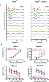
Loss of FMRP alters the central processing of auditory signals. A, Plots of the ABR thresholds for wild-type mice (N = 17 animals) and Fmr1−/y mice (N = 28 animals). B, ABR wave recordings from WT and Fmr1−/y mice stimulated at 11.3 kHz at different sound levels (35–90 dB). Arrows indicate wave IV of the ABR response. C, D, Plots of the amplitudes (C) and latencies (D) of ABR waves I and IV as a function of sound levels (40–90 dB). The magnitude of wave I is decreased for high sound levels (p = 0.0002) only, and there is a significant increase in wave IV amplitude in Fmr1−/y mice at most sound levels tested (p < 0.0001) without significant change in the ABR latencies. Error bars indicate the mean ± SEM. In C, asterisks indicate statistical significance (two-way ANOVA followed by Bonferroni–Dunn post-test).
Figure 2.
Figure 2.
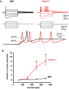
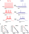
MNTB neurons from Fmr1−/y mice are hyperexcitable. A, Current-clamp recordings of action potentials in response to a series of sustained hyperpolarizing and depolarizing current pulses (200 ms square current pulses, −250 to +350 pA). Note the repetitive firing and the shorter latency firing of MNTB neurons from Fmr1−/y mice compared with the WT mice, which never fired more than one or two action potentials. B, Group data showing a significant increase in number of action potentials evoked in neurons from Fmr1−/y mice in response to currents of increasing amplitude (Asterisks indicate statistical significance. p = 0.002, Student's unpaired t test with Welch's correction).
Figure 3.
Figure 3.
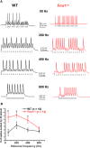
MNTB neurons from Fmr1−/y mice fire more action potentials in response to repetitive stimulation. A, Current-clamp recordings of action potentials in response to repetitive stimulation with brief current pulses (1 nA, 0.3 ms, 20 stimuli) applied at rates from 50 to 600 Hz. B, Group data showing that MNTB neurons from Fmr1−/y mice fire more action potentials at all stimulus frequencies up to 400 Hz. Asterisks indicate significant differences (two-way ANOVA followed by Holm–Sidak post-test).
Figure 4.
Figure 4.
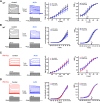
High- and low-threshold potassium currents are altered in MNTB neurons of Fmr1−/y mice. A, Representative whole-cell patch-clamp recordings of high-threshold potassium current evoked by holding the membrane potential at −40 mV for 2 min before stepping to test potentials between −30 and +60 mV in 10 mV increments. Middle, Plot shows that the amplitude of high-potassium current was significantly higher in MNTB neurons from Fmr1−/y mice compared with wild-type mice (two-way ANOVA, p < 0.0001). Right, Plot shows the corresponding normalized conductance that was not significantly (p = 0.16) different between WT and Fmr1−/y mice. B, Representative whole-cell patch-clamp recordings of low-threshold potassium current by holding the membrane potential at −80 mV before stepping to test potentials from −70 to +60 mV in 10 mV increments. Middle, Plots show that the peak amplitude of low-potassium current was significantly reduced in Fmr1−/y mice compared with WT mice (p < 0.0001, two-way ANOVA). Plot on the right shows a significant shift in voltage dependence of the low-threshold K+ conductance in Fmr1−/y mice (p = 0.01, Student's unpaired t test with Welch's correction). In the I–V plots, asterisks indicate statistical significance in post-test results for multiple comparisons (Holm–Sidak method).
Figure 5.
Figure 5.
AUT2 decreases high-threshold potassium current and increases low-threshold potassium current of MNTB neurons from Fmr1−/y mice. A, Representative traces of high-threshold potassium current evoked by holding the membrane potential at −40 mV and stepping to test potentials from −30 and +60 mV in 10 mV increments before and after the addition of 10 μm AUT2. The chemical structure of AUT2 is shown above the right-hand current traces. Middle, Group data for the effects of AUT2 on current amplitude at different voltages. Right, group data for conductance-voltage relations normalized to maximal value at +60 mV (G/G+60), indicating that AUT2 did not produce a significant change of the voltage-dependent activation of high-threshold potassium currents. B, Representative whole-cell patch-clamp recordings of low-threshold potassium current evoked by holding the membrane potential at −80 mV and applying test potentials from −70 to +60 mV in 10 mV increments before and after addition of AUT2 (10 μm). Panels at center and right show group data for current amplitudes and conductance–voltage relations, demonstrating that AUT2 significantly increased the low-threshold potassium currents in Fmr1−/y mice. Asterisks in A and B indicate statistical significance in post-test results of the simple effect test for multiple comparison (Holm–Sidak method).
Figure 6.
Figure 6.
Addition of AUT2 produces a left shift in the voltage dependence of inactivation of high-threshold potassium currents in wild-type and Fmr1−/y mice. A, Standard voltage-clamp protocol to assess the inactivation of K+ currents. Cells were held at potentials between −100 and +10 mV for 30 s before a test pulse to +40 mV. B, Representative traces of the outward current recorded in neurons from Fmr1−/y mice at a test potential of +40 mV before and after AUT2 (10 μm). C, D, Plots of steady-state inactivation as a function of a 30 s prepulse to potentials between −100 and +10 mV in Fmr1−/y (C) and WT (D) mice.
Figure 7.
Figure 7.
AUT2 fails to alter high- and low-threshold K+ currents in the presence of the Kv3 channel blocker TEA. A–D, Voltage-clamp recordings of the high-threshold (A, C) and low-threshold (B, D) potassium currents in wild-type and in Fmr1−/y mice. Panels on the right show the current–voltage and the conductance–voltage (normalized to maximal value at +60 mV; G/G+60) curves for high- and low-threshold K+ currents before and after AUT2, in the presence of the Kv3 channels blocker TEA (1 mm). AUT2 failed to alter the magnitude and the conductance of both high- and low-threshold potassium currents in these conditions.
Figure 8.
Figure 8.
AUT2 reduces the firing rate of MNTB neurons of Fmr1−/y mice. A, Current-clamp recordings of action potentials evoked by 20 consecutive stimuli of intracellular current pulses (2 nA, 0.3 ms) in MNTB neurons applied at 50, 200, or 300 Hz before and after application of AUT2 (10 μm). B, Plots of the numbers of action potentials evoked by repetitive stimulation at 50, 200, and 300 Hz in different experiments before and after AUT2 application. AUT2 decreased the number of evoked action potentials in MNTB neurons at all frequencies tested (n = 3, N = 3 animals; p < 0.0001, two-way ANOVA followed by Holm–Sidak post-test).
Figure 9.
Figure 9.
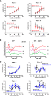
AUT2 rescues the auditory function of Fmr1−/y mice. A, Representative ABR traces recorded from Fmr1−/y mice before and 20 min after intraperitoneal injection of AUT2 (30 mg/kg). Arrows indicate wave IV of the ABR response. B, Plots of the amplitudes and latencies of ABR waves I and IV as a function of sound level before and after injection of AUT2. The group data show a significant decrease in the amplitude of ABR wave IV after AUT2 injection (p = 0.001) with no change in the amplitude of wave I (p = 0.3). In B, asterisks indicate statistical significance (two-way ANOVA followed by Holm–Sidak post-test).
Figure 10.
Figure 10.
Administration of vehicle does not alter ABR waves in Fmr1−/y mice, and AUT2 does not alter auditory function in wild-type mice. A, Plots of the amplitudes and latencies of ABR waves I and IV as a function of sound level before and after the administration of vehicle to Fmr1−/y mice. No statistical differences in the magnitude of wave I and IV (p = 0.99 for both) or in the latencies of wave I and IV (p = 0.98 and p = 0.90, respectively) were found after vehicle injection (two-way ANOVA followed by Holm–Sidak post-test). B, Representative ABR traces recorded from WT mice before and 20 min after intraperitoneal injection of AUT2 (30 mg/kg). Traces represent responses to sounds delivered at 80, 85, and 90 dB. C, Plots of the amplitudes and latencies of ABR waves I and IV as a function of sound level before and after injection of AUT2. The group data show no significant changes in the amplitude of ABR waves I and IV (p = 0.92, p = 0.3 respectively) after AUT2 injection. Similarly, AUT2 did not change the latencies of waves I and IV (p = 0.8 and p = 0.46, respectively, two-way ANOVA by Holm–Sidak post-test).

Similar articles

Cited by

References

    1. Akil O, Oursler AE, Fan K, Lustig LR (2016) Mouse auditory brainstem response testing. Bio Protoc 6:e1768. 10.21769/BioProtoc.1768 - DOI - PMC - PubMed
    1. Aloisi E, Le Corf K, Dupuis J, Zhang P, Ginger M, Labrousse V, Spatuzza M, Georg Haberl M, Costa L, Shigemoto R, Tappe-Theodor A, Drago F, Vincenzo Piazza P, Mulle C, Groc L, Ciranna L, Catania MV, Frick A (2017) Altered surface mGluR5 dynamics provoke synaptic NMDAR dysfunction and cognitive defects in Fmr1 knockout mice. Nat Commun 8:1103. 10.1038/s41467-017-01191-2 - DOI - PMC - PubMed
    1. Arinami T, Sato M, Nakajima S, Kondo I (1988) Auditory brain-stem responses in the fragile X syndrome. Am J Hum Genet 43:46–51. - PMC - PubMed
    1. Bhattacharjee A, Gan L, Kaczmarek LK (2002) Localization of the Slack potassium channel in the rat central nervous system. J Comp Neurol 454:241–254. 10.1002/cne.10439 - DOI - PubMed
    1. Bhattacharjee A, von Hehn CA, Mei X, Kaczmarek LK (2005) Localization of the Na+-activated K+ channel Slick in the rat central nervous system. J Comp Neurol 484:80–92. 10.1002/cne.20462 - DOI - PubMed

Publication types

MeSH terms