. 2015;2(4):448-472.
doi: 10.3934/matersci.2015.4.448. Epub 2015 Nov 16.

Biomedical diagnosis perspective of epigenetic detections using alpha-hemolysin nanopore

Affiliations

Biomedical diagnosis perspective of epigenetic detections using alpha-hemolysin nanopore

Yong Wang et al. AIMS Mater Sci. 2015.

Abstract

The α-hemolysin nanopore has been studied for applications in DNA sequencing, various single-molecule detections, biomolecular interactions, and biochips. The detection of single molecules in a clinical setting could dramatically improve cancer detection and diagnosis as well as develop personalized medicine practices for patients. This brief review shortly presents the current solid state and protein nanopore platforms and their applications like biosensing and sequencing. We then elaborate on various epigenetic detections (like microRNA, G-quadruplex, DNA damages, DNA modifications) with the most widely used alpha-hemolysin pore from a biomedical diagnosis perspective. In these detections, a nanopore electrical current signature was generated by the interaction of a target with the pore. The signature often was evidenced by the difference in the event duration, current level, or both of them. An ideal signature would provide obvious differences in the nanopore signals between the target and the background molecules. The development of cancer biomarker detection techniques and nanopore devices have the potential to advance clinical research and resolve health problems. However, several challenges arise in applying nanopore devices to clinical studies, including super low physiological concentrations of biomarkers resulting in low sensitivity, complex biological sample contents resulting in false signals, and fast translocating speed through the pore resulting in poor detections. These issues and possible solutions are discussed.

Keywords: DNA damage; DNA hydroxymethylation; DNA methylation; G-quadruplex; abasic site; cancer biomarker; guanine oxidation; microRNA; nanopore device; α-hemolysin.


Conflict of interest statement

Conflict of Interest All authors declare no conflict of interest.

Figures

Figure 1.
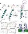
Figure 1.. microRNA detections in the lung cancer patients.
a, Molecular diagram of a microRNA (red) bound to a probe (green) with signal tags. b, The unzipping signature (left) and the molecule pathways of the dsDNA unzipping signature (right) in the nanopore. c, The frequencies of miR-155 signature events (f155) from six healthy individuals (#1 to #6) and six patients with lung cancer (#7 to #12) in the presence of spiked-in synthetic miR-39. d The frequencies of spike-in miR-39 signature events detected all the samples that were used in c. The patient conditions were the following: #7, metastatic squamous lung carcinoma; #8, recurrent small-cell cancer; #9, early-stage small-cell carcinoma, status post-chemotherapy and -radiation; #10, early-stage small-cell cancer, status post-chemotherapy; #11, late-stage non-small cell carcinoma, status post-resection and -chemotherapy; #12, late-stage adenocarcinoma, status post-chemotherapy. Reprinted with permission from reference [65].
Figure 2.
Figure 2.. Cationic peptide probe-based interference-free detection of microRNAs in the nanopore.
The probe includes: a polycationic polymer lead (peptide, blue) and a capture domain (PNA, green). The capture domain can hybridize with the target microRNA (red). The miRNA·probe complex is drawn into the nanopore by the applied voltage at the pore (trans) opening, while all other negatively charged free nucleic acids without the probe binding (gray) were electrophoretically expelled away from the pore. Reprinted with permission from reference [66].
Figure 3.
Figure 3.. Barcode tagged probe through click chemistry for nanopore ionic current modulation and multiplex detection of mircroRNAs in the nanopore.
Each PEG-labeled DNA probe can target a specific microRNA and generate a specific nanopore current profile. Upon being captured in the nanopore, the PEG on the probe specifically regulates the nanopore current profile, thereby generating a signature for target identification. Reprinted with permission from reference [68].
Figure 4.
Figure 4.. Interactions between G-quadruplex [5’-TAGGG(TTAGGG)3TT-3’] subtypes and the alpha-hemolysin reveal the size-dependent properties of the protein nanopore.
a, Space-filling models of G-quadruplexes interacting with the nanopore constructed using different PBD structures, b-d, Stick models of the proposed interaction mechanisms and current-time traces yielded by (b) the hybrid folds (50 mM KCl, 950 mM LiCl, 25 mM Tris, pH 7.9), (c) the basket folds (1 M NaCl, 25 mM Tris, pH 7.9), and (d) the propeller folds (20 mM KCl, 5 M LiCl, 25 mM Tris, pH 7.9). All current traces were recorded under 120 mV (trans vs. cis). I and IM values are indicated as a percentage of the open-channel current Io. Reprinted with permission from reference [75].
Figure 5.
Figure 5.. Detection of thrombin-binding aptamer (TBA, GGTTGGTGTGGTTGG) with metal ions in the nanopore.
a, the sequence and structure of TBA G-quadruplex (left) and the two G-tetrad planes in the TBA G-quadruplex formed by guanines at the position 1, 6, 10 and 15, and the bottom one by guanine 2, 5, 11 and 14 (right). A cation in between is coordinated with eight carbonyls. b, Schemes of the current trace showing characteristic signature blocks. c, Long-lived block for capturing a single G-quadruplex in the nanocavity enclosed by the α-hemolysin nanopore; d, The long block terminal spike produced by translocation of the unfolded G-quadruplex through the β-barrel. The long-lived block with an ending spike served as the nanopore signature for the folded form of TBA. e, Short-lived block formed by translocation of linear form TBA. Reprinted with permission from reference [78].
Figure 6.
Figure 6.. Detection of guanine oxidation in the human telomere repeat sequence.
Oxidation of a 120-mer portion of the human telomere repeat sequence (Q5) with 1O2 to yield 8-oxo-7,8-dihydroguanine (OG) that was labeled with 18c6 followed by nanopore detection and quantification. a, Reaction scheme for oxidation of guanine to yield OG and the labeling reaction of OG by 18c6 in the presence of K2IrBr6. b, Model of the Q5 strand in biologically relevant salts (hybrid G4) followed by oxidation labeling and refolding in NH4Cl (100 mM) and LiCl (2 M) electrolyte to yield the propeller fold. c, More than 50% of the events contained pulse-like current modulations: i.e., ~35% of the events presented one modulation, ~14% two modulations, and ~5% three modulations. Nanopore recordings were conducted in 25 mM Tris, pH 7.9, 100 mM NH4Cl, and 2 M LiCl at 25 °C. Reprinted with permission from reference [84].
Figure 7.
Figure 7.. Detection of abasic site in the β-barrel of the nanopore. Individual i-t traces of AP-18c6 in homopolymeric strands.
a, Identification of single AP-18c6 adduct. Sample i-t traces for mono adduct (120 mV trans vs. cis). b, Identification of two AP-18c6 adducts. Sample i-t traces for bis adducts (120 mV trans vs. cis). DNA was captured into the nanopore from 5’ entry. The pulse like nanopore signature for AP-18c6 was generated for the AP detection. Reprinted with permission from reference [98].
Figure 8.
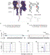
Figure 8.. Abasic site detection in the latch zone of the nanopore by monitoring the UDG enzyme activity for dsDNA in the nanopore.
Top scheme: Left: The structure of dsDNA with a 5’- poly(T)24 tail within WT α-HL. The box indicates the location of the uracil (U) base or the abasic site (AP). Right: Scheme of the UDG hydrolysis reaction. The α-HL structure was taken from pdb 7AHL. DNA structure is shown on a 1:1 scale with α-HL. a, Sequence of the starting material formed by a 41-mer U-containing strand hybridized to a 17-mer strand. b, Sequence of the product containing AP. c, d, Sample current-time (i-t) traces for blockages generated by the U duplex (c) or the AP duplex (d) in individual experiments. The blue and red lines indicate the current blockage levels used to determine the duplex identity. e, f, g, Histograms of current blockage levels for the U duplex (e), AP duplex (f) and for a mixture of U and AP duplexes (g, mole ratio ~2:1). Single-nucleotide recognition was achieved between the U-containing duplex (a, c, and e) and the AP-containing duplex (b, d, and f) based on a ~2 pA difference in blockage current levels of the unzipping events in a 14 μM DNA, 150 mM buffered KCl solution at −120 mV. Reprinted with permission from reference [99].
Figure 9.
Figure 9.. Molecule dynamic simulations revealed a cation binding site in the C-C mismatch and unstable hmc-C pairing. The DNA duplex is in the “stick” presentation, and the two backbones are illustrated as yellow and green belts respectively.
Potassium ions that neutralize the entire simulation system are shown as tan balls. Water in a cubic box (78.5 × 78.5 × 78.5 Å3) is shown transparently. (b) A snap-shot of pairing between two cytosine bases. The dashed circle highlights the binding site for a cation. (c) A snap-shot of hmC-C pairing before the pairing was broken. (d-f) Time-dependent distances between the N3 atom of one base and the N4 atom of the other base, in C-C(d), mC-C(e) and hmC-C(f) mismatches. Reprinted with permission from reference [120].
Figure 10.
Figure 10.. Methylation detection by designing a mercury interstrand lock.
(a), (b) and (c) compared the duration of short and long signature blocks for targets Tp16-1 (a), Tpl6-2 (b) and Tpl6-3 (c) detected by four probes PC6, PC8, PC14 and PC16. The duration of signature blocks allowed determining of the methylation status for each of four CpG cytosines. The DNA sequences of the three p16 fragments containined bases 1, 2 and 3 mC in the DNA strand. C can be converted to U by bisulfite treatment and then form a U-T pair which can be stabilized by a mercury ion evidenced by the prolonged duration (gray bar). However, mC can not be converted, so it forms a mC-T pair which can not be stabilized by a mercury ion, therefore only short blocks were observed (white bar). All traces were recorded at +130 mV in 1 M KCl and 10 mM Tris (pH 7.4). Reprinted with permission from reference [127].

Similar articles

Cited by

References

    1. Dupont C, Armant DR, Brenner CA (2009) Epigenetics: Definition, mechanisms and clinical perspective. Semin Reprod Med 27: 351–357. - PMC - PubMed
    1. Dawson MA, Kouzarides T (2012) Cancer epigenetics: From mechanism to therapy. Cell 150: 12–27. - PubMed
    1. Esteller M (2008) Molecular origins of cancer: Epigenetics in cancer. New Engl J Med 358: 1148–1159+1096. - PubMed
    1. Laird PW (2005) Cancer epigenetics. Hum Mol Genet 14: R65–R76. - PubMed
    1. Okugawa Y, Grady WM, Goel A (2015) Epigenetic Alterations in Colorectal Cancer: Emerging Biomarkers. Gastroenterology 149: 1204–1225e1212. - PMC - PubMed

LinkOut - more resources