High TNFRSF14 and low BTLA are associated with poor prognosis in Follicular Lymphoma and in Diffuse Large B-cell Lymphoma transformation
- PMID: 30918139
- PMCID: PMC6528140
- DOI: 10.3960/jslrt.19003
High TNFRSF14 and low BTLA are associated with poor prognosis in Follicular Lymphoma and in Diffuse Large B-cell Lymphoma transformation
Abstract
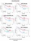
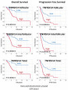
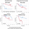
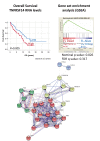
The microenvironment influences the behavior of follicular lymphoma (FL) but the specific roles of the immunomodulatory BTLA and TNFRSF14 (HVEM) are unknown. Therefore, we examined their immunohistochemical expression in the intrafollicular, interfollicular and total histological compartments in 106 FL cases (57M/49F; median age 57-years), and in nine relapsed-FL with transformation to DLBCL (tFL). BTLA expression pattern was of follicular T-helper cells (TFH) in the intrafollicular and of T-cells in the interfollicular compartments. The mantle zones were BTLA+ in 35.6% of the cases with similar distribution of IgD. TNFRSF14 expression pattern was of neoplastic B lymphocytes (centroblasts) and "tingible body macrophages". At diagnosis, the averages of total BTLA and TNFRSF14-positive cells were 19.2%±12.4STD (range, 0.6%-58.2%) and 46.7 cells/HPF (1-286.5), respectively. No differences were seen between low-grade vs. high-grade FL but tFL was characterized by low BTLA and high TNFRSF14 expression. High BTLA correlated with good overall survival (OS) (total-BTLA, Hazard Risk=0.479, P=0.022) and with high PD-1 and FOXP3+Tregs. High TNFRSF14 correlated with poor OS and progression-free survival (PFS) (total-TNFRSF14, HR=3.9 and 3.2, respectively, P<0.0001), with unfavorable clinical variables and higher risk of transformation (OR=5.3). Multivariate analysis including BTLA, TNFRSF14 and FLIPI showed that TNFRSF14 and FLIPI maintained prognostic value for OS and TNFRSF14 for PFS. In the GSE16131 FL series, high TNFRSF14 gene expression correlated with worse prognosis and GSEA showed that NFkB pathway was associated with the "High-TNFRSF14/dead-phenotype".In conclusion, the BTLA-TNFRSF14 immune modulation pathway seems to play a role in the pathobiology and prognosis of FL.
Keywords: BTLA; Follicular lymphoma; TNFRSF14 (HVEM); immune microenvironment; transformed follicular lymphoma.
Conflict of interest statement
Figures


Similar articles
-
High expression of the inhibitory receptor BTLA in T-follicular helper cells and in B-cell small lymphocytic lymphoma/chronic lymphocytic leukemia.Am J Clin Pathol. 2009 Oct;132(4):589-96. doi: 10.1309/AJCPPHKGYYGGL39C. Am J Clin Pathol. 2009. PMID: 19762537
-
Loss of the HVEM Tumor Suppressor in Lymphoma and Restoration by Modified CAR-T Cells.Cell. 2016 Oct 6;167(2):405-418.e13. doi: 10.1016/j.cell.2016.08.032. Epub 2016 Sep 29. Cell. 2016. PMID: 27693350 Free PMC article.
-
TNFRSF14 aberrations in follicular lymphoma increase clinically significant allogeneic T-cell responses.Blood. 2016 Jul 7;128(1):72-81. doi: 10.1182/blood-2015-10-679191. Epub 2016 Apr 21. Blood. 2016. PMID: 27103745 Free PMC article. Clinical Trial.
-
The Tumor Microenvironment in Follicular Lymphoma: Its Pro-Malignancy Role with Therapeutic Potential.Int J Mol Sci. 2021 May 19;22(10):5352. doi: 10.3390/ijms22105352. Int J Mol Sci. 2021. PMID: 34069564 Free PMC article. Review.
-
Follicular Lymphoma: The Role of the Tumor Microenvironment in Prognosis.J Clin Exp Hematop. 2016;56(1):1-19. doi: 10.3960/jslrt.56.1. J Clin Exp Hematop. 2016. PMID: 27334853 Free PMC article. Review.
Cited by
-
The role of ARL4C in predicting prognosis and immunotherapy drug susceptibility in pan-cancer analysis.Front Pharmacol. 2023 Dec 20;14:1288492. doi: 10.3389/fphar.2023.1288492. eCollection 2023. Front Pharmacol. 2023. PMID: 38178862 Free PMC article.
-
Patient-derived lymphoma spheroids integrating immune tumor microenvironment as preclinical follicular lymphoma models for personalized medicine.J Immunother Cancer. 2023 Oct;11(10):e007156. doi: 10.1136/jitc-2023-007156. J Immunother Cancer. 2023. PMID: 37899130 Free PMC article.
-
The role of the BTLA-HVEM complex in the pathogenesis of breast cancer.Breast Cancer. 2024 May;31(3):358-370. doi: 10.1007/s12282-024-01557-7. Epub 2024 Mar 14. Breast Cancer. 2024. PMID: 38483699 Review.
-
Beyond the anti-PD-1/PD-L1 era: promising role of the BTLA/HVEM axis as a future target for cancer immunotherapy.Mol Cancer. 2023 Aug 30;22(1):142. doi: 10.1186/s12943-023-01845-4. Mol Cancer. 2023. PMID: 37649037 Free PMC article. Review.
-
Early progression and transformation of a splenic diffuse red pulp small B-cell lymphoma with NOTCH1, ARID2, CREBBP, and TNFRSF14 gene mutations.Leuk Res Rep. 2023 Aug 22;20:100384. doi: 10.1016/j.lrr.2023.100384. eCollection 2023. Leuk Res Rep. 2023. PMID: 37664441 Free PMC article.

