

Imaging of neuroinflammation in migraine with aura: A [11C]PBR28 PET/MRI study
- PMID: 30918090
- PMCID: PMC6511078
- DOI: 10.1212/WNL.0000000000007371
Imaging of neuroinflammation in migraine with aura: A [11C]PBR28 PET/MRI study
Abstract
Objective: To determine if migraine with aura is associated with neuroinflammation, which has been suggested by preclinical models of cortical spreading depression (CSD) as well as imaging of human pain conditions.
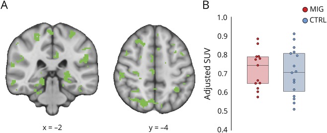
Methods: Thirteen migraineurs with aura and 16 healthy controls received integrated PET/MRI brain scans with [11C]PBR28, a radioligand that binds to the 18 kDa translocator protein, a marker of glial activation. Standardized uptake value ratio (SUVR) was compared between groups, and regressed against clinical variables, using region of interest and whole-brain voxelwise analyses.
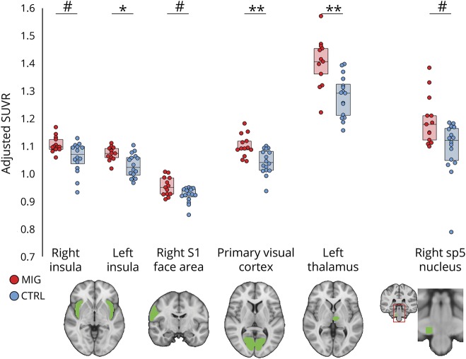
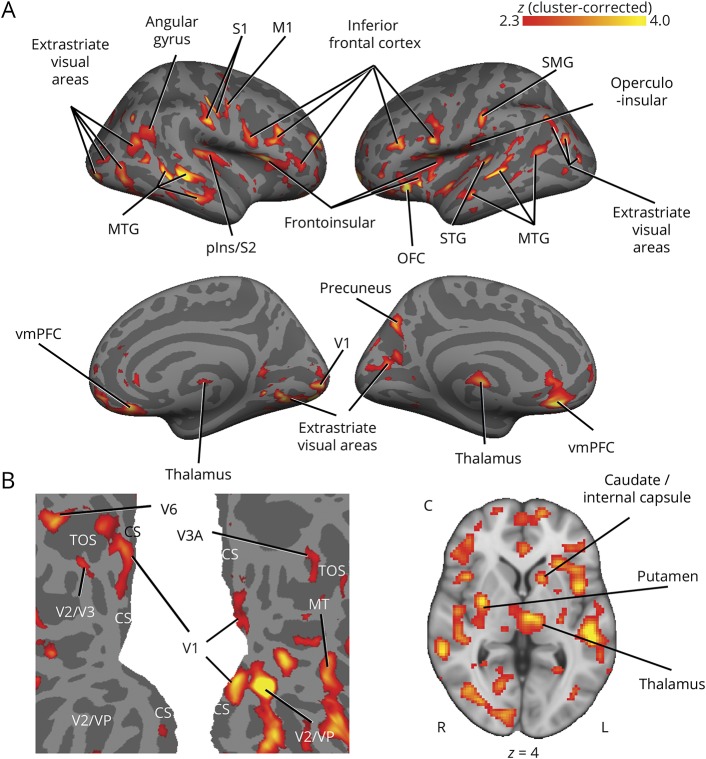
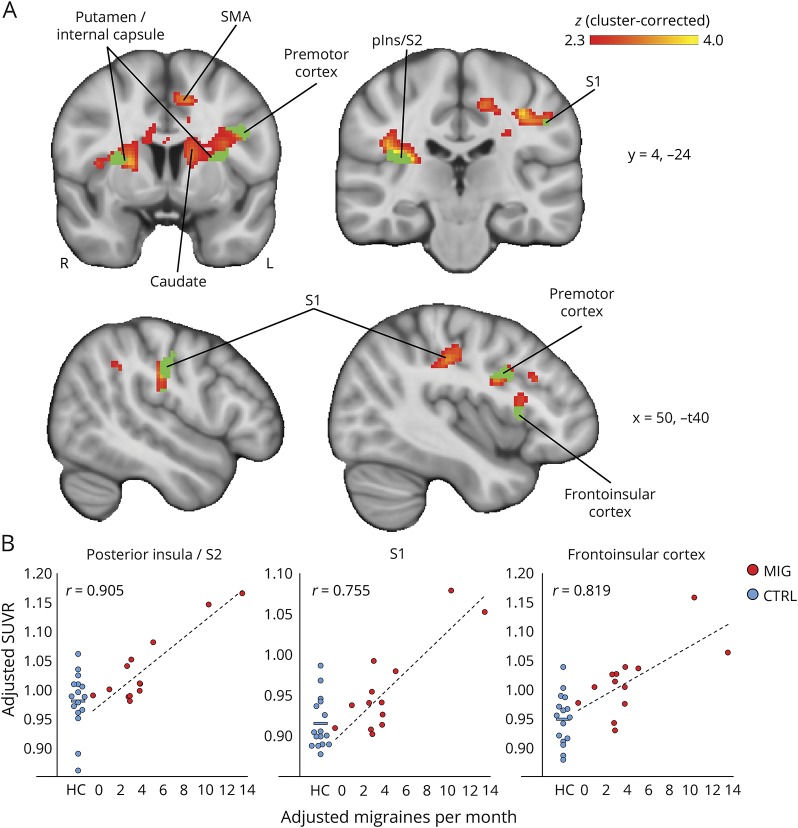
Results: Compared to healthy controls, migraineurs demonstrated SUVR elevations in nociceptive processing areas (e.g., thalamus and primary/secondary somatosensory and insular cortices) as well as in areas previously shown to be involved in CSD generation (visual cortex). SUVR levels in frontoinsular cortex, primary/secondary somatosensory cortices, and basal ganglia were correlated with frequency of migraine attacks.
Conclusions: These findings demonstrate that migraine with aura is associated with neuroimmune activation/neuroinflammation, and support a possible link between CSD and glial activation, previously observed in animals.
© 2019 American Academy of Neurology.
Figures
Similar articles
-
Extra-Axial Inflammatory Signal in Parameninges in Migraine with Visual Aura.Ann Neurol. 2020 Jun;87(6):939-949. doi: 10.1002/ana.25731. Epub 2020 Apr 22. Ann Neurol. 2020. PMID: 32239542 Free PMC article.
-
Brain glial activation in fibromyalgia - A multi-site positron emission tomography investigation.Brain Behav Immun. 2019 Jan;75:72-83. doi: 10.1016/j.bbi.2018.09.018. Epub 2018 Sep 14. Brain Behav Immun. 2019. PMID: 30223011 Free PMC article.
-
Neuroimmune signatures in chronic low back pain subtypes.Brain. 2022 Apr 29;145(3):1098-1110. doi: 10.1093/brain/awab336. Brain. 2022. PMID: 34528069 Free PMC article.
-
Neuroimaging clues of migraine aura.J Headache Pain. 2019 Apr 3;20(1):32. doi: 10.1186/s10194-019-0983-2. J Headache Pain. 2019. PMID: 30943894 Free PMC article. Review.
-
Cortical excitability in migraine: Contributions of magnetic resonance imaging.Rev Neurol (Paris). 2021 Sep;177(7):809-815. doi: 10.1016/j.neurol.2021.07.008. Epub 2021 Jul 28. Rev Neurol (Paris). 2021. PMID: 34332777 Review.
Cited by
-
Migraine and neuroinflammation: the inflammasome perspective.J Headache Pain. 2021 Jun 10;22(1):55. doi: 10.1186/s10194-021-01271-1. J Headache Pain. 2021. PMID: 34112082 Free PMC article. Review.
-
Efficacy and safety of transesophageal ultrasound-guided patent foramen ovale closure for migraine in adolescents.Front Pediatr. 2023 Nov 15;11:1296825. doi: 10.3389/fped.2023.1296825. eCollection 2023. Front Pediatr. 2023. PMID: 38046679 Free PMC article.
-
The effect of P2X7 antagonism on subcortical spread of optogenetically-triggered cortical spreading depression and neuroinflammation.J Headache Pain. 2024 Jul 24;25(1):120. doi: 10.1186/s10194-024-01807-1. J Headache Pain. 2024. PMID: 39044141 Free PMC article.
-
The Characteristics of White Matter Hyperintensities in Patients With Migraine.Front Pain Res (Lausanne). 2022 Jun 20;3:852916. doi: 10.3389/fpain.2022.852916. eCollection 2022. Front Pain Res (Lausanne). 2022. PMID: 35794956 Free PMC article.
-
[11C]PBR28 radiotracer kinetics are not driven by alterations in cerebral blood flow.J Cereb Blood Flow Metab. 2021 Nov;41(11):3069-3084. doi: 10.1177/0271678X211023387. Epub 2021 Jun 23. J Cereb Blood Flow Metab. 2021. PMID: 34159823 Free PMC article.
References
-
- Murray CJ, Vos T, Lozano R, et al. . Disability-adjusted life years (DALYs) for 291 diseases and injuries in 21 regions, 1990-2010: a systematic analysis for the Global Burden of Disease Study 2010. Lancet 2012;380:2197–2223. - PubMed
-
- Pietrobon D, Moskowitz MA. Pathophysiology of migraine. Annu Rev Physiol 2013;75:365–391. - PubMed
-
- Karatas H, Erdener SE, Gursoy-Ozdemir Y, et al. . Spreading depression triggers headache by activating neuronal Panx1 channels. Science 2013;339:1092–1095. - PubMed
MeSH terms
Grants and funding
- R21 NS087472/NS/NINDS NIH HHS/United States
- P01 AT009965/AT/NCCIH NIH HHS/United States
- S10 RR026666/RR/NCRR NIH HHS/United States
- R01 AT007550/AT/NCCIH NIH HHS/United States
- R01 NS094306/NS/NINDS NIH HHS/United States
- UL1 RR025758/RR/NCRR NIH HHS/United States
- R61 AT009306/AT/NCCIH NIH HHS/United States
- R21 NS082926/NS/NINDS NIH HHS/United States
- R01 NS095937/NS/NINDS NIH HHS/United States
- R01 AR064367/AR/NIAMS NIH HHS/United States
- S10 OD023517/OD/NIH HHS/United States
- UL1 TR000170/TR/NCATS NIH HHS/United States
- UL1 TR001102/TR/NCATS NIH HHS/United States
LinkOut - more resources
Full Text Sources
Medical
Miscellaneous