Review
. 2019 Mar 15:2:103.
doi: 10.1038/s42003-019-0318-5. eCollection 2019.

Bridging human chaperonopathies and microbial chaperonins

Affiliations
Review

Bridging human chaperonopathies and microbial chaperonins

Everly Conway de Macario et al. Commun Biol. .

Abstract

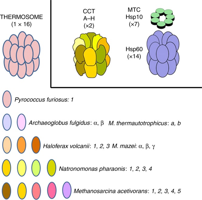
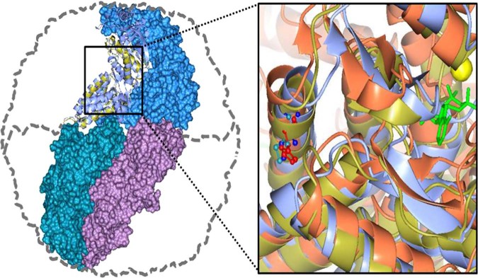
Chaperonins are molecular chaperones that play critical physiological roles, but they can be pathogenic. Malfunctional chaperonins cause chaperonopathies of great interest within various medical specialties. Although the clinical-genetic aspects of many chaperonopathies are known, the molecular mechanisms causing chaperonin failure and tissue lesions are poorly understood. Progress is necessary to improve treatment, and experimental models that mimic the human situation provide a promising solution. We present two models: one prokaryotic (the archaeon Pyrococcus furiosus) with eukaryotic-like chaperonins and one eukaryotic (Chaetomium thermophilum), both convenient for isolation-study of chaperonins, and report illustrative results pertaining to a pathogenic mutation of CCT5.


Conflict of interest statement

The authors declare no competing interests.

Figures

Fig. 1
Fig. 1
Chaperonins of Group I and II in archaea and humans. Top left: archaeal CCT complex or thermosome; top center the CCT chaperonin of Group II complex typically resident of the eukaryotic-cell cytosol; and top right the chaperonin of Group I (Hsp60 or Cpn60) complex (with Hsp10 on top) characteristic of bacteria and eukaryotic-cell mitochondria. Archaeal species vary in their content of chaperonin genes–proteins from only one through a maximum (at least from what we know at the present time) of five. These subunits are variously designated with Arabic numbers, English letters, or Greek letters. As far as we know, they all form a hexadecameric megadalton sized complex, the thermosome, of the type shown on top to the left, which is an example of a hexadecamer found in archaea encoding only one chaperonin subunit, e.g., P. furiosus. The composition of hexadecamers in all archaea that have two or more subunits is not yet fully elucidated. M. thermautotrophicus, Methanothermobacter thermautotrophicus ΔH, previously known as Methanobacterium thermoautotrophicum ΔH. M. mazei, Methanosarcina mazei. (Reproduced with permission from ref. , published under the CC-BY license)
Fig. 2
Fig. 2
Human CCT5 and P. furiosus Pf-Cpn (Pf-CD1) superposed onto the crystal structure of Thermococcus strain KS-1 subunit α (1Q3Q). Left: The Pf-CD1 graphic representation (gold ribbon) was obtained in Swiss-Model (http://swissmodel.expasy.org/) and superposed onto the crystal structure of KS-1 subunit α (monomers are displayed in marine blue, violet, and deep teal colors as surface, and in cyan color as ribbon). The whole hexadecamer double-ring structure is depicted by a dotted line. Right: Magnified image of the superposed structures of the Pf-CD1 (gold ribbon) and Human CCT5 (orange ribbon) onto the KS-1 α subunit crystal structure (1Q3Q; cyan ribbon). Side chains of isoleucine at 138 of Pf-CD1 (blue), isoleucine at 138 of KS-1 subunit α (deep teal), and histidine at 147 of human CCT5 (red) are represented as ball and stick. AMP-PNP (β,γ-Imidoadenosine 5′-triphosphate lithium salt hydrate; green stick) and magnesium ion (yellow ball). (Reproduced with permission from ref. , published under the CC-BY license)
Fig. 3
Fig. 3
a The pathogenic mutant causes a loss of ATPase activity evidenced at various temperatures. Pf-CD1 (red squares), Pf-H (green triangles), and Pf-R (purple stars). The results shown are mean values (±standard deviations) and the experiments were carried out in triplicate. See ref. . b Comparative analyses of protective capacity of mutant and wild type chaperonins. Protection from heat denaturation of malate dehydrogenase (MDH) by mixed oligomers of Pf-CD1 (red square), Pf-H (green triangle), and Pf-R (purple star) at 37 °C (top left panel) and 42 °C (top right panel); and protection of MDH at 37 °C (bottom left panel) and shrimp alkaline phosphatase (SAP) at 50 °C (bottom right panel) by pure hexadecamers. Negative control (no chaperonin added), i.e., MDH, or SAP in bottom right panel, alone: blue diamond. The results shown are mean values (±SD) of triplicate experiments. See ref. . c The pathogenic mutant fails to disperse amyloid fibrils. Dispersion of amyloid fibrils by archaeal Pf-CD1 (top row of panels), partial dispersion by Pf-H (middle row of panels), and no dispersion by Pf-R (bottom row of panels). Atomic force microscopy (AFM) of bovine insulin amyloid fibrils treated with Cpn and Mg++, and ATP. Control panels, no added chaperonin. Scale bar: 250 nm. (Reproduced with permission from ref. , published under the CC-BY license)
Fig. 4
Fig. 4
Schematic representation of the asymmetric conformational changes in the CCT revealed by diffracted X-ray tracking (DXT) and using the C. thermophilum model. a Internal motions of group II chaperonins by DXT. b Asymmetric conformational changes in the CCT. Subunits are colored according to nucleotide affinity as follows: red, high; blue, low. The conformational change proceeds in a sequential manner owing to the asymmetric consumption of ATP by the eukaryotic group II chaperonin. Firstly, ATP binds to the high ATP affinity hemisphere and induces the conformational change, in less than one second. Subsequently, the low-ATP affinity hemisphere changes to the closed conformation. Finally, the ring rotates in counter clockwise direction. (Reproduced with permission from ref. , published under the CC-BY license)

Similar articles

Cited by

References

    1. Horwich AL, Fenton WA, Chapman E, Farr GW. Two families of chaperonin: physiology and mechanism. Annu. Rev. Cell. Dev. Biol. 2007;23:115–145. doi: 10.1146/annurev.cellbio.23.090506.123555. - DOI - PubMed
    1. Cappello F, et al. Hsp60 expression, new locations, functions and perspectives for cancer diagnosis and therapy. Cancer Biol. Ther. 2008;7:801–809. doi: 10.4161/cbt.7.6.6281. - DOI - PubMed
    1. Henderson B, Fares MA, Lund PA. Chaperonin 60: a paradoxical, evolutionarily conserved protein family with multiple moonlighting functions. Biol. Rev. Camb. Philos. Soc. 2013;88:955–987. doi: 10.1111/brv.12037. - DOI - PubMed
    1. Kim YE, Hipp MS, Bracher A, Hayer-Hartl M, Hartl FU. Molecular chaperone functions in protein folding and proteostasis. Annu. Rev. Biochem. 2013;82:323–355. doi: 10.1146/annurev-biochem-060208-092442. - DOI - PubMed
    1. Macario, A. J. L., Conway de Macario, E. & Cappello, F. The Chaperonopathies. Diseases with Defective Molecular Chaperones. (Springer, Dordrecht, Heidelberg, New York, London, 2013). http://link.springer.com/book/10.1007%2F978-94-007-4667-1.

Publication types

MeSH terms