. 2019 Apr:42:157-173.
doi: 10.1016/j.ebiom.2019.03.033. Epub 2019 Mar 22.

Tau antibody chimerization alters its charge and binding, thereby reducing its cellular uptake and efficacy

Affiliations

Tau antibody chimerization alters its charge and binding, thereby reducing its cellular uptake and efficacy

Erin E Congdon et al. EBioMedicine. 2019 Apr.

Abstract

Background: Bringing antibodies from pre-clinical studies to human trials requires humanization, but this process may alter properties that are crucial for efficacy. Since pathological tau protein is primarily intraneuronal in Alzheimer's disease, the most efficacious antibodies should work both intra- and extracellularly. Thus, changes which impact uptake or antibody binding will affect antibody efficacy.

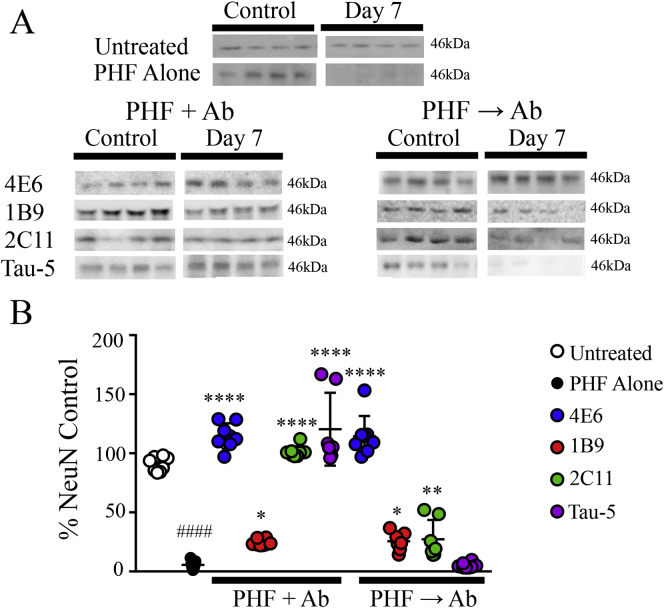
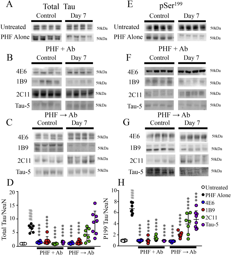
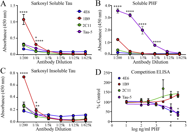
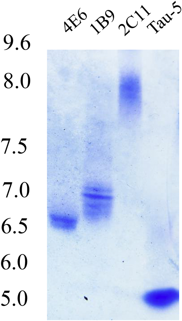
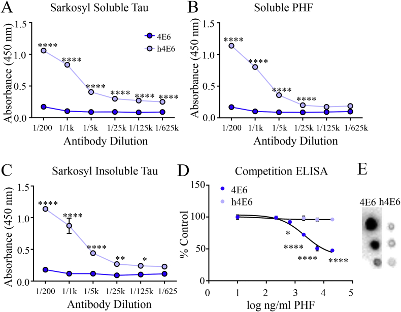
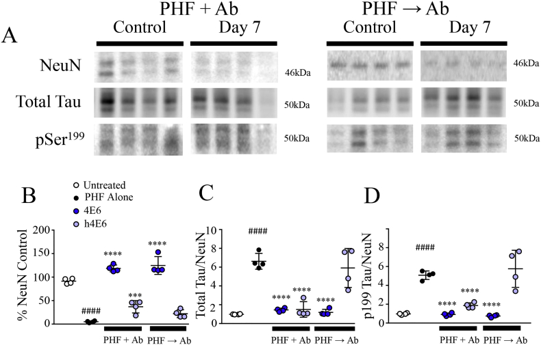
Methods: Initially, we examined four tau mouse monoclonal antibodies with naturally differing charges. We quantified their neuronal uptake, and efficacy in preventing toxicity and pathological seeding induced by human-derived pathological tau. Later, we generated a human chimeric 4E6 (h4E6), an antibody with well documented efficacy in multiple tauopathy models. We compared the uptake and efficacy of unmodified and chimeric antibodies in neuronal and differentiated neuroblastoma cultures. Further, we analyzed tau binding using ELISA assays.

Findings: Neuronal uptake of tau antibodies and their efficacy strongly depends on antibody charge. Additionally, their ability to prevent tau toxicity and seeding of tau pathology does not necessarily go together. Particularly, chimerization of 4E6 increased its charge from 6.5 to 9.6, which blocked its uptake into human and mouse cells. Furthermore, h4E6 had altered binding characteristics despite intact binding sites, compared to the mouse antibody. Importantly, these changes in uptake and binding substantially decreased its efficacy in preventing tau toxicity, although under certain conditions it did prevent pathological seeding of tau.

Conclusions: These results indicate that efficacy of chimeric/humanized tau antibodies should be thoroughly characterized prior to clinical trials, which may require further engineering to maintain or improve their therapeutic potential. FUND: National Institutes of Health (NS077239, AG032611, R24OD18340, R24OD018339 and RR027990, Alzheimer's Association (2016-NIRG-397228) and Blas Frangione Foundation.

Keywords: Alzheimer's disease; Antibody engineering; Immunotherapy; Neuroblastoma; Neuron; Tau protein; Vaccine development.


Figures

Fig. 1
Fig. 1
Prevention of PHF induced toxicity, as measured by LDH levels, depends on antibody and dosing method. JNPL3 primary neurons were exposed to 10 μg/ml of human- derived PHF and 1 μg/ml tau monoclonal antibody in one of two dosing methods. In the first, PHF and antibody were added simultaneously. In the second, PHF was added, followed 24 h later by the antibody. Media was collected from day 0 control cells and those 7 days post treatment and analyzed using a commercially available LDH assay kit. An additional group of cells was left untreated for 7 days and compared to their own day 0 controls (shown in white). Significant antibody, treatment group and interaction effects were seen using a two-way ANOVA (p < .0001). Addition of PHF significantly increased LDH levels relative to day 7 untreated control cells (120% above control, p = .0017). In the PHF + Ab paradigm, 4E6, 2C11 and Tau-5 significantly blocked PHF toxicity (20% above, 87 of, and 14% above control, p = .006, p < .0001, and p = .005). Using the PHF → Ab dosing method, only 4E6 prevented PHF toxicity (25% above control, p = .01) and Tau-5 enhanced it with LDH levels 293% above that of internal controls and 173% above PHF alone (p < .0001 for both). All samples are included in the graph with mean ± SD. ##p ≤ .01, **p ≤ .01, ****p ≤ .0001, #difference compared to untreated cells, *difference compared to PHF alone treated cells.
Fig. 2
Fig. 2
Prevention of PHF induced toxicity, as measured by NeuN levels, depends on antibody and dosing method. Lysate from the JNPL3 neuronal cultures used in LDH experiments were collected at day 0 and 7 days after the final treatment. An additional group of cells was left untreated for 7 days, then compared to their own day 0 controls, to account for normal changes in NeuN (shown in white). All samples were normalized using control cells prepared from the same animal. A. Blots from day 0 controls, day 7 untreated, as well as PHF and tau mAb treated cells were incubated with a commercially available NeuN antibody (Cell Signaling) and developed to show levels in cells treated with PHF and 1B9, 2C11, and Tau-5. B. A two-way ANOVA revealed significant antibody, treatment group and interaction effects (p < .0001). Exposure to PHF induced substantial toxicity (a 95% decrease in NeuN, p < .0001). Under PHF + Ab conditions, all four antibodies tested prevented this toxicity (15% above, 24% of, 99% of and 20% above their internal day 1 control (set at 100%) for 4E6, 1B9, 2C11 and Tau-5, p < .0001, p = .01, p < .0001, and p < .0001). In the PHF → Ab paradigm, 4E6, 1B9 and 2C11 were able to prevent the PHF induced toxicity (114%, 25%, and 27% of control respectively, p < .0001, p = .01, and p = .005). (Control and day 7 samples were run on the same blot). All data points are included in the graph with mean ± SD. ####, ****p ≤ .0001, #difference compared to untreated cells, *difference compared to PHF alone treated cells.
Fig. 3
Fig. 3
Dosing method affects antibody efficacy in preventing increased intracellular tau. In addition to NeuN levels, samples from the same JNPL3 primary neuronal cultures were assayed for total and phospho-tau. To account for any loss of cells due to PHF toxicity, total and phospho tau levels were normalized using NeuN. A-C. Immunoblots were incubated with commercially available pan-tau (Dako). Control cells prepared from the same animal were also collected at day 0 prior to treatment. A second group of untreated cells were collected after 7 days and also compared to their day 0 samples to account for normal changes in tau levels (shown in white). Developed blots show total tau levels in cells treated with PHF and either 4E6, 1B9, 2C11 or Tau-5 in PHF + Ab and PHF → Ab paradigm. Data from 4E6 experiments is included to serve as a positive control. D. A two-way ANOVA showed significant antibody, treatment group and interaction effects (p < .0001 for all). PHF added to cultures led to an increase in tau levels in the remaining cells over 7 days (5.6 fold above control, p < .0001) compared to 7 day untreated samples (shown in white). Under the PHF + Ab condition, all four antibodies, 4E6, 1B9, 2C11 and Tau-5, significantly prevented increased tau levels compared to PHF alone (Tau/NeuN ratio 1.4 for 4E6, 2.0 for 1B9, 0.90 for 2C11, and 1.4 for Tau-5, p < .0001 for all, with untreated control at 1.0). Under the PHF → Ab paradigm only 4E6, and 1B9 had significantly lower tau levels compared to PHF alone (Tau/NeuN 1.1 and 1.5, p < .0001 for both). E-G. Immunoblots showing pSer199 tau levels in day 0 controls, untreated cells, and cells treated with PHF and either 4E6, 1B9, 2C11 or Tau-5 in PHF + Ab and PHF → Ab paradigm. H. A two-way ANOVA of pSer199 revealed significant antibody, treatment group and interaction effects (p < .0001). PHF alone increased intracellular phospho-tau (5.7 fold above control, p < .0001) compared to untreated cells. In the PHF + Ab condition, all four antibodies lowered pSer199 levels (pSer199/NeuN 0.93 for 4E6, 0.94 for 1B9, 1.3 for 2C11, and 0.91 for Tau-5, p < .0001 for all, with untreated control at 1.0). In the PHF → Ab paradigm, all of the antibodies also had significantly lower phospho-tau levels compared to PHF alone (pSer199/NeuN 0.8 4E6, 2.0 1B9, 3.9 2C11 and 4.7 Tau-5, p < .0001, p < .0001, p < .0001, and p = .002) (Control and day 7 samples were run on the same blot) All data points are included in the graph with mean ± SD. ***p ≤ .001, ####, ****p ≤ .0001, #difference compared to untreated cells, *difference compared to PHF alone treated cells.
Fig. 4
Fig. 4
Monoclonal antibodies vary in their tau binding. Assay plates were coated with one of three different tau fractions prepared from the same AD patient, sarkosyl soluble tau, soluble PHF tau and sarkosyl insoluble tau. A. When plates were coated with sarkosyl soluble tau 1B9 had significantly higher binding than all other antibodies at the 1/200 (p < .0001 for all) and 1/1000 dilutions (p < .0001 for 4E6 and 2C11 and p = 0.012 for Tau-5). B. Tau-5 had significantly higher binding to soluble PHF compared to 1B9 and 2C11 from 1/200–1/25 k (p < .0001 for all). C. On plates coated with sarkosyl insoluble tau, 1B9 again had significantly higher binding than all other antibodies at the 1/200 dilution (p < .0001 for all), and was significantly higher than 4E6 and 2C11 at 1/1000 (p = .003, 0.04). D. A competitive ELISA was performed by incubating each antibody with increasing concentrations of soluble PHF prior to addition to the plate. A two-way ANOVA revealed significant antibody and interaction effects (p < .0001 and p = .002). Of the four, only 4E6 and Tau-5 showed a significant decrease in binding. 4E6 had significantly reduced binding to the plate at the two highest PHF concentrations (50 and 47% of control, p = .03 and p = .02) and Tau-5 at the highest concentration (34% of control value, p ≤ .03) indicating that these two may preferentially bind soluble PHF species. All points and columns are mean ± SD. *p ≤ .05, **p ≤ .01, ***p ≤ .001, ****p ≤ .0001.
Fig. 5
Fig. 5
Antibodies differ in their uptake into neurons. Primary neurons prepared from day 0 JNPL3 pups were incubated with 1 μg/ml tau monoclonal antibodies 4E6, 1B9, 2C11, or Tau-5 for 24 h. Following this period, cells were fixed and stained with a rabbit pan tau antibody (Dako) to visualize the cells. 4E6, 1B9, 2C11 and Tau-5 were detected using a fluorescent anti-mouse secondary. A-C. Confocal images showing uptake of 4E6. The antibody is readily taken up by the neurons. D-L. Confocal images showing uptake of 1B9, 2C11, and Tau-5. Few cells show antibody positive puncta inside the cells. M. Quantitation of antibody uptake for each of the antibodies examined. Multiple images were collected and the percentage of colocalization between the cell stain and the antibody puncta per image were calculated using Image J. Colocalization analysis showed an overall significant difference in antibody uptake (One-way ANOVA, p < .0001). All three antibodies showed significantly reduced neuronal uptake with colocalization at 18%, 9% and 2% of the values seen for 4E6 (p < .0001 for all, with 4E6 values set at 100%). N. Uptake results were confirmed using immunoblotting. Neurons were incubated for 24 h with 1 μg/ml of 4E6, 1B9, 2C11 or Tau-5. Cell lysate was then collected and run on a western blot. Membranes were probed with mouse secondary antibody to detect tau mAbs. Bands were visible in cells treated with 4E6, but not in blots made from those treated with other tau mAbs. O. A dot blot was then used to compare binding of mouse secondary to each of the antibodies. Samples were diluted to 200 μg/ml and 1 μl of each mAb was spotted onto the membrane, which was then incubated with HRP-conjugated mouse secondary and developed. 4E6 shows the lowest signal, with Tau-5 higher and 1B9 and 2C11 the strongest. All data points are included in the graph with mean ± SD. ****p ≤ .0001.
Fig. 6
Fig. 6
Tau antibodies have differing isoelectric points. Tau monoclonal antibodies 4E6, 1B9, 2C11 and Tau-5 were run on an isoelectric focusing gel. 4E6 has an IEP of 6.5. 1B9 has a neutral IEP and 2C11 is more basic (7.0 and approximately 8.0 respectively), while Tau-5 is acidic with an IEP near 5.0.
Fig. 7
Fig. 7
Increasing IEP reduces antibody uptake without affecting tau peptide binding. Because the tau mAbs used recognize different epitopes, and in the case of 2C11 are of a different IgG isotype, we examined the effects of direct modification of a single antibody. An aliquot of 4E6 was cationized with hexamethylenediamine (HMD) to raise its IEP. A. Cationization resulted in several bands ranging from 6.5–7.0 on an isoelectric focusing gel. B. JNPL3 neurons were incubated with 10 or 20 μg/ml of 4E6 or the cationized 4E6 (cat4E6 in the figure) for 24 h. Intraneuronal antibody levels were detected using Western blotting. At both 10 and 20 μg/ml, a 0.5 change in IEP significantly reduced the uptake of 4E6 into the neurons (23% and 16%, p = .04 and p = .01 respectively). C. Serial dilutions of 4E6 and cat4E6 (1, 0.25, 0.0625 mg/ml) were spotted onto a nitrocellulose membrane and detected with HRP-conjugated mouse secondary (1:5000). Similar reactivity was seen with both antibodies. D, E. ELISA plates were coated with tau peptides representing the C-terminal either phosphorylated at Ser396/404 or non-phosphorylated. Cationization did not significantly alter antibody binding. All data points are included in panel B with mean ± SD, graphs in D and E show mean ± SD. *p ≤ .05, **p ≤ .01.
Fig. 8
Fig. 8
Human chimerization reduces 4E6 uptake and increases IEP. A. When run on an IEF gel, unmodified 4E6 has an IEP of 6.5, whereas human chimerization increases this to 9.6. Samples were run on the same gel and images cropped to remove unrelated samples. B-D. Primary neurons prepared from JNPL3 mice were incubated with 1 μg/ml CypHer 5 labeled 4E6 for 24 h. In live cell images the antibody is visible as bright puncta which fill the cell bodies, indicating that neurons readily take up the antibody. E-G. However, in JNPL3 neurons incubated with 1 μg/ml CypHer 5 labeled human chimeric 4E6 (h4E6) show substantially reduced uptake via live cell imaging. Few cells contain fluorescent puncta and the overall signal is dimmer. H-J. Human neuroblastoma cells (SH-SY5Y) were incubated with 20 μg/ml CypHer 5 labeled 4E6 for 3 h. Results were similar to those seen in neurons, with high levels of uptake in live cell images. K-M. Uptake was greatly reduced using CypHer 5 labeled h4E6 in the neuroblastoma cells, with relatively few puncta visible. N. Neuronal uptake of the human chimeric antibody was quantified by calculating the percentage of each image containing fluorescent signal using Image J. This signal was significantly reduced in h4E6 treated neurons compared to unmodified 4E6 (95% reduction in mouse primary neurons, 91% in human neuroblastoma (p < .0001). All data points are included in the graph with mean ± SD. ****p ≤ .0001.
Fig. 9
Fig. 9
Human chimerization of 4E6 alters its tau binding. A-C. ELISA plates were coated with sarkosyl soluble, solubilized PHF and sarkosyl insoluble tau. Serial dilutions of unmodified and human chimeric 4E6 were added and binding to each fraction quantified. h4E6 showed similar levels of binding to each fraction, and was significantly higher than mouse 4E6 at every dilution with sarkosyl soluble tau and soluble PHF (p < .0001). h4E6 also had significantly higher binding to sarkosyl insoluble tau at every dilution except 1/625 k (p ≤ .0001-0.02). D. A competitive ELISA was performed to assess binding to soluble PHF. Significant antibody, PHF dose and interaction effects were seen using a two-way ANOVA (p < .0001 for all). Reduced binding to the plate was seen at all but the lowest PHF concentration with 4E6 (8–53% reduction, p ≤ .03–0.0001). In contrast, h4E6 did not show a reduction in binding to the assay plate when pre-incubated with PHF, indicating that it preferentially binds insoluble species. E. Serial dilutions of 4E6 and h4E6 (1, 0.25, 0.062 mg/ml) were spotted onto nitrocellulose membrane and incubated with the same HRP-conjugated mouse and human secondary antibodies used in the ELISA assay at equal concentrations. The h4E6 samples showed lower reactivity, either the result of reduced secondary antibody or ECL developer binding. Thus, the higher signal obtained in ELISA assays with h4E6 is not the result of increased secondary antibody affinity. All points and columns are mean ± SD. *p ≤ .05, **p ≤ .01, ****p ≤ .0001.
Fig. 10
Fig. 10
Chimerization impairs efficacy of 4E6 in preventing PHF induced toxicity and tau seeding. To determine how the changes in IEP resulting from human chimerization affect the efficacy of 4E6, JNPL3 neurons were incubated with 10 μg/ml PHF and 1 μg/ml of antibody in the dosing paradigms described above. A. Samples from day 0 control cells, and those treated with 10 μg/ml PHF and 1 μg/ml h4E6 in the PHF + Ab and PHF → Ab paradigms, were assayed for NeuN, total and phospho-tau. Immunoblots were probed using commercially available antibodies. As above, a set of untreated cells were collected at day 7 and compared to their own day 0 controls (shown in white). B. There was a significant antibody effect by two-way ANOVA (p < .0001), when NeuN levels were quantified. Addition of PHF resulted in increased toxicity relative to untreated cells (p < .0001). Under both dosing paradigms unmodified 4E6 significantly reduced PHF-induced toxicity (p < .0001). Under PHF + Ab dosing conditions, h4E6 was able to prevent some of the PHF toxicity (36% of control, p = .0005), but it was ineffective under the PHF → Ab paradigm. C. There were also significant overall antibody, dosing method and interaction effects when corrected total tau levels were quantified using a two-way ANOVA (p < .0001, p = .0003, and p < .0001). As with toxicity data, 4E6 prevented total tau increase in both treatment groups (p < .0001). In the PHF + Ab condition, h4E6 prevented the accumulation of tau in the remaining cells (Tau/NeuN ratio 1.49, p < .0001). D. A two-way ANOVA using phosphorylated tau levels found an overall significant antibody, treatment group and interaction effects (p < .0001, p = .0016, and p < .0001). Again, 4E6 treated cells had significantly lower phospho-tau levels compared to PHF alone under both conditions (p < .0001). Significantly lower corrected phospho-tau levels were recorded using h4E6 in the PHF + Ab paradigm (pSer199/NeuN 1.8, p < .0001), but not in the PHF → Ab paradigm. All data points are included in the graph with mean ± SD. ####, ***p≤ 0.001, ****p ≤ .0001, #difference compared to untreated cells, *difference compared to PHF alone treated cells.

Similar articles

Cited by

References

    1. Sigurdsson E.M. Tau immunotherapies for Alzheimer's disease and related tauopathies: progress and potential pitfalls. J Alzheimers Dis. 2018;64(s1) S555-S65. - PMC - PubMed
    1. Congdon E.E., Sigurdsson E.M. Tau-targeting therapies for Alzheimer disease. Nat Rev Neurol. 2018;14:399–415. - PMC - PubMed
    1. van der Kleij H., Charles N., Karimi K., Mao Y.K., Foster J., Janssen L. Evidence for neuronal expression of functional Fc (epsilon and gamma) receptors. J Allergy Clin Immunol. 2010;125(3):757–760. - PMC - PubMed
    1. Kam T.I., Song S., Gwon Y., Park H., Yan J.J., Im I. FcgammaRIIb mediates amyloid-beta neurotoxicity and memory impairment in Alzheimer's disease. J Clin Invest. 2013;123(7):2791–2802. - PMC - PubMed
    1. Nakamura K., Hirai H., Torashima T., Miyazaki T., Tsurui H., Xiu Y. CD3 and immunoglobulin G Fc receptor regulate cerebellar functions. Mol Cell Biol. 2007;27(14):5128–5134. - PMC - PubMed

MeSH terms