Targeting senescent cells alleviates obesity-induced metabolic dysfunction
- PMID: 30907060
- PMCID: PMC6516193
- DOI: 10.1111/acel.12950
Targeting senescent cells alleviates obesity-induced metabolic dysfunction
Abstract
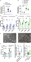
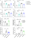
Adipose tissue inflammation and dysfunction are associated with obesity-related insulin resistance and diabetes, but mechanisms underlying this relationship are unclear. Although senescent cells accumulate in adipose tissue of obese humans and rodents, a direct pathogenic role for these cells in the development of diabetes remains to be demonstrated. Here, we show that reducing senescent cell burden in obese mice, either by activating drug-inducible "suicide" genes driven by the p16Ink4a promoter or by treatment with senolytic agents, alleviates metabolic and adipose tissue dysfunction. These senolytic interventions improved glucose tolerance, enhanced insulin sensitivity, lowered circulating inflammatory mediators, and promoted adipogenesis in obese mice. Elimination of senescent cells also prevented the migration of transplanted monocytes into intra-abdominal adipose tissue and reduced the number of macrophages in this tissue. In addition, microalbuminuria, renal podocyte function, and cardiac diastolic function improved with senolytic therapy. Our results implicate cellular senescence as a causal factor in obesity-related inflammation and metabolic derangements and show that emerging senolytic agents hold promise for treating obesity-related metabolic dysfunction and its complications.
Keywords: adipogenesis; aging; cellular senescence; dasatinib; quercetin; senolytics; type 2 diabetes.
© 2019 The Authors. Aging Cell published by the Anatomical Society and John Wiley & Sons Ltd.
Conflict of interest statement
J.L.K., T.T., T.P., A.K.P., Y.Z., M.X., J.C., and M.D. have a financial interest related to this research. Patents on senolytic drugs are held by Mayo Clinic. This research has been reviewed by the Mayo Clinic and Buck Institute Conflict of Interest Review Boards and was conducted in compliance with Mayo Clinic and Buck Institute Conflict of Interest policies. None of the other authors has a relevant conflict of financial interest.
Figures


Similar articles
-
Cellular Senescence in Diabetes Mellitus: Distinct Senotherapeutic Strategies for Adipose Tissue and Pancreatic β Cells.Front Endocrinol (Lausanne). 2022 Mar 31;13:869414. doi: 10.3389/fendo.2022.869414. eCollection 2022. Front Endocrinol (Lausanne). 2022. PMID: 35432205 Free PMC article. Review.
-
Senolytics decrease senescent cells in humans: Preliminary report from a clinical trial of Dasatinib plus Quercetin in individuals with diabetic kidney disease.EBioMedicine. 2019 Sep;47:446-456. doi: 10.1016/j.ebiom.2019.08.069. Epub 2019 Sep 18. EBioMedicine. 2019. PMID: 31542391 Free PMC article.
-
Senolytic drugs, dasatinib and quercetin, attenuate adipose tissue inflammation, and ameliorate metabolic function in old age.Aging Cell. 2023 Feb;22(2):e13767. doi: 10.1111/acel.13767. Epub 2023 Jan 13. Aging Cell. 2023. PMID: 36637079 Free PMC article.
-
Transient metabolic improvement in obese mice treated with navitoclax or dasatinib/quercetin.Aging (Albany NY). 2020 Jun 25;12(12):11337-11348. doi: 10.18632/aging.103607. Epub 2020 Jun 25. Aging (Albany NY). 2020. PMID: 32584785 Free PMC article.
-
Cellular senescence: Implications for metabolic disease.Mol Cell Endocrinol. 2017 Nov 5;455:93-102. doi: 10.1016/j.mce.2016.08.047. Epub 2016 Aug 31. Mol Cell Endocrinol. 2017. PMID: 27591120 Free PMC article. Review.
Cited by
-
Cellular senescence in cancer: from mechanisms to detection.Mol Oncol. 2021 Oct;15(10):2634-2671. doi: 10.1002/1878-0261.12807. Epub 2020 Oct 22. Mol Oncol. 2021. PMID: 32981205 Free PMC article. Review.
-
Inflammation/bioenergetics-associated neurodegenerative pathologies and concomitant diseases: a role of mitochondria targeted catalase and xanthophylls.Neural Regen Res. 2021 Feb;16(2):223-233. doi: 10.4103/1673-5374.290878. Neural Regen Res. 2021. PMID: 32859768 Free PMC article. Review.
-
Evaluating causality of cellular senescence in non-alcoholic fatty liver disease.JHEP Rep. 2021 May 1;3(4):100301. doi: 10.1016/j.jhepr.2021.100301. eCollection 2021 Aug. JHEP Rep. 2021. PMID: 34113839 Free PMC article. Review.
-
Systemic overexpression of C-C motif chemokine ligand 2 promotes metabolic dysregulation and premature death in mice with accelerated aging.Aging (Albany NY). 2020 Oct 26;12(20):20001-20023. doi: 10.18632/aging.104154. Epub 2020 Oct 26. Aging (Albany NY). 2020. PMID: 33104522 Free PMC article.
-
Senolytic Therapy: A Potential Approach for the Elimination of Oncogene-Induced Senescent HPV-Positive Cells.Int J Mol Sci. 2022 Dec 8;23(24):15512. doi: 10.3390/ijms232415512. Int J Mol Sci. 2022. PMID: 36555154 Free PMC article. Review.
References
-
- Christopher, L. J. , Cui, D. , Wu, C. , Luo, R. , Manning, J. A. , Bonacorsi, S. J. , … Iyer, R. A. (2008). Metabolism and disposition of dasatinib after oral administration to humans. Drug Metabolism and Disposition: the Biological Fate of Chemicals, 36, 1357–1364. 10.1124/dmd.107.018267 - DOI - PubMed
Publication types
MeSH terms
Substances
Grants and funding
- R01 AG053832/AG/NIA NIH HHS/United States
- BB/I020748/1/BB_/Biotechnology and Biological Sciences Research Council/United Kingdom
- BB/K019260/1/BB_/Biotechnology and Biological Sciences Research Council/United Kingdom
- F30 AG046061/AG/NIA NIH HHS/United States
- R01 CA233790/CA/NCI NIH HHS/United States
- 24009/CRUK_/Cancer Research UK/United Kingdom
- R01 AG013925/AG/NIA NIH HHS/United States
- R24 AG044396/AG/NIA NIH HHS/United States
- P01 AG031736/AG/NIA NIH HHS/United States
- K23 DK109134/DK/NIDDK NIH HHS/United States
- R01 HL141819/HL/NHLBI NIH HHS/United States
- P30 DK050456/DK/NIDDK NIH HHS/United States
- R37 AG013925/AG/NIA NIH HHS/United States
- P30 CA016058/CA/NCI NIH HHS/United States
- P01 AG041122/AG/NIA NIH HHS/United States
- R01 AG058812/AG/NIA NIH HHS/United States
- BB/M023389/1/BB_/Biotechnology and Biological Sciences Research Council/United Kingdom
- T32 GM065841/GM/NIGMS NIH HHS/United States
- K99 AG051661/AG/NIA NIH HHS/United States
- R00 AG051661/AG/NIA NIH HHS/United States
LinkOut - more resources
Full Text Sources
Other Literature Sources
Medical
Molecular Biology Databases

