Comparative Study
. 2019 Mar 6:9:43.
doi: 10.3389/fcimb.2019.00043. eCollection 2019.

A Pathogen and a Non-pathogen Spotted Fever Group Rickettsia Trigger Differential Proteome Signatures in Macrophages

Affiliations
Comparative Study

A Pathogen and a Non-pathogen Spotted Fever Group Rickettsia Trigger Differential Proteome Signatures in Macrophages

Pedro Curto et al. Front Cell Infect Microbiol. .

Abstract

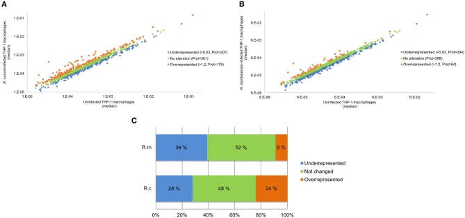
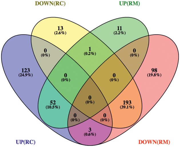
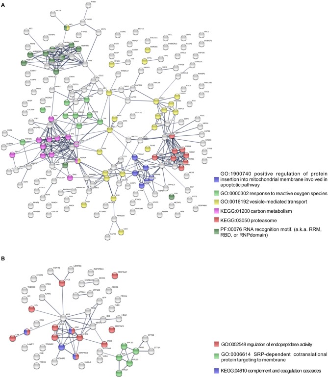
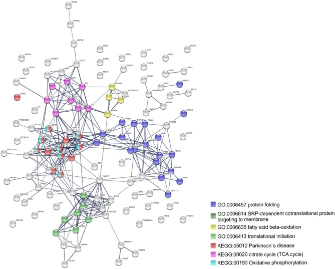
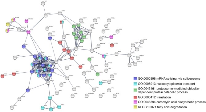
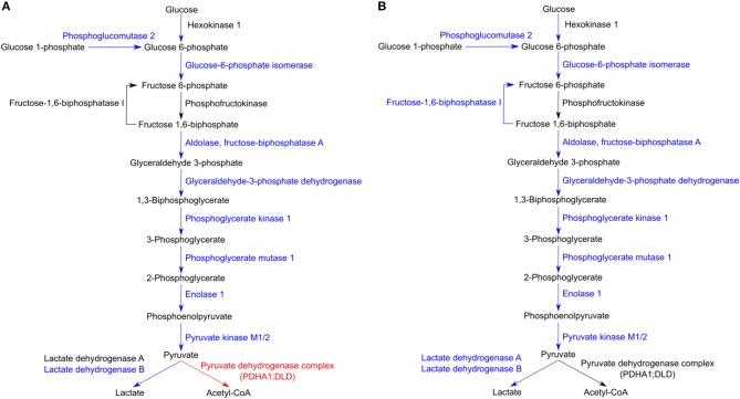
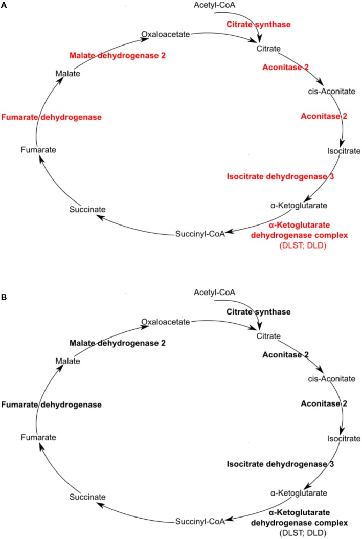
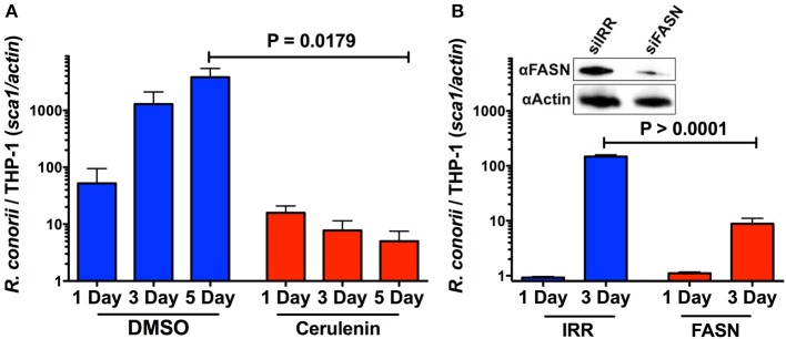
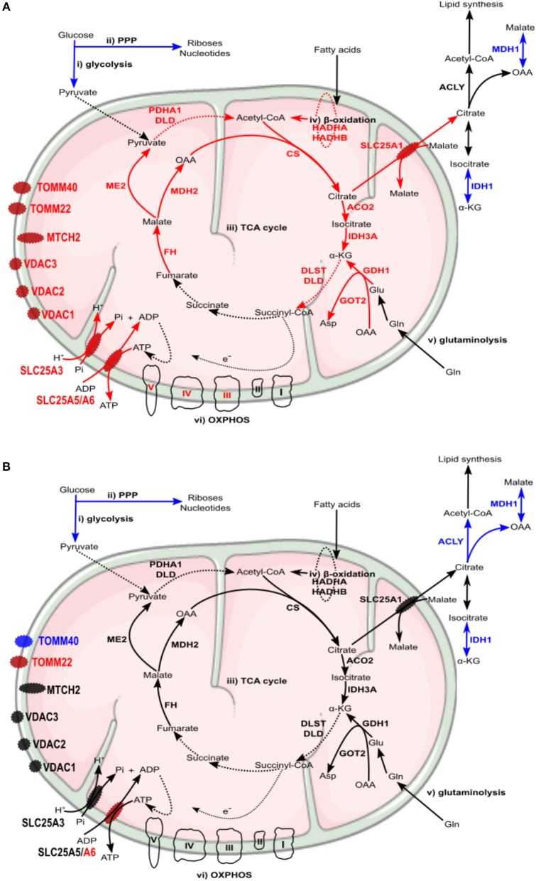
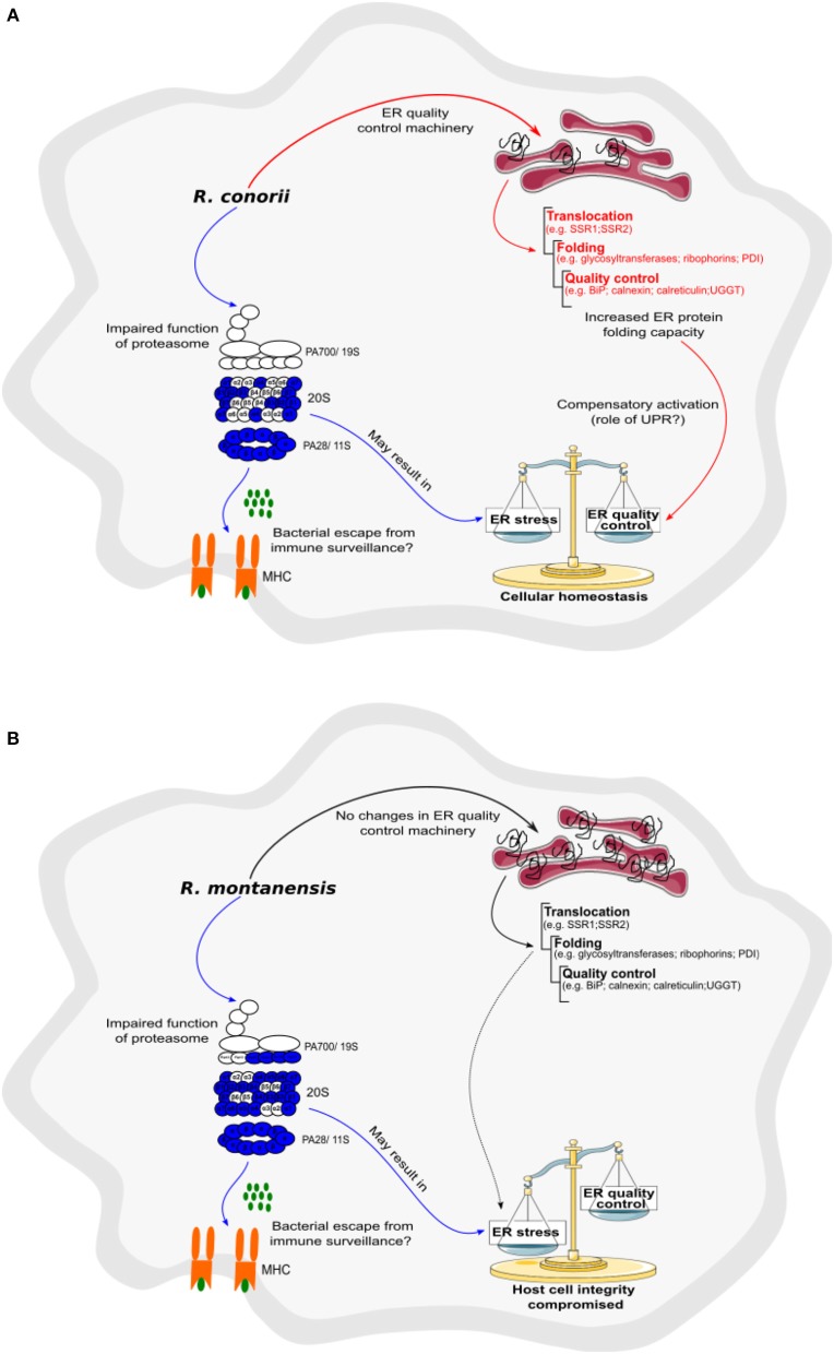
We have previously reported that Rickettsia conorii and Rickettsia montanensis have distinct intracellular fates within THP-1 macrophages, suggesting that the ability to proliferate within macrophages may be a distinguishable factor between pathogenic and non-pathogenic Spotted fever group (SFG) members. To start unraveling the molecular mechanisms underlying the capacity (or not) of SFG Rickettsia to establish their replicative niche in macrophages, we have herein used quantitative proteomics by SWATH-MS to profile the alterations resulted by the challenge of THP-1 macrophages with R. conorii and R. montanensis. We show that the pathogenic, R. conorii, and the non-pathogenic, R. montanensis, member of SFG Rickettsia trigger differential proteomic signatures in macrophage-like cells upon infection. R. conorii specifically induced the accumulation of several enzymes of the tricarboxylic acid cycle, oxidative phosphorylation, fatty acid β-oxidation, and glutaminolysis, as well as of several inner and outer membrane mitochondrial transporters. These results suggest a profound metabolic rewriting of macrophages by R. conorii toward a metabolic signature of an M2-like, anti-inflammatory activation program. Moreover, several subunits forming the proteasome and immunoproteasome are found in lower abundance upon infection with both rickettsial species, which may help bacteria to escape immune surveillance. R. conorii-infection specifically induced the accumulation of several host proteins implicated in protein processing and quality control in ER, suggesting that this pathogenic Rickettsia may be able to increase the ER protein folding capacity. This work reveals novel aspects of macrophage-Rickettsia interactions, expanding our knowledge of how pathogenic rickettsiae explore host cells to their advantage.

Keywords: Rickettsia conorii; Rickettsia montanensis; SWATH-MS; host-pathogen interactions; macrophages; metabolic reprogramming; protein processing pathways; spotted fever group Rickettsia.


Figures

Figure 1
Figure 1
Overall analysis of R. conorii- and R. montanensis-induced changes in global proteome of THP-1 macrophages. (A,B) Scatterplot representation of changes in protein abundance of THP-1 macrophages upon infection with R. conorii (A) and R. montanensis (B). The 746 quantified proteins that were confidently quantified in all 3 experimental conditions were plotted and considered altered when a change of at least 20% in abundance (fold change ≤ 0.83 or fold change ≥ 1.2) was observed between infected and uninfected conditions. Proteins that were considered to decrease, not change or increase its abundance upon infection are represented in blue, green and orange, respectively. (C) Bar chart displaying the percentage (out of the 746 quantified proteins that were confidently quantified) of host proteins that were considered to decrease, not change or increase its abundance upon infection with both R. conorii and R. montanensis. See also Supplementary Table 2.
Figure 2
Figure 2
Venn diagram depicting the number and distribution of host proteins that changed their abundance upon infection with R. conorii or R. montanensis. Host proteins that change their abundance in the same direction (increase or decrease abundance) upon infection with both R. conorii and R. montanensis are considered to be a common response to infection. On the other hand, host proteins that change their abundance in only one infection condition, but show unchanged protein levels in the other, are considered to be a species-specific host response. DOWN(RC), yellow—proteins that are in decreased abundance in R. conorii-infected THP-1 macrophages compared to uninfected THP-1 macrophages; UP(RC), blue—proteins that are in increased abundance in R. conorii-infected THP-1 macrophages compared to uninfected THP-1 macrophages; DOWN(RM), red—proteins that are in decreased abundance in R. montanensis-infected THP-1 macrophages compared to uninfected THP-1 macrophages; UP(RM), green—proteins that are in increased abundance in R. montanensis-infected THP-1 macrophages compared to uninfected THP-1 macrophages. Individual information about the proteins in each group of the Venn diagram can be found in Supplementary Table 3. Venn diagrams were obtained using VENNY 2.1 (http://bioinfogp.cnb.csic.es/tools/venny/).
Figure 3
Figure 3
Protein network analysis of common responses to infection with both R. conorii and R. montanensis. (A,B) Clustering of protein-protein interaction networks for the 193 and 52 host proteins commonly altered between infection conditions, found with reduced abundance (A) or increased abundance (B), respectively. List of the individual host proteins for each independent analysis can be found in Supplementary Table 3. The analysis was carried out with STRING 10.5 (http://string-db.org/) using high confidence (0.7) score. Nodes are represented with different colors according to their categorization in gene ontology (GO) terms, KEGG pathways or PFAM protein domains.
Figure 4
Figure 4
Clustering of host proteins that specifically increase their abundance upon infection with R. conorii. Protein-protein interaction network for the 123 host proteins with increased abundance upon infection with R. conorii, but unchanged levels upon infection with R. montanensis. List of the individual host proteins for each independent analysis can be found in Supplementary Table 3. The analysis was carried out with STRING 10.5 (http://string-db.org/) using high confidence (0.7) score. Nodes are represented with different colors according to their categorization in gene ontology (GO) terms or KEGG pathways.
Figure 5
Figure 5
Clustering of host proteins that specifically decrease their abundance upon infection with R. montanensis. Protein-protein interaction network for the 98 host proteins with decreased abundance upon infection with R. montanensis, but unchanged protein levels upon infection with R. conorii. List of the individual host proteins for each independent analysis can be found in Supplementary Table 3. The analysis was carried out with STRING 10.5 (http://string-db.org/) using high confidence (0.7) score. Nodes are represented with different colors according to their categorization in gene ontology (GO) terms or KEGG pathways.
Figure 6
Figure 6
Host glycolytic enzymes found in reduced abundance upon infection of THP-1 macrophages with both rickettsial species. (A,B) Infection of THP-1 macrophages with either R. conorii (A) or R. montanensis (B) resulted in a decrease in the abundance of several host glycolytic enzymes at 24 h post-infection. UniProt accession number and the respective fold change upon infection can be found in Table 3 for each respective enzyme. Enzymes with unchanged or decreased protein levels, when compared to uninfected cells, are represented in black and blue, respectively. Red represents enzymes of the pyruvate dehydrogenase complex found accumulated in R. conorii-infected cells (Table 3).
Figure 7
Figure 7
Rickettsia conorii, but not R. montanensis, infection increased the abundance of several enzymes of the TCA cycle in THP-1 macrophages. Infection of THP-1 macrophages with either R. conorii (A) or R. montanensis (B) resulted in alterations in the abundance of several host TCA cycle enzymes at 24 h post-infection. UniProt accession number and the respective fold change upon infection can be found in Table 3 for each respective enzyme. Enzymes with unchanged or increased protein levels, when compared to uninfected cells, are represented in black and red, respectively.
Figure 8
Figure 8
Inhibition of FASN function and expression inhibits the ability of R. conorii to proliferate within THP-1 cells. (A) Quantitative PCR analysis of gDNA from PMA differentiated THP-1 cells infected with R. conorii Malish7 in the presence of DMSO (vehicle control) or FASN inhibitor, cerulenin, at 24 h, 3, and 5 days-post treatment. (B, Inset) Western immunoblot analysis confirms the inhibition of FASN protein expression from samples isolated at 3 days-post transfection when compared to control RNA treated cells (siIRR). An immunoblot for Actin is used to control for equal protein loading in each lane. Quantitative PCR analysis of gDNA from PMA differentiated THP-1 cells transfected with siRNA against FASN or a control RNA and infected with R. conorii Malish7 at an MOI of 2. Samples were analyzed at 24 h and 3 days post infection. Data is representative of two independent experiments with each condition in triplicate. Statistical analysis was performed by One-Way ANOVA with a Dunnett's post-hoc test for pairwise comparison. Statistical significance (p < 0.05).
Figure 9
Figure 9
Rickettsia conorii and R. montanensis trigger a differential metabolic signature in THP-1 macrophages. (A,B) Prediction model of alterations in host cell metabolism, based on changes in the abundance of host proteins induced by infection of THP-1 macrophages with R. conorii (A) or R. montanensis (B) Increase/decrease in the abundance of host enzymes are predicted to contribute with increase/decrease in activity for the respective biological enzymatic activity and are represented in red and blue, respectively. Enzymes quantified in our analysis but with no alteration in abundance upon infection are represented in black (A) In R. conorii-infected THP-1 macrophages, glycolysis (i) and pentose phosphate pathway (ii) are predicted to be reduced at 24 h post-infection. This may impact pyruvate production from glycolysis as well as production of riboses, nucleotides and ROS from PPP, globally contributing to reduce pro-inflammatory signals. Several TCA cycle enzymes (iii) were found overrepresented upon infection, suggesting an increase in TCA cycle activity. Acetyl-CoA production from fatty-acid β-oxidation (iv) and glutamine anaplerosis (v) may contribute to replenish the TCA cycle which may result in a sustained ATP production via oxidative phosphorylation (vi). Accumulation of several inner and outer membrane transporters is suggestive of a metabolic configuration with higher needs in metabolic supply and demand. (B) In R. montanensis-infected THP-1 macrophages, pyruvate production from glycolysis (i) is also predicted to be reduced at this time of infection. However, in contrast with R. conorii, unchanged levels of enzymes of the TCA cycle (iii), fatty-acid β-oxidation (iv), glutaminolysis (v), and proteins from the respiratory complex (vi) found in R. montanensis-infected cells, together with no alterations observed in most of the quantified mitochondrial transporters, suggests very distinct metabolic requirements between infection conditions (see Tables 3, 4 for details).
Figure 10
Figure 10
Rickettsia conorii, but not R. montanensis, may be able to restore host cell homeostasis by increasing ER folding capacity. (A,B) Prediction model of alterations in proteasome and protein processing in endoplasmic reticulum activity, based on changes in the abundance of host proteins, that are induced by the infection of THP-1 macrophages with R. conorii (A) or R. montanensis (B). Increase/decrease in the abundance of host enzymes are predicted to contribute with increase/decrease in activity and are represented in red and blue, respectively. Increase, no alteration, or decrease in the abundance of host enzymes are predicted to contribute with increase, unchanged or decrease activity for the respective biological function and are represented in red, black, and blue arrows, respectively. (A) In R. conorii-infected THP-1 macrophages, several proteasome and immunoproteasome activator subunits are underrepresented at 24 h post-infection, which may lead to a decrease in antigen peptide generation, and subsequent decrease in antigen presentation by MHC complex Type I, and bacterial evasion from immune system surveillance. Decrease in proteasome activity may lead to an accumulation of misfolded proteins in ER, inducing ER stress. However, R. conorii specifically increases the abundance of several ER proteins involved protein translocation, folding, and quality control, which may be a compensatory mechanism activated by the UPR. (B) In R. montanensis-infected THP-1 macrophages, several proteasome and immunoproteasome activator subunits are also underrepresented at 24 h post-infection, which may lead to a decrease in antigen peptide generation (ii), and subsequent antigen presentation by MHC class I and bacteria evasion from immune system surveillance. Decrease in proteasome activity may lead to an accumulation of misfolded proteins and induction of ER stress. However, in contrast with R. conorii, R. montanensis infection did not result in increased levels of ER quality control components, likely resulting in the inability of R. montanensis to restore host cell homeostasis (see Table 5 for details).

Similar articles

Cited by

References

    1. Abu Kwaik Y., Bumann D. (2015). Host delivery of favorite meals for intracellular pathogens. PLoS Pathog. 11:e1004866. 10.1371/journal.ppat.1004866 - DOI - PMC - PubMed
    1. Ammerman N. C., Beier-Sexton M., Azad A. F. (2008). Laboratory maintenance of Rickettsia rickettsii. Curr. Protoc. Microbiol. 3:3A5. 10.1002/9780471729259.mc03a05s11 - DOI - PMC - PubMed
    1. Anjo S. I., Santa C., Manadas B. (2015). Short GeLC-SWATH: a fast and reliable quantitative approach for proteomic screenings. Proteomics 15, 757–762. 10.1002/pmic.201400221 - DOI - PubMed
    1. Anjo S. I., Santa C., Manadas B. (2017). SWATH-MS as a tool for biomarker discovery: from basic research to clinical applications. Proteomics 17, 3–4. 10.1002/pmic.201600278 - DOI - PubMed
    1. Ashida H., Kim M., Sasakawa C. (2014). Exploitation of the host ubiquitin system by human bacterial pathogens. Nat. Rev. Microbiol. 12, 399–413. 10.1038/nrmicro3259 - DOI - PubMed

Publication types