. 2019 Mar 20;17(1):27.
doi: 10.1186/s12964-019-0339-1.

Recombinant RGD-disintegrin DisBa-01 blocks integrin αvβ3 and impairs VEGF signaling in endothelial cells

Affiliations

Recombinant RGD-disintegrin DisBa-01 blocks integrin αvβ3 and impairs VEGF signaling in endothelial cells

Taís M Danilucci et al. Cell Commun Signal. .

Abstract

Background: Integrins mediate cell adhesion, migration, and survival by connecting the intracellular machinery with the surrounding extracellular matrix. Previous studies demonstrated the interaction between αvβ3 integrin and VEGF type 2 receptor (VEGFR2) in VEGF-induced angiogenesis. DisBa-01, a recombinant His-tag fusion, RGD-disintegrin from Bothrops alternatus snake venom, binds to αvβ3 integrin with nanomolar affinity blocking cell adhesion to the extracellular matrix. Here we present in vitro evidence of a direct interference of DisBa-01 with αvβ3/VEGFR2 cross-talk and its downstream pathways.

Methods: Human umbilical vein (HUVECs) were cultured in plates coated with fibronectin (FN) or vitronectin (VN) and tested for migration, invasion and proliferation assays in the presence of VEGF, DisBa-01 (1000 nM) or VEGF and DisBa-01 simultaneously. Phosphorylation of αvβ3/VEGFR2 receptors and the activation of intracellular signaling pathways were analyzed by western blotting. Morphological alterations were observed and quantified by fluorescence confocal microscopy.

Results: DisBa-01 treatment of endothelial cells inhibited critical steps of VEGF-mediated angiogenesis such as migration, invasion and tubulogenesis. The blockage of αvβ3/VEGFR2 cross-talk by this disintegrin decreases protein expression and phosphorylation of VEGFR2 and β3 integrin subunit, regulates FAK/SrC/Paxillin downstream signals, and inhibits ERK1/2 and PI3K pathways. These events result in actin re-organization and inhibition of HUVEC migration and adhesion. Labelled-DisBa-01 colocalizes with αvβ3 integrin and VEGFR2 in treated cells.

Conclusions: Disintegrin inhibition of αvβ3 integrin blocks VEGFR2 signalling, even in the presence of VEGF, which impairs the angiogenic mechanism. These results improve our understanding concerning the mechanisms of pharmacological inhibition of angiogenesis.

Keywords: Angiogenesis; Cross-talk; DisBa-01; Disintegrin; Extracellular matrix; VEGFR2; αvβ3 integrin.


Conflict of interest statement

Author’s information

All authors are from the Laboratory of Biochemistry and Molecular Biology, Department of Physiological Sciences, Federal University of São Carlos at São Carlos, São Paulo State, Brazil.

Ethics approval and consent to participate

Not applicable.

Consent for publication

All authors have read this manuscript and approved for the submission.

Competing interests

The authors declare that they have no competing interests.

Publisher’s Note

Springer Nature remains neutral with regard to jurisdictional claims in published maps and institutional affiliations.

Figures
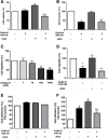
Fig. 1
Fig. 1
DisBa-01 effects on VEGF-induced HUVEC viability, invasion, migration and adhesion. a Cells were treated with DisBa-01 (1000 nM), VEGF (10 ng/mL) or both proteins in DMEM supplemented with 0.5% FBS followed by 24 h of incubation. Cell viability was measured by spectrophotometry at 540 nm after incubation with MTT. b HUVECs (2 × 105 cells/well) were treated with 1000 nM DisBa-01 and/or VEGF (10 ng/mL) on serum-free DMEM for 30 min at 4 °C. Cells were pipetted into the Boyden’s chamber and then it was inserted on well containing DMEM 10% FBS. The negative control comprised of serum-free DMEM on the wells. Invasion was allowed to occur for 18 h at 37 °C. Cell nuclei were stained with DAPI (0.7 ng/μl). Quantification of invasive cells was measured by automated cell counting. c-d For the migration assay, HUVECs (1 × 105 cells/well) were exposed to DisBa-01 (1, 10, 100 and 1000 nM), VEGF (10 ng/mL) or VEGF plus DisBa-01 (1000 nM) and immediately inserted into the Boyden’s chamber. The chambers were immersed in 10% FBS medium and allowed to migrate for 6 h at 37 °C. Control chambers were inserted in serum-free medium. Cell nuclei were stained with DAPI (0.7 ng/μl) and cell migration was measured by automated cell counting. e-f HUVECs (1 × 105 cells/well) were treated with DisBa-01 (1000 nM) and/or VEGF (10 ng/mL) and were immediately incubated (37 °C, 1 h) in fibronectin and vitronectin precoated-wells. Negative control was comprised of wells coated with 2% BSA. Cell nuclei were stained with DAPI (0.7 ng/μl) and quantification of adhesion cells was measured by automated cell counting. Results represent the average of three independent experiments in triplicate. Values of *p < 0.05 were significantly different when compared to untreated (a), treated with DisBa-01 (b), or with VEGF (c)
Fig. 2
Fig. 2
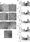
DisBa-01 inhibits HUVEC tubulogenesis. HUVECs (3 × 104 cells/well) were treated for 30 min with VEGF (10 ng/mL), DisBa-01 (1, 10, 100 and 1000 nM) or VEGF plus DisBa-01 (1000 nM) in DMEM containing 0.5% FBS and then seeded on a solidified Matrigel. The plate was placed in a humidified CO2 incubator at 37 °C for 14 h to allow the formation of tubes. a Photos (40x magnification) were obtained from a representative experiment (n = 3). The results were expressed as b Total length (μm2), c Number of mashes, d Number of nodes, e Number of master junctions and f Angiogenesis Score (analysed area x tube length x total of branches). Images were photographed using the AxionVision Rel.4.8 software of a Vert.A1 microscope (Zeiss) and analysed using the Angiogenesis Analyzer plugin for ImageJ software (version 1.51n). Results represent the average of three independent experiments in triplicate. Values of *p < 0.05 were significantly different when compared to untreated (a), DisBa-01 (b) and VEGF (c) groups
Fig. 3
Fig. 3
DisBa-01 decreases VEGFR2 protein content. a Analysis of VEGFR2 protein content by western blot. HUVECs (5 × 105 cells/well) were seeded in 6-well plates and left to adhere on an incubator at 37 °C, 5% CO2, overnight, followed by a period of 24 h of starvation at serum-free medium. Cells were treated with 1 ml of DMEM supplemented with 10% FBS and either DisBa-01 (1000 nM), VEGF (10 ng/mL) or a co-treatment and incubated for 1 and 24 h at 37 °C, 5% CO2, followed by cell lysis. Twenty micrograms of protein from cell lysates were separated on SDS-PAGE. Blots were probed with VEGFR2 antibody and GAPDH antibody was used to normalize analysis. Bands corresponding to all proteins were quantified by densitometry using the ImageJ FIJI program. Bar graph shows the mean ± SE of VEGFR2/GAPDH expression from three independent experiments. b VEGFR2 mRNA (KDR) expression. HUVECs (5 × 105/well) were seeded in 6-well plates containing DMEM and 10% FBS, followed by a 24-h starvation period on serum-free medium. Cells were treated with DisBa-01 (1000 nM) and/or VEGF (10 ng/mL) for 24 h followed by lysis and RNA isolation. Quantitative RT-PCR was carried out using specific primers to human KDR (VEGFR2) and GAPDH (housekeeping). Bar graph shows the mean ± SE of VEGFR2 expression from three independent experiments. Values of *p < 0.05 were significantly different when compared to untreated (a), DisBa-01 (b) and VEGF (c) groups
Fig. 4
Fig. 4
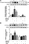
DisBa-01 inhibits VEGFR2 and β3 phosphorylation after VEGF stimulation. HUVECs (5 × 105 cells/well) were seeded in 6-well plates and left to adhere at 37 °C, 5% CO2, overnight, followed by a period of 24 h of starvation in serum-free medium. Cells were treated with 1 ml of DMEM supplemented with 10% FBS and DisBa-01 (1000 nM), VEGF (10 ng/mL) or a co-treatment and incubated for 1 and 24 h at 37 °C, 5% CO2, followed by cell lysis. Twenty micrograms of protein from cell lysates were resolved by SDS-PAGE. Blots were probed with antibodies to a P-TY1054 + TY1059 VEGFR2, to b P-Ty773β3 and GAPDH, this last to normalize loading. Bands corresponding to all proteins were quantified by densitometry using the ImageJ FIJI program. Bar graph shows the mean ± SE of phosphorylated VEGFR2/GAPDH and β3/GAPDH expression from three independent experiments. Values of *p < 0.05 were significantly different when compared to untreated (a), DisBa-01 (b) and VEGF (c) groups
Fig. 5
Fig. 5
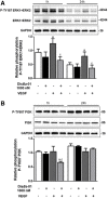
DisBa-01 inhibits ERK1/2 and PI3K phosphorylation. HUVECs (5 × 105 cells/well) were seeded in 6-well plates and left to adhere at 37 °C, 5% CO2, overnight, followed by a period of 24 h of starvation at serum-free medium. Cells were treated with 1 ml of DMEM supplemented with 10% FBS and either DisBa-01 (1000 nM), VEGF (10 ng/mL) or a co-treatment and incubated for 1 and 24 h at 37 °C, 5% CO2, followed by cell lysis. Twenty micrograms of protein from the cell lysate were separated on SDS-PAGE. Blots were probed with antibodies to a P-TY187 ERK1 + ERK2 and anti-ERK1 + ERK2; to b P-TY607 PI3K and anti-PI3K and GAPDH, this last used to normalize loading. Bands corresponding to all proteins were quantified by densitometry using the ImageJ FIJI program. Bar graph shows the mean ± SE of phosphorylated ERK1 + ERK2/ERK1 + ERK2/GAPDH and PI3K/PI3K/GAPDH expression from three independent experiments. Values of *p < 0.05 were significantly different when compared to untreated (a), DisBa-01 (b) and VEGF (c) groups
Fig. 6
Fig. 6
DisBa-01 promotes FAK, Src and paxillin phosphorylation. HUVECs (5 × 105 cells/well) were seeded in 6-well plates and left to adhere at 37 °C, 5% CO2, overnight, followed by a period of 24 h of starvation at serum-free medium. Cells were treated with 1 ml of DMEM supplemented with 10% FBS and either DisBa-01 (1000 nM), VEGF (10 ng/mL) or a co-treatment and incubated for 1 and 24 h at 37 °C, 5% CO2, followed by cell lysis. Twenty micrograms of protein from the cell lysate were separated on SDS-PAGE. Blots were probed with antibodies to a P-Y397 FAK and anti-FAK; to b P-TY418 Src; to c phospho LIM1 Paxilin; and GAPDH, this last used to normalize loading. Bands corresponding to all proteins were quantified by densitometry using the ImageJ FIJI program. Bar graph shows the mean ± SE of phosphorylated pFAK/FAK/GAPDH, pSrc/GAPDH and pLIM1Paxilin/GAPDH expression from three independent experiments. Values of *p < 0.05 were significantly different when compared to untreated (a), DisBa-01 (b) and VEGF (c) groups
Fig. 7
Fig. 7
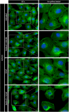
DisBa-01 induces morphological changes in endothelial cells. HUVECs (3 × 104 cells/well) were plated in a 96-well microplate previously coated with FN (1 μg/cm2), in serum-free DMEM and incubated overnight at 37 °C, 5% CO2. Cells were exposed to VEGF (10 ng/mL), DisBa-01 (1000 nM) and VEGF plus DisBa-01 for 30 min in DMEM 10% FBS. Cell nuclei were stained with DAPI (0.7 ng/μl) and cytoplasm was stained with Alexa Fluor™ 488 phalloidin for 10 min. Images were observed with 60x magnification. Representative images were obtained from three independent experiments. Scale bar = 50 μm (left panel) and 20 μm (right panel)
Fig. 8
Fig. 8
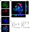
DisBa-01 colocalizes with VEGFR2 and αvβ3. a Representative confocal images of triple stained HUVECs cells cultured in FN coated plates: αvβ3 (Green), DisBa-01 (Red), and VEGFR2 (Blue) in separated and merged channels. White = triple colocalization. b Orthogonal view of Z-stack projections showing the colocalization of αvβ3 integrin, DisBa-01 and VEGFR2 receptor. c Measurement of colocalization coefficients (tM1 and Pearson) of DisBa-01 with VEGFR2 and αvβ3. Results represent the average of n = 10 cells from three independent experiments. Slides were analyzed by confocal microscopy and pictures were taken using 63x magnification. Scale bar = 5 μm

Similar articles

Cited by

References

    1. Folkman J. Angiogenesis in cancer, vascular, rheumatoid and other disease. Nat Med. 1995;1:27–31. doi: 10.1038/nm0195-27. - DOI - PubMed
    1. Folkman J. Angiogenesis: an organizing principle for drug discovery? Nat Rev Drug Discov. 2007;6:273–286. doi: 10.1038/nrd2115. - DOI - PubMed
    1. Kerbel R. Tumor angiogenesis. N Engl J Med. 2008;358:2039–2049. doi: 10.1056/nejmra0706596. - DOI - PMC - PubMed
    1. De S, Razorenova O, McCabe NP, et al. VEGF-integrin interplay controls tumor growth and vascularization. Proc Natl Acad Sci U S A. 2005;102:7589–7594. doi: 10.1073/pnas.0502935102. - DOI - PMC - PubMed
    1. Ferrara N, Gerber HP, LeCouter J. The biology of VEGF and its receptors. Nat Med. 2003;9:669–676. doi: 10.1038/nm0603-669. - DOI - PubMed

Publication types

MeSH terms

LinkOut - more resources