Membrane trafficking in osteoclasts and implications for osteoporosis
- PMID: 30837319
- PMCID: PMC6490703
- DOI: 10.1042/BST20180445
Membrane trafficking in osteoclasts and implications for osteoporosis
Abstract
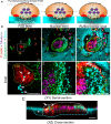
Osteoclasts are large multinucleated cells exquisitely adapted to resorb bone matrix. Like other eukaryotes, osteoclasts possess an elaborate ensemble of intracellular organelles through which solutes, proteins and other macromolecules are trafficked to their target destinations via membrane-bound intermediaries. During bone resorption, membrane trafficking must be tightly regulated to sustain the structural and functional polarity of the osteoclasts' membrane domains. Of these, the ruffled border (RB) is most characteristic, functioning as the osteoclasts' secretory apparatus. This highly convoluted organelle is classically considered to be formed by the targeted fusion of acidic vesicles with the bone-facing plasma membrane. Emerging findings disclose new evidence that the RB is far more complex than previously envisaged, possessing discrete subdomains that are serviced by several intersecting endocytic, secretory, transcytotic and autophagic pathways. Bone-resorbing osteoclasts therefore serve as a unique model system for studying polarized membrane trafficking. Recent advances in high-resolution microscopy together with the convergence of genetic and cell biological studies in humans and in mice have helped illuminate the major membrane trafficking pathways in osteoclasts and unmask the core molecular machinery that governs these distinct vesicle transport routes. Among these, small Rab GTPases, their binding partners and members of the endocytic sorting nexin family have emerged as critical regulators. This mini review summarizes our current understanding of membrane trafficking in osteoclasts, the key molecular participants, and discusses how these transport machinery may be exploited for the development of new therapies for metabolic disorders of bone-like osteoporosis.
Keywords: Rab GTPases; membrane trafficking; osteoclast; osteoporosis; secretory lysosomes; sorting nexins.
© 2019 The Author(s).
Conflict of interest statement
The Authors declare that there are no competing interests associated with the manuscript.
Figures


Similar articles
-
Vesicular trafficking in osteoclasts.Semin Cell Dev Biol. 2008 Oct;19(5):424-33. doi: 10.1016/j.semcdb.2008.08.004. Epub 2008 Aug 14. Semin Cell Dev Biol. 2008. PMID: 18768162 Review.
-
Rab GTPases in Osteoclastic Bone Resorption and Autophagy.Int J Mol Sci. 2020 Oct 16;21(20):7655. doi: 10.3390/ijms21207655. Int J Mol Sci. 2020. PMID: 33081155 Free PMC article. Review.
-
Endocytic pathway from the basal plasma membrane to the ruffled border membrane in bone-resorbing osteoclasts.J Cell Sci. 1997 Aug;110 ( Pt 15):1767-80. doi: 10.1242/jcs.110.15.1767. J Cell Sci. 1997. PMID: 9264464
-
Intracellular membrane trafficking pathways in bone-resorbing osteoclasts revealed by cloning and subcellular localization studies of small GTP-binding rab proteins.Biochem Biophys Res Commun. 2002 May 10;293(3):1060-5. doi: 10.1016/S0006-291X(02)00326-1. Biochem Biophys Res Commun. 2002. PMID: 12051767
-
Intracellular membrane trafficking in bone resorbing osteoclasts.Microsc Res Tech. 2003 Aug 15;61(6):496-503. doi: 10.1002/jemt.10371. Microsc Res Tech. 2003. PMID: 12879417 Review.
Cited by
-
Improvement of polydopamine-loaded salidroside on osseointegration of titanium implants.Chin Med. 2022 Feb 21;17(1):26. doi: 10.1186/s13020-022-00569-9. Chin Med. 2022. PMID: 35189918 Free PMC article.
-
Inflammatory activation of the FcγR and IFNγR pathways co-influences the differentiation and activity of osteoclasts.Front Immunol. 2022 Sep 6;13:958974. doi: 10.3389/fimmu.2022.958974. eCollection 2022. Front Immunol. 2022. PMID: 36148242 Free PMC article.
-
Sotrastaurin, a PKC inhibitor, attenuates RANKL-induced bone resorption and attenuates osteochondral pathologies associated with the development of OA.J Cell Mol Med. 2020 Aug;24(15):8452-8465. doi: 10.1111/jcmm.15404. Epub 2020 Jul 11. J Cell Mol Med. 2020. PMID: 32652826 Free PMC article.
-
A Review of Signaling Transduction Mechanisms in Osteoclastogenesis Regulation by Autophagy, Inflammation, and Immunity.Int J Mol Sci. 2022 Aug 30;23(17):9846. doi: 10.3390/ijms23179846. Int J Mol Sci. 2022. PMID: 36077242 Free PMC article. Review.
-
Investigation of long non-coding RNA expression profiles in patients with post-menopausal osteoporosis by RNA sequencing.Exp Ther Med. 2020 Aug;20(2):1487-1497. doi: 10.3892/etm.2020.8881. Epub 2020 Jun 11. Exp Ther Med. 2020. PMID: 32742382 Free PMC article.
References
-
- Väänänen H.K., Zhao H., Mulari M. and Halleen J.M. (2000) The cell biology of osteoclast function. J. Cell Sci. 113, 377–381 PMID: - PubMed
Publication types
MeSH terms
Substances
LinkOut - more resources
Full Text Sources
Medical

