Review
. 2019 Mar 29;9(3):180256.
doi: 10.1098/rsob.180256.

Pro-apoptotic and pro-proliferation functions of the JNK pathway of Drosophila: roles in cell competition, tumorigenesis and regeneration

Affiliations
Review

Pro-apoptotic and pro-proliferation functions of the JNK pathway of Drosophila: roles in cell competition, tumorigenesis and regeneration

Noelia Pinal et al. Open Biol. .

Abstract

The Jun N-terminal kinase (JNK) is a member of the mitogen-activated protein kinase family. It appears to be conserved in all animal species where it regulates important physiological functions involved in apoptosis, cell migration, cell proliferation and regeneration. In this review, we focus on the functions of JNK in Drosophila imaginal discs, where it has been reported that it can induce both cell death and cell proliferation. We discuss this apparent paradox in the light of recent findings and propose that the pro-apoptotic and the pro-proliferative functions are intrinsic properties of JNK activity. Whether one function or another is predominant depends on the cellular context.

Keywords: Jun N-terminal kinase; apoptosis; cell competition; regeneration; tumorigenesis.


Conflict of interest statement

We declare we have no competing interests.

Figures
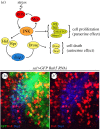
Figure 1.
Figure 1.
Autocrine and paracrine functions of JNK. (a) Activation and maintenance of the function of the JNK pathway. After an initiation event (irradiation, heat shock), the high ROS levels produced activate JNK. In turn, JNK activates the pro-apoptotic genes rpr and hid that suppress the activity of the apoptosis inhibitor diap1. The loss of diap1 function permits the activation of the apical caspase Dronc and subsequently of the effector caspases Drice and Dcp1, which causes the death of JNK-expressing cells; an autocrine effect. The fact that Dronc further stimulates JNK activity results in an amplification loop, necessary for complete apoptotic response to stress. Besides, JNK-expressing cells have the capacity of sending proliferative signals to neighbour cells, a paracrine effect likely achieved by upregulation of other signalling pathways like JAK/STAT, Wg and Dpp. In normal circumstances, the prompt death of JNK-expressing cells makes the proliferative signalling inconsequential, but it may become prominent if the apoptosis machinery is compromised. Besides the stimulation by Dronc, JNK also has the property of self-maintenance, due to a loop generated by the transcriptional activation of mol, a DUOX factor that increases the levels of ROS and thus sustains JNK activity. (b,b′) Fragment of a sal>Rab5i GFP wing disc triply labelled with GFP, Dcp1 (blue) and BrdU (red). The GFP cells are defective in Rab5 function and the majority are in apoptosis as indicated by the blue (Dcp1) staining (b′). Close to the dying cells, there is an accumulation of BrdU labelled cells, indicating they are actively dividing. The image illustrates both the autocrine (promoting cell death) and paracrine (promoting cell proliferation) functions of JNK.
Figure 2.
Figure 2.
‘Loser’ cells are eliminated by JNK activation. (a) Region of the wing disc showing clones of scrib cells 72 h after induction (GFP) being eliminated by cell competition. These cells activate JNK as indicated by the presence of Mmp1 (red, a′), a known JNK target and are in apoptosis as shown by Dcp1 staining (blue, a″). Note the fragmentation of many of the clones. (b) Clones of scrib cells 72 h after induction in which JNK is blocked by the expression of its negative regulator the phosphatase Puc (GFP). Note that there is no Mmp1 (red) or Dcp1 (blue) in the clones. These clones do not enter apoptosis and appear to grow normally. (c) A clone of scrib cells 72 h after induction (GFP) showing the adventitious presence of ROS, indicated by the label with dihydroethidium (DHE), not seen in the rest of the disc.
Figure 3.
Figure 3.
The group protection mechanism to evade cell competition. (a,b) Wing discs expressing Rab5 RNAi in the pouch region (aa′) or in a clone (bb′) labelled with GFP (green). Many of the Rab5 RNAi cells that are located at the border, in contact with surrounded wild-type cells, are in apoptosis as indicated by the Dcp1 label (red, a″ and b″). The Rab5 RNAi cells inside the domain that are not in contact with wild-type cells are beyond the range of cell competition and can continue proliferating.
Figure 4.
Figure 4.
Overgrowth induced by persistent JNK activity. (a,b) Wing discs expressing in the posterior compartment the RHGmiRNA construct that supresses the activity of the pro-apoptotic genes rpr, hid and grim, labelled with GFP (green). JNK activity is monitored by staining with the matrix metaloprotease1 (Mmp1). The disc in (a,a′) has not been irradiated and shows no ectopic JNK activity nor overgrowth. The disc in (b,b′) is of the same genotype but was irradiated (3000 R) 72 h before fixation. Note the presence of numerous regions with Mmp1 expression and also the overgrowth of the posterior compartment. (c,d) Wing discs expressing the oncogenic form of Ras (RasV12) in the posterior compartment. The A/P border is demarcated by the expression of cubitus interruptus (ci, green) that labels the anterior compartment. The disc in (c) is not irradiated and presents neither ectopic JNK activity nor overgrowth. The disc in (d) is of the same genotype but was irradiated 72 h before fixation. Note persistent JNK activity (red) and overgrowth of the posterior compartment. The regions of interest are outlined in yellow.
Figure 5.
Figure 5.
JNK implication in the regeneration process. (a) Scheme illustrating the ablation procedure. The sd-Gal4 line is expressed in all the appendage cells (wing proper and hinge) of the wing disc. Larvae of the sd-Gal4>UAS-rpr Gal80TS genotype are kept at 17°C, a temperature at which there is no rpr activity because the Gal80TS dominant suppressor of Gal4 is active at that temperature. When shifted to 29°C, the Gal80TS is inactivated, allowing Gal4 and rpr activity. In the experiments shown in the figure, the ablation period was of 40 h and started at the end of the second larval instar. A shift back to 17°C stops ablation and allows recovery for the remaining of the development. Adult flies emerge after the treatment and the effect can be inspected in the adult structures. (b) Photograph of a wing and notum of a wild-type fly (wt). (c) Photograph of an adult fly subjected during the larval period to the 40 h ablation procedure described in (a) and showing a notum duplication (arrow). (d) Photograph of an adult fly of sd-Gal4>UAS-rpr Gal80TS UAS-puc genotype subjected to 40 h ablation following the same procedure. In this genotype, JNK activity is blocked, only in the appendage cells where the ablation is induced, by the expression of the phosphatase Puckered (puc). Note the absence of a duplicated notum. The magnification of (b) is lower than that of (c) and (d).

Similar articles

Cited by

References

    1. Bosch M, Serras F, Martin-Blanco E, Baguna J. 2005. JNK signaling pathway required for wound healing in regenerating Drosophila wing imaginal discs. Dev. Biol. 280, 73–86. (10.1016/j.ydbio.2005.01.002) - DOI - PubMed
    1. Kanda H, Miura M. 2004. Regulatory roles of JNK in programmed cell death. J. Biochem. 136, 1–6. (10.1093/jb/mvh098) - DOI - PubMed
    1. Tournier C, et al. 2000. Requirement of JNK for stress-induced activation of the cytochrome c-mediated death pathway. Science 288, 870–874. (10.1126/science.288.5467.870) - DOI - PubMed
    1. Sabapathy K, Hochedlinger K, Nam SY, Bauer A, Karin M, Wagner EF. 2004. Distinct roles for JNK1 and JNK2 in regulating JNK activity and c-Jun-dependent cell proliferation. Mol. Cell. 15, 713–725. (10.1016/j.molcel.2004.08.028) - DOI - PubMed
    1. Xia Y, Karin M. 2004. The control of cell motility and epithelial morphogenesis by Jun kinases. Trends Cell Biol. 14, 94–101. (10.1016/j.tcb.2003.12.005) - DOI - PubMed

Publication types

MeSH terms

Substances

LinkOut - more resources