Review
. 2019 Aug;108(2):624-635.
doi: 10.1016/j.athoracsur.2019.01.054. Epub 2019 Mar 2.

In Search of the Ideal Valve: Optimizing Genetic Modifications to Prevent Bioprosthetic Degeneration

Affiliations
Review

In Search of the Ideal Valve: Optimizing Genetic Modifications to Prevent Bioprosthetic Degeneration

Benjamin Smood et al. Ann Thorac Surg. 2019 Aug.

Abstract

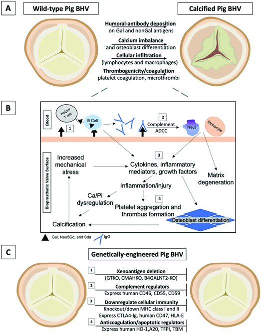
Background: Bioprosthetic heart valves undergo structural degeneration and calcification. Similarities exist in the histopathologic features of explanted bioprosthetic valves and rejected pig tissues and organs after xenotransplantation into nonhuman primates. The development of more durable bioprosthetic valves, namely from genetically modified pigs, could negate the need for the insertion of mechanical prostheses in children and young adults with the requirement for life-long anticoagulation and might avoid the need for reoperation in elderly patients.

Methods: We reviewed the literature (MedlinePlus, PubMed, Google Scholar) through September 1, 2018, under four key terms: (1) bioprosthetic heart valves, (2) xenograft antigens, (3) immunologic responses to bioprosthetic valves, and (4) genetic modification of xenografts.

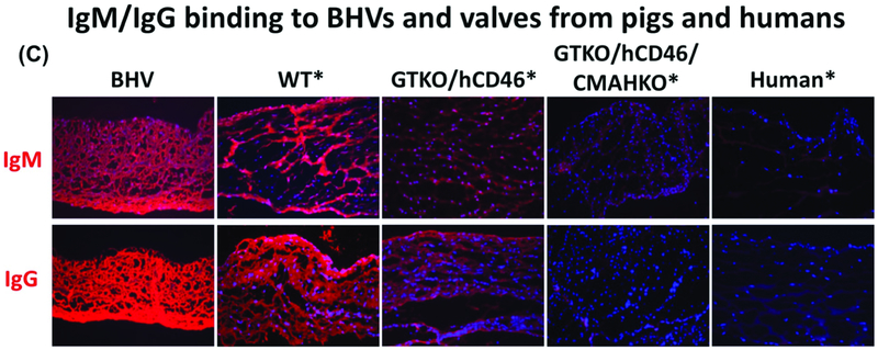
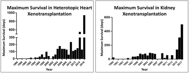
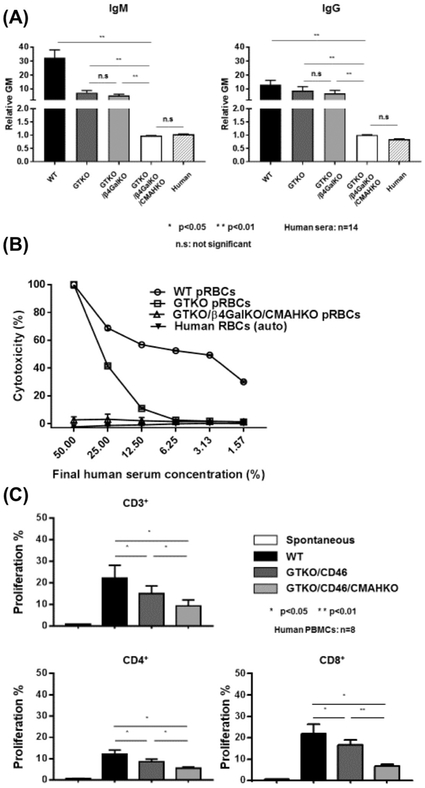
Results: Advances in tissue and organ xenotransplantation have elucidated important immunologic barriers that provide innovative approaches to prevent structural degeneration of bioprosthetic heart valves. The current evidence suggests that bioprosthetic valves derived from genetically modified pigs lacking xenogeneic antigens (namely Gal, Neu5Gc, and Sda), termed triple-knockout pigs, would function considerably longer than current wild-type (genetically unmodified) porcine valves in human recipients.

Conclusions: Preclinical and clinical studies to determine the safety and efficacy of triple-knockout porcine bioprosthetic valves will likely establish that they are more resistant to human immune responses and thus less susceptible to structural degeneration.


Figures

Figure 1.
Figure 1.. Overview of immunologic responses and proposed genetic modifications to combat bioprosthetic valve degeneration and calcification.
(A) Mechanisms of degeneration and calcification in wild-type (genetically-unmodified) pig BHVs, and (B) a schematic overview of immune-mediated degeneration and calcification. (C) Established genetic modifications that may inhibit these processes and improve commercial BHV durability (see Table 1 for feasibility, advantages, and disadvantages). (1) Xenoantigens are retained despite BHV fixation and decellularization. Genetic deletion of three key xenoantigens, Gal, Neu5Gc, and Sda (GTKO, CMAHKO, β4GALNT2-KO, respectively) prevents most antibody-dependent cell-mediated cytotoxicity (ADCC) and complement activation. ADCC results from host immune cells (e.g., macrophages) that bind antibodies through their Fc receptors. Host antigen-presenting cells, including B cells and dendritic cells, opsonize xenoantigens for presentation to helper T cells, which activate B cell maturation, class switching, and additional antibody production. (2) Transgenic expression of human complement regulators further attenuates the innate immune response by preventing activation of the cell-killing membrane attack complex. Products of the complement cascade also activate neutrophil and macrophage infiltration, promoting inflammation and phagocytosis that degenerates the surrounding matrix. (3) Macrophages, neutrophils, and lymphocytes play a major role in cellular immunity against implanted foreign materials. Preventing recruitment and activation could inhibit the production of inflammatory cytokines that incite fibroblast and valve interstitial cell osteoblastic differentiation. Genetically-engineered porcine BHVs express proteins that may reduce macrophage activity (hCD47), cytotoxicity (CTLA4-Ig), and natural killer cell immunity (HLA-E). Knockdown (or knockout) of MHC molecules (swine leukocyte antigens) reduces porcine protein presentation and recognition required for the innate and adaptive cellular immune responses. (4) Similarly, transgenic expression of anti-apoptotic (A20 and HO-1) or anti-coagulation proteins (TBM, ECPR, CD39) can minimize cell destruction and vascular inflammation that serves as a nidus for calcium nucleation and thrombus formation, respectively. Deposition of calcium orthophosphates alter the hemodynamics and increase mechanical stress, further disrupting the homeostatic environment of the bioprosthetic valve and surrounding tissues. (2-4) It is important to note that glutaraldehyde-fixed pig BHVs may not retain biologic activity comparable to fresh pig tissues; therefore, expression of these molecules may be negated in pretreated valves. A20 = tumor necrosis factor-alpha-induced protein 3; β4GALNT2-KO = Sda knockout; BHV = bioprosthetic heart valve; CD39 = ectonucleoside triphosphate diphosphohydrolase-1; CD46 = membrane cofactor protein; CD55 = decay-accelerating factor; CD59 = protectin or membrane inhibitor of reactive lysis; CMAHKO = Neu5Gc-knockout; CTLA4-Ig = cytotoxic T-lymphocyte antigen 4-Ig; ECPR = endothelial protein C receptor; GTKO = Gal-knockout; hCD47 = human integrin-associated protein; HO-1 = hemeoxygenase-1; MHC = major histocompatibility complex; Neut = neutrophil; TBM = thrombomodulin; TFPI = tissue factor pathway inhibitor.
Figure 2:
Figure 2:. Xenoantigen expression and human antibody binding on human, wild-type pig, and genetically-modified pig valves.
Glutaraldehyde fixation of wild-type (WT) and three different BHVs does not eliminate structural glycoprotein/xenoantigen components, as indicated by immunofluorescence staining. Gal and Neu5Gc are likely to be, at least partially, responsible for a xenogeneic host response. (A) Immunofluorescence staining for Gal (red) on various valves. In BHVs, Gal expression is still quite strong compared to WT pig valves, whereas GTKO, GTKO/hCD46/CMAHKO, and human tissue are negative. Of note, BHVs stain positive for nuclei (blue; DAPI). While BHVs are considered ‘devitalized’, the nuclear remnants may continue to act as a source of xenoantigens. (B) Neu5Gc epitopes (red) are not decreased in BHVs compared to WT or GTKO/hCD46 valves. In comparison, Neu5Gc was not detected in GTKO/hCD46/CMAHKO pigs, or human valves. (C) Valves were incubated with human serum for 1 hour, and stained with biotin-conjugated anti-human IgM and IgG antibodies, followed by incubation with Alexa Fluor 647-conjugated streptavidin. Immunofluorescence staining of human IgM and IgG (red) binding did not differ between BHV and WT tissues, but decreased to indicate reduced binding to GTKO/hCD46 pig tissue. Antibody binding was further decreased on GTKO/hCD46/CMAHKO pig valves, which look comparable to human valves. (Magnification x200. *Tissue was fixed with 0.2% glutaraldehyde for 48 hours prior to staining. BHV = Bioprosthetic heart valve; BHV1 = Carpentier Edwards Model 2605 bovine-stented supra-annular valve [Edwards Lifesciences, Irvine, CA]; BHV2 = Medtronic Mosaic 305 porcine-stented Cinch valve [Medtronic, Minneapolis, MN]; BHV3 = Medtronic Freestyle 995 Porcine-stentless valve.) (Modified from Lee and colleagues [35] with permission from John Wiley and Sons)
Figure 3:
Figure 3:. Survival after pig organ transplantation in nonhuman primates, 1986-2017.
(Left) After heterotopic (non-life-supporting) pig heart xenotransplantation (in the abdomen) in nonhuman primates, maximum survival has improved from <8 hours in 1986 to 945 days. (Right) After life-supporting pig kidney xenotransplantation, maximum survival has improved from 23 days in 1989 to >1 year. NB. Organ transplantation from genetically-engineered pigs expressing a human complement-regulatory protein was first reported in 1995, and organ transplantation from GTKO pigs was first reported in 2005. GTKO pigs expressing both human complement- and coagulation-regulatory proteins were first tested in 2011. (Reviewed in reference [51])
Figure 4:
Figure 4:
(A) Human IgM (left) and IgG (right) antibody binding to wild-type (WT), Gal knockout (GTKO), Gal/Sda double-knockout (GTKO/β4GalKO), and Gal, Sda, Neu5Gc triple-knockout (GTKO/β4GalKO/CMAHKO) pig red blood cells (pRBCs). Binding to TKO pig RBCs was not significantly different from human IgM and IgG binding to human RBCs of blood type O. (B) Pooled human serum complement-dependent cytotoxicity (hemolysis) to WT, GTKO, double-knockout, and triple-knockout pig RBCs. Cytotoxicity of the same serum to autologous human O RBCs was tested as a control. (C) Human T cell proliferative response to WT, GTKO/hCD46, and GTKO/hCD46/CMAHKO pig peripheral blood mononuclear cells (PBMCs) in mixed leukocyte reaction. (Triple-knockout pig PBMCs were not available to us at the time.)
Figure 5:
Figure 5:. Biomechanical properties of triple-knockout porcine pericardium are similar to those of wild-type pig pericardium
Uniaxial stress test of wild-type (WT) and triple-knockout (TKO) pig pericardium (n = 3/group). (A) Pericardium was harvested from 6 WT and 6 TKO pigs and tested for stress strain. (B) Box plot of the maximum stress (10.56±4.39 MPa and 13.54 ± 2.61 MPa for the WT and TKO, respectively). (C) Box plot of the strain at the maximum stress (WT was 74.62±12.43%, TKO was 67.81±6.44%). (NS = not significant; TKO = triple-knockout (GTKO/β4GalNT2-KO/CMAHKO); WT = wild-type) (Reproduced from Zhang and colleagues [37] with permission from Elsevier)

Comment in

Similar articles

Cited by

References

    1. D'agostino RS, Jacobs JP, Badhwar V, et al. The Society of Thoracic Surgeons Adult Cardiac Surgery Database: 2018 Update on Outcomes and Quality. Ann Thorac Surg 2018;105:15–23. - PubMed
    1. Head S, Mevlut C, Kappetein P. Mechanical versus bioprosthetic aortic valve replacement. Eur Heart J 2017;38:2183–2191. - PubMed
    1. Goldstone AJ, Chiu P, Baiocchi M, et al. Mechanical or biologic prostheses for aortic-valve and mitral-valve replacement. N Engl J Med 2017;377:1847–1857. - PMC - PubMed
    1. Cohn LH, Collins JJ, Disesa VJ, et al. Fifteen-year experience with 1678 Hancock porcine bioprosthetic heart valve replacements. Ann Surg 1989;210:435–442. - PMC - PubMed
    1. Jamieson W, Munro A, Miyagishima R, Allen P, Burr LH,Tyers GF. Carpentier-Edwards standard porcine bioprosthesis: clinical performance to seventeen years. Ann Thorac Surg 1995;60:999–1006. - PubMed

Publication types