. 2019 Feb 26;129(6):2293-2304.
doi: 10.1172/JCI126428.

Ferroptotic cell death and TLR4/Trif signaling initiate neutrophil recruitment after heart transplantation

Affiliations

Ferroptotic cell death and TLR4/Trif signaling initiate neutrophil recruitment after heart transplantation

Wenjun Li et al. J Clin Invest. .

Abstract

Non-apoptotic forms of cell death can trigger sterile inflammation through the release of danger-associated molecular patterns, which are recognized by innate immune receptors. However, despite years of investigation the mechanisms which initiate inflammatory responses after heart transplantation remain elusive. Here, we demonstrate that ferrostatin-1 (Fer-1), a specific inhibitor of ferroptosis, decreases the level of pro-ferroptotic hydroperoxy-arachidonoyl-phosphatidylethanolamine, reduces cardiomyocyte cell death and blocks neutrophil recruitment following heart transplantation. Inhibition of necroptosis had no effect on neutrophil trafficking in cardiac grafts. We extend these observations to a model of coronary artery ligation-induced myocardial ischemia reperfusion injury where inhibition of ferroptosis resulted in reduced infarct size, improved left ventricular systolic function, and reduced left ventricular remodeling. Using intravital imaging of cardiac transplants, we uncover that ferroptosis orchestrates neutrophil recruitment to injured myocardium by promoting adhesion of neutrophils to coronary vascular endothelial cells through a TLR4/TRIF/type I IFN signaling pathway. Thus, we have discovered that inflammatory responses after cardiac transplantation are initiated through ferroptotic cell death and TLR4/Trif-dependent signaling in graft endothelial cells. These findings provide a platform for the development of therapeutic strategies for heart transplant recipients and patients, who are vulnerable to ischemia reperfusion injury following restoration of coronary blood flow.

Keywords: Inflammation; Neutrophils; Organ transplantation; Transplantation.


Conflict of interest statement

Conflict of interest: DK serves on The Scientific Advisory Board of and receives research support from Compass Therapeutics. DK and KJL have a pending patent entitled “Compositions and methods for detecting CCR2 receptors” (US patent application no. 15/611,577).

Figures

Figure 1
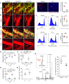
Figure 1. Ferroptosis regulates cell death and promotes neutrophil recruitment in injured hearts.
Intravital 2-photon imaging of neutrophil (green) behavior in (A) control WT cardiac grafts, (B) after treatment of recipient mice with Nec-1, (C) in RIPK3-deficient donor hearts, and (D) after administration of Fer-1 to heart recipients. Vessels were labeled red after injection of quantum dots. n = 4 per experimental group. Scale bars: 30 μm (AD). (E) Intravascular rolling velocities of neutrophils, (F) density of neutrophils, and (G) percentage of extravasated neutrophils in the experimental conditions displayed in AD. Data in EG represent the mean ± SEM. *P < 0.05, **P < 0.01, and ***P < 0.001, by 1-way ANOVA followed by post hoc Tukey’s multiple comparisons test. (H) Neutrophil recruitment per minute to coronary veins in control cardiac grafts and after treatment of recipient mice with Fer-1. (I) Cardiomyocyte death determined by ethidium bromide in cardiac grafts after treatment of recipient mice with vehicle or Fer-1. n = 4 per experimental group. Original magnification, ×200. (J) Flow cytometric assessment of death (DAPI+) of fibroblasts and endothelial cells in B6 cardiac grafts after transplantation into vehicle- or Fer-1–treated syngeneic recipient mice. n = 6 per experimental group. (K) LC-MS/MS assessment of pro-ferroptotic PE molecular species. MS spectrum of PE obtained from WT mice. Inset: MS spectrum in the range of m/z from 798.49 to 798.58. The content of PE(38:4)+OO molecular species (HOO-AA-PE, ferroptotic cell death signal) in cardiac grafts after treatment of recipient mice with vehicle or Fer-1. Data in HK represent the mean ± SEM. *P < 0.05, by 2-sided Mann-Whitney U test.
Figure 2
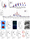
Figure 2. Ferroptosis regulates cardiomyocyte cell death and LV remodeling following myocardial infarction.
(A) Measurement of total CK activity in control hearts and hearts subjected to 30 minutes of ischemia followed by 30 minutes of reperfusion using Langendorff preparations. Mice were treated with either vehicle control or Fer-1 two hours before harvesting. n = 5 per group. (B) Serial measurement of CK activity following reperfusion in control hearts and vehicle- and Fer-1–treated hearts subjected to IRI. n = 5 per group. *P < 0.05 versus control; **P < 0.05 versus other groups, by 2-sided Mann-Whitney U test. (C) Arachidonic acid metabolites measured by LC-MS/MS in control hearts and vehicle- and Fer-1–treated hearts subjected to IRI. Data are displayed as box-and-whisker plots. Lines indicate the mean value. *P < 0.05 versus control; **P < 0.05 versus other groups; #P < 0.05 versus the vehicle IRI group. (D) TTC staining and measurement of infarct area in hearts obtained from vehicle- and Fer-1–treated mice 48 hours following 90 minutes of IRI. *P < 0.05 compared with vehicle control. Original magnification, ×10. (E) Ly6G immunostaining and quantification 48 hours following 90 minutes of IRI in vehicle- and Fer-1–treated hearts. Blue indicates DAPI staining. Original magnification, ×200. *P < 0.05 versus vehicle control. (F) Echocardiography of vehicle- and Fer-1–treated mice 4 weeks following 90 minutes of IRI. Yellow dashed line indicates the akinetic area. (GI) Quantification of LV ejection fraction, LV diastolic and systolic volumes, and akinetic area in vehicle- and Fer-1–treated mice 4 weeks after IRI. The akinetic area is expressed as a percentage of the LV area. *P < 0.05 versus vehicle control, by Mann-Whitney U test. (J) Picrosirius red staining (red) and infarct size quantification 4 weeks following 90 minutes of IRI in vehicle- and Fer-1–treated hearts. Lines represent the mean. *P < 0.05, by 2-sided Mann-Whitney U test.
Figure 3
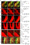
Figure 3. Vascular endothelial TLR4 expression regulates neutrophil recruitment to injured hearts via Trif signaling.
Intravital 2-photon imaging of neutrophil (green) behavior in (A) control WT, (B) TLR4-deficient, (C) CD14-deficient, and (D) Trif-deficient cardiac grafts. Vessels were labeled red after injection of quantum dots. n = 4 per experimental group. (E) Intravascular rolling velocities of neutrophils, (F) density of neutrophils, and (G) percentage of extravasated neutrophils in the experimental conditions displayed in AD. Intravital 2-photon imaging of neutrophil (green) behavior in hearts that lacked TLR4 expression on (H) vascular endothelial cells (Tie2-Cre TLR4fl/fl) or (I) cardiomyocytes (Myh6-Cre TLR4fl/fl). (J) Intravascular rolling velocities, (K) density of neutrophils, and (L) percentage of extravasated neutrophils in the experimental conditions displayed in H and I as well as control TLR4-floxed heart grafts. Data in EG and JL represent the mean ± SEM. *P < 0.05, **P < 0.01, and ***P < 0.001, by 1-way ANOVA followed by post hoc Tukey’s multiple comparisons test. Scale bars: 30 μm.
Figure 4
Figure 4. Ferroptosis promotes neutrophil recruitment to injured hearts through type I IFN signaling.
Intravital 2-photon imaging of neutrophil (green) behavior in (A) control WT, (B) IFNAR-deficient, and (C) IRF3/-5/-7 triple–deficient heart grafts as well as after transplantation into Fer-1–treated recipients that received (D) vehicle or (E) recombinant type I IFN (rIFN-α) at the time of implantation. Vessels were labeled red after injection of quantum dots. n = 4 per experimental group. (F) Intravascular rolling velocities, (G) density of neutrophils, and (H) percentage of extravasated neutrophils in the experimental conditions displayed in AE. Data in FH represent the mean ± SEM. *P < 0.05, **P < 0.01, and ***P < 0.001, by 1-way ANOVA followed by post hoc Tukey’s multiple comparisons test. Scale bars: 30 μm.
Figure 5
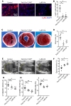
Figure 5. Neutrophilic infiltration contributes to cardiac damage.
(A) Ly6G immunostaining and (B) quantification 4 days following 90 minutes of closed-chest IRI in the hearts of control (TLR4fl/fl), Tie2-Cre TLR4fl/fl, and neutrophil-depleted (Ly6G Ab) mice. Blue indicates DAPI staining; red indicates Ly6G Ab staining. Scale bar: 40 μm. (C) TTC staining and (D) measurement of infarct area in hearts obtained from control (TLR4fl/fl), Tie2-Cre TLR4fl/fl, and neutrophil-depleted (Ly6G Ab) mice 4 days following 90 minutes of closed-chest IRI. Original magnification, ×10. (E) Echocardiography of control (TLR4fl/fl), Tie2-Cre TLR4fl/fl, and neutrophil-depleted (Ly6G Ab) mice 4 days following 90 minutes of IRI. Yellow dashed line indicates the akinetic area. (FI) Quantification of LV ejection fraction, LV diastolic and systolic volumes, and akinetic area in control (TLR4fl/fl), Tie2-Cre TLR4fl/fl, and neutrophil-depleted (Ly6G Ab) mice 4 days following 90 minutes of IRI. The akinetic area is expressed as a percentage of the LV area. n = 4–5 per experimental group. Lines represent the mean. *P < 0.05 and **P < 0.01, by 1-way ANOVA followed by post hoc Tukey’s multiple comparisons test.

Similar articles

Cited by

  • Sterile inflammation in thoracic transplantation.
    Frye CC, Bery AI, Kreisel D, Kulkarni HS. Frye CC, et al. Cell Mol Life Sci. 2021 Jan;78(2):581-601. doi: 10.1007/s00018-020-03615-7. Epub 2020 Aug 17. Cell Mol Life Sci. 2021. PMID: 32803398 Free PMC article. Review.
  • Innate immunity in lung transplantation.
    Shepherd HM, Gauthier JM, Li W, Krupnick AS, Gelman AE, Kreisel D. Shepherd HM, et al. J Heart Lung Transplant. 2021 Jul;40(7):562-568. doi: 10.1016/j.healun.2021.03.017. Epub 2021 Mar 26. J Heart Lung Transplant. 2021. PMID: 34020867 Free PMC article. Review.
  • A Focused Review on Primary Graft Dysfunction after Clinical Lung Transplantation: A Multilevel Syndrome.
    Van Slambrouck J, Van Raemdonck D, Vos R, Vanluyten C, Vanstapel A, Prisciandaro E, Willems L, Orlitová M, Kaes J, Jin X, Jansen Y, Verleden GM, Neyrinck AP, Vanaudenaerde BM, Ceulemans LJ. Van Slambrouck J, et al. Cells. 2022 Feb 21;11(4):745. doi: 10.3390/cells11040745. Cells. 2022. PMID: 35203392 Free PMC article. Review.
  • Roles of Ferroptosis in Cardiovascular Diseases.
    Guo Y, Zhang W, Zhou X, Zhao S, Wang J, Guo Y, Liao Y, Lu H, Liu J, Cai Y, Wu J, Shen M. Guo Y, et al. Front Cardiovasc Med. 2022 May 23;9:911564. doi: 10.3389/fcvm.2022.911564. eCollection 2022. Front Cardiovasc Med. 2022. PMID: 35677693 Free PMC article. Review.
  • Effects of Ferroptosis on the Metabolome in Cardiac Cells: The Role of Glutaminolysis.
    Rodríguez-Graciani KM, Chapa-Dubocq XR, Ayala-Arroyo EJ, Chaves-Negrón I, Jang S, Chorna N, S Maskrey T, Wipf P, Javadov S. Rodríguez-Graciani KM, et al. Antioxidants (Basel). 2022 Jan 29;11(2):278. doi: 10.3390/antiox11020278. Antioxidants (Basel). 2022. PMID: 35204160 Free PMC article.

References

    1. Kobashigawa J, et al. Report from a consensus conference on primary graft dysfunction after cardiac transplantation. J Heart Lung Transplant. 2014;33(4):327–340. doi: 10.1016/j.healun.2014.02.027. - DOI - PubMed
    1. Nicoara A, et al. Primary graft dysfunction after heart transplantation: Incidence, trends, and associated risk factors. Am J Transplant. 2018;18(6):1461–1470. doi: 10.1111/ajt.14588. - DOI - PubMed
    1. Kreisel D, et al. Bcl3 prevents acute inflammatory lung injury in mice by restraining emergency granulopoiesis. J Clin Invest. 2011;121(1):265–276. doi: 10.1172/JCI42596. - DOI - PMC - PubMed
    1. Ma XL, Tsao PS, Lefer AM. Antibody to CD-18 exerts endothelial and cardiac protective effects in myocardial ischemia and reperfusion. J Clin Invest. 1991;88(4):1237–1243. doi: 10.1172/JCI115427. - DOI - PMC - PubMed
    1. McDonald B, et al. Intravascular danger signals guide neutrophils to sites of sterile inflammation. Science. 2010;330(6002):362–366. doi: 10.1126/science.1195491. - DOI - PubMed

Publication types

MeSH terms