

T-Cell-Driven Inflammation as a Mediator of the Gut-Brain Axis Involved in Parkinson's Disease
- PMID: 30828335
- PMCID: PMC6384270
- DOI: 10.3389/fimmu.2019.00239
T-Cell-Driven Inflammation as a Mediator of the Gut-Brain Axis Involved in Parkinson's Disease
Abstract
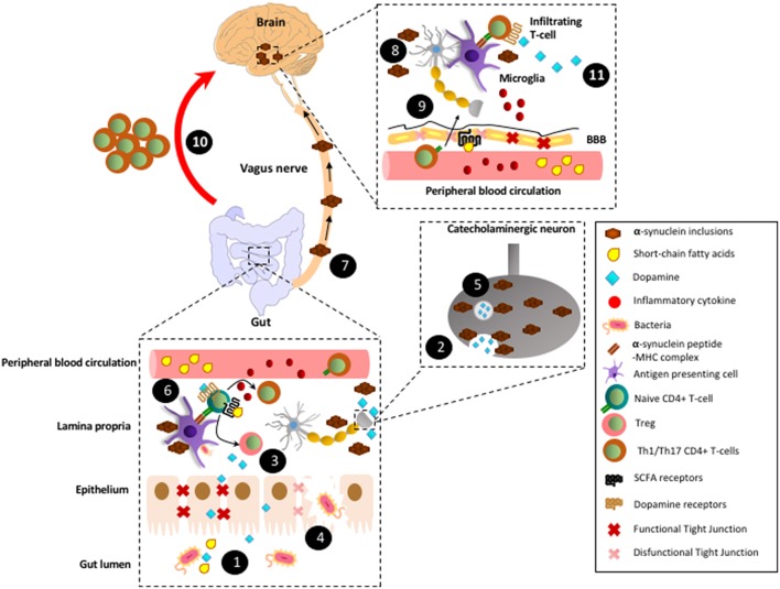
Parkinson's disease (PD) is a neurodegenerative disorder affecting mainly the dopaminergic neurons of the nigrostriatal pathway, a neuronal circuit involved in the control of movements, thereby the main manifestations correspond to motor impairments. The major molecular hallmark of this disease corresponds to the presence of pathological protein inclusions called Lewy bodies in the midbrain of patients, which have been extensively associated with neurotoxic effects. Importantly, different research groups have demonstrated that CD4+ T-cells infiltrate into the substantia nigra of PD patients and animal models. Moreover, several studies have consistently demonstrated that T-cell deficiency results in a strong attenuation of dopaminergic neurodegeneration in animal models of PD, thus indicating a key role of adaptive immunity in the neurodegenerative process. Recent evidence has shown that CD4+ T-cell response involved in PD patients is directed to oxidised forms of α-synuclein, one of the main constituents of Lewy bodies. On the other hand, most PD patients present a number of non-motor manifestations. Among non-motor manifestations, gastrointestinal dysfunctions result especially important as potential early biomarkers of PD, since they are ubiquitously found among confirmed patients and occur much earlier than motor symptoms. These gastrointestinal dysfunctions include constipation and inflammation of the gut mucosa and the most distinctive pathologic features associated are the loss of neurons of the enteric nervous system and the generation of Lewy bodies in the gut. Moreover, emerging evidence has recently shown a pivotal role of gut microbiota in triggering the development of PD in genetically predisposed individuals. Of note, PD has been positively correlated with inflammatory bowel diseases, a group of disorders involving a T-cell driven inflammation of gut mucosa, which is strongly dependent in the composition of gut microbiota. Here we raised the hypothesis that T-cell driven inflammation, which mediates dopaminergic neurodegeneration in PD, is triggered in the gut mucosa. Accordingly, we discuss how structural components of commensal bacteria or how different mediators produced by gut-microbiota, including short-chain fatty acids and dopamine, may affect the behaviour of T-cells, triggering the development of T-cell responses against Lewy bodies, initially confined to the gut mucosa but later extended to the brain.
Keywords: CD4+ T-cell mediated immunity; Parkinson's disease; gut microbiota; gut-brain axis; neo-antigens; neuroinflammation.
Figures
Similar articles
-
Brain-gut-microbiota axis in Parkinson's disease.World J Gastroenterol. 2015 Oct 7;21(37):10609-20. doi: 10.3748/wjg.v21.i37.10609. World J Gastroenterol. 2015. PMID: 26457021 Free PMC article. Review.
-
Altered gut microbiota and intestinal permeability in Parkinson's disease: Pathological highlight to management.Neurosci Lett. 2019 Nov 1;712:134516. doi: 10.1016/j.neulet.2019.134516. Epub 2019 Sep 24. Neurosci Lett. 2019. PMID: 31560998 Review.
-
Microbiome-Gut-Brain Axis and Toll-Like Receptors in Parkinson's Disease.Int J Mol Sci. 2018 Jun 6;19(6):1689. doi: 10.3390/ijms19061689. Int J Mol Sci. 2018. PMID: 29882798 Free PMC article. Review.
-
The gut microbiome in Parkinson's disease: A culprit or a bystander?Prog Brain Res. 2020;252:357-450. doi: 10.1016/bs.pbr.2020.01.004. Epub 2020 Mar 5. Prog Brain Res. 2020. PMID: 32247371 Review.
-
The immunology of Parkinson's disease.Semin Immunopathol. 2022 Sep;44(5):659-672. doi: 10.1007/s00281-022-00947-3. Epub 2022 Jun 8. Semin Immunopathol. 2022. PMID: 35674826 Free PMC article. Review.
Cited by
-
The gut microbiota-inflammation-brain axis in end-stage renal disease: perspectives from default mode network.Theranostics. 2019 Oct 18;9(26):8171-8181. doi: 10.7150/thno.35387. eCollection 2019. Theranostics. 2019. PMID: 31754388 Free PMC article.
-
Correlation of Decreased Serum Pituitary Adenylate Cyclase-Activating Polypeptide and Vasoactive Intestinal Peptide Levels With Non-motor Symptoms in Patients With Parkinson's Disease.Front Aging Neurosci. 2021 Sep 8;13:689939. doi: 10.3389/fnagi.2021.689939. eCollection 2021. Front Aging Neurosci. 2021. PMID: 34566619 Free PMC article.
-
Cross-talk between T-cells and gut-microbiota in neurodegenerative disorders.Neural Regen Res. 2019 Dec;14(12):2091-2092. doi: 10.4103/1673-5374.262582. Neural Regen Res. 2019. PMID: 31397345 Free PMC article. No abstract available.
-
α-Synuclein-containing erythrocytic extracellular vesicles: essential contributors to hyperactivation of monocytes in Parkinson's disease.J Neuroinflammation. 2022 Feb 22;19(1):53. doi: 10.1186/s12974-022-02413-1. J Neuroinflammation. 2022. PMID: 35193594 Free PMC article.
-
Rotenone aggravates PD-like pathology in A53T mutant human α-synuclein transgenic mice in an age-dependent manner.Front Aging Neurosci. 2022 Aug 8;14:842380. doi: 10.3389/fnagi.2022.842380. eCollection 2022. Front Aging Neurosci. 2022. PMID: 36004003 Free PMC article.
References
Publication types
MeSH terms
LinkOut - more resources
Full Text Sources
Medical
Research Materials