

In Vivo Rodent Models of Type 2 Diabetes and Their Usefulness for Evaluating Flavonoid Bioactivity
- PMID: 30823474
- PMCID: PMC6470730
- DOI: 10.3390/nu11030530
In Vivo Rodent Models of Type 2 Diabetes and Their Usefulness for Evaluating Flavonoid Bioactivity
Erratum in
-
Correction: Fang et al. In Vivo Rodent Models of Type 2 Diabetes and Their Usefulness for Evaluating Flavonoid Bioactivity. Nutrients 2019, 11, 530.Nutrients. 2023 Jun 26;15(13):2881. doi: 10.3390/nu15132881. Nutrients. 2023. PMID: 37447402 Free PMC article.
Abstract
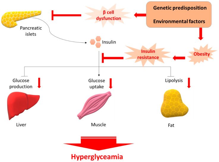
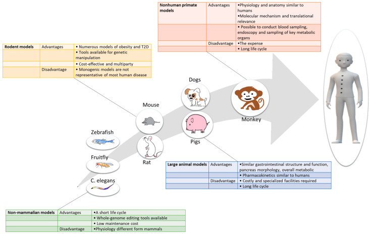
About 40% of the world's population is overweight or obese and exist at risk of developing type 2 diabetes mellitus (T2D). Obesity is a leading pathogenic factor for developing insulin resistance (IR). It is well established that IR and a progressive decline in functional β-cell mass are hallmarks of developing T2D. In order to mitigate the global prevalence of T2D, we must carefully select the appropriate animal models to explore the cellular and molecular mechanisms of T2D, and to optimize novel therapeutics for their safe use in humans. Flavonoids, a group of polyphenols, have drawn great interest for their various health benefits, and have been identified in naturally occurring anti-diabetic compounds. Results from many clinical and animal studies demonstrate that dietary intake of flavonoids might prove helpful in preventing T2D. In this review, we discuss the currently available rodent animal models of T2D and analyze the advantages, the limitations of each T2D model, and highlight the potential anti-diabetic effects of flavonoids as well as the mechanisms of their actions.
Keywords: animal model; antioxidant; flavonoid; type 2 diabetes.
Conflict of interest statement
The authors declare no conflict of interest. The funders had no role in the design of the study; in the collection, analyses, or interpretation of data; in the writing of the manuscript, and in the decision to publish the results.
Figures
Similar articles
-
Dietary Flavonoids in the Prevention of T2D: An Overview.Nutrients. 2018 Mar 31;10(4):438. doi: 10.3390/nu10040438. Nutrients. 2018. PMID: 29614722 Free PMC article. Review.
-
Recent advances in understanding the anti-diabetic actions of dietary flavonoids.J Nutr Biochem. 2013 Nov;24(11):1777-89. doi: 10.1016/j.jnutbio.2013.06.003. Epub 2013 Sep 9. J Nutr Biochem. 2013. PMID: 24029069 Free PMC article. Review.
-
Diabetes and branched-chain amino acids: What is the link?J Diabetes. 2018 May;10(5):350-352. doi: 10.1111/1753-0407.12645. Epub 2018 Feb 13. J Diabetes. 2018. PMID: 29369529
-
Dietary Flavonoids and Insulin Signaling in Diabetes and Obesity.Cells. 2021 Jun 11;10(6):1474. doi: 10.3390/cells10061474. Cells. 2021. PMID: 34208379 Free PMC article. Review.
-
The Use of Natural Compounds as a Strategy to Counteract Oxidative Stress in Animal Models of Diabetes Mellitus.Int J Mol Sci. 2021 Jun 29;22(13):7009. doi: 10.3390/ijms22137009. Int J Mol Sci. 2021. PMID: 34209800 Free PMC article. Review.
Cited by
-
Application of Animal Models in Diabetic Cardiomyopathy.Diabetes Metab J. 2021 Mar;45(2):129-145. doi: 10.4093/dmj.2020.0285. Epub 2021 Mar 25. Diabetes Metab J. 2021. PMID: 33813812 Free PMC article. Review.
-
Liraglutide versus pramlintide in protecting against cognitive function impairment through affecting PI3K/AKT/GSK-3β/TTBK1 pathway and decreasing Tau hyperphosphorylation in high-fat diet- streptozocin rat model.Pflugers Arch. 2024 May;476(5):779-795. doi: 10.1007/s00424-024-02933-0. Epub 2024 Mar 27. Pflugers Arch. 2024. PMID: 38536493 Free PMC article.
-
The Effect of Chrysin-Loaded Phytosomes on Insulin Resistance and Blood Sugar Control in Type 2 Diabetic db/db Mice.Molecules. 2020 Nov 24;25(23):5503. doi: 10.3390/molecules25235503. Molecules. 2020. PMID: 33255372 Free PMC article.
-
A Selective Role of Dietary Anthocyanins and Flavan-3-ols in Reducing the Risk of Type 2 Diabetes Mellitus: A Review of Recent Evidence.Nutrients. 2019 Apr 13;11(4):841. doi: 10.3390/nu11040841. Nutrients. 2019. PMID: 31013914 Free PMC article. Review.
-
Impact of flavonol extracts derived from green tea or targeted flavonols as secondary ingredients on intestinal glucose transport.J Food Sci Technol. 2022 Apr;59(4):1317-1325. doi: 10.1007/s13197-021-05140-2. Epub 2021 May 26. J Food Sci Technol. 2022. PMID: 35250057 Free PMC article.
References
Publication types
MeSH terms
Substances
LinkOut - more resources
Full Text Sources
Medical
