Review
. 2019 Feb 22;11(2):190.
doi: 10.3390/v11020190.

Novel Approaches for The Development of Live Attenuated Influenza Vaccines

Affiliations
Review

Novel Approaches for The Development of Live Attenuated Influenza Vaccines

Pilar Blanco-Lobo et al. Viruses. .

Abstract

Influenza virus still represents a considerable threat to global public health, despite the advances in the development and wide use of influenza vaccines. Vaccination with traditional inactivate influenza vaccines (IIV) or live-attenuated influenza vaccines (LAIV) remains the main strategy in the control of annual seasonal epidemics, but it does not offer protection against new influenza viruses with pandemic potential, those that have shifted. Moreover, the continual antigenic drift of seasonal circulating influenza viruses, causing an antigenic mismatch that requires yearly reformulation of seasonal influenza vaccines, seriously compromises vaccine efficacy. Therefore, the quick optimization of vaccine production for seasonal influenza and the development of new vaccine approaches for pandemic viruses is still a challenge for the prevention of influenza infections. Moreover, recent reports have questioned the effectiveness of the current LAIV because of limited protection, mainly against the influenza A virus (IAV) component of the vaccine. Although the reasons for the poor protection efficacy of the LAIV have not yet been elucidated, researchers are encouraged to develop new vaccination approaches that overcome the limitations that are associated with the current LAIV. The discovery and implementation of plasmid-based reverse genetics has been a key advance in the rapid generation of recombinant attenuated influenza viruses that can be used for the development of new and most effective LAIV. In this review, we provide an update regarding the progress that has been made during the last five years in the development of new LAIV and the innovative ways that are being explored as alternatives to the currently licensed LAIV. The safety, immunogenicity, and protection efficacy profile of these new LAIVs reveal their possible implementation in combating influenza infections. However, efforts by vaccine companies and government agencies will be needed for controlled testing and approving, respectively, these new vaccine methodologies for the control of influenza infections.

Keywords: immunogenicity; influenza inactivated vaccine (IIV); influenza reverse genetics; influenza vaccines; influenza virus; live-attenuated influenza vaccine (LAIV); protection efficacy; recombinant influenza virus.


Conflict of interest statement

The authors declare no conflict of interest.

Figures

Figure 1
Figure 1
Influenza A virus (IAV) virion structure and genome organization. A) Virion structure: IAV is an eight-segmented, negative-sense, single-stranded RNA enveloped virus surrounded by a lipid bilayer that contains three viral glycoproteins: hemagglutinin (HA), responsible for binding to sialic acid receptors, entry into the cell and fusion of the viral envelop with the endosome; neuraminidase (NA), which removes sialic acids, allowing for viral release from infected cells; and, the ion channel matrix 2 (M2) protein, which is responsible for the acidification of the virion following endocytosis, and viral assembly. Under the viral envelope, there is a protein layer that is made of the matrix 1 (M1) protein, which is involved in virion assembly and budding. The nuclear export protein (NEP) is found inside the viral particle and it is required for the nuclear export of the eight viral ribonucleoprotein (vRNP) complexes from the nucleus to the cytoplasm at the late stages of viral replication. The vRNP complexes, which are present in the core of the virion, are made of the negative-sense, single-stranded viral (v)RNAs packed by the viral nucleoprotein (NP) and the three subunits (PB2, PB1, and PA) of the RNA-dependent RNA polymerase (RdRp) complex that are responsible for viral RNA genome replication and gene transcription in the nuclei of infected cells. IAV proteins and their schematic representation are shown at the bottom. B) Genome organization: The IAV genome is made of eight single-stranded, negative-sense, vRNA segments (PB2, PB1, PA, HA, NP, NA, M, and NS). White boxes represent packaging signals that are responsible for the selective packaging of each vRNA segment into the virion. Numbers represent the nucleotide lengths of each of the 3′ and 5′ packaging signals in each of the vRNAs. Each vRNA is flanked by the 3’and 5´ non-coding regions (NCRs, black lines) recognized by the viral RdRp for viral genome replication and gene transcription.
Figure 2
Figure 2
Production of the inactivated influenza vaccine (IIV): To prepare IIV, 10–11 days old chicken embryonated eggs are infected with two IAVs: the candidate virus recommended by the WHO (top, gray) and a high-growth virus (bottom, black). Reassortant viruses are harvested from the allantoic fluid of infected eggs 2–3 days post-infection and the appropriate reassortant virus to be included in the IIV containing the HA and NA viral segments from the WHO candidate virus (gray) and the six internal segments (PB2, PB1, PA, NP, M, and non-structural (NS)) of the high-growth virus (black) is selected by amplification in the presence of antibodies against the HA and NA glycoproteins of the high-growth virus. Genomic composition of the reassortant virus must be confirmed by sequencing. The selected virus to be used in the IIV is chemically inactivated with β-propiolactone (β-PL) or formalin, concentrated, purified for vaccine production, and then administrated intramuscularly (IM).
Figure 3
Figure 3
Generation of the live-attenuated influenza vaccine (LAIV): The LAIV is produced by co-infection of 10–11 days old chicken embryonated eggs with the candidate virus recommended by the WHO (top, gray) and the A/Ann Arbor/6/60 H2N2 (bottom, black) Master Donor Virus (MDV). After 2–3 days post-infection, the appropriate reassortant seed virus containing the six internal segments (PB2, PB1, PA, NP, M, and NS) derived from the MDV (black) and the two glycoprotein (HA and NA) segments from the recommended WHO circulating strain (gray) is selected by amplification at low temperatures in the presence of antibodies against the MDV, HA, and NA. The selected LAIV is then administrated intranasally (IN). Amino acid substitutions in the PB2 (N265S), PB1 (K391E, E581G, and A661T) and NP (D34G) viral segments responsible for the attenuated (att), temperature sensitive (ts), and cold-adapted (ca) phenotype of the MDV A/Ann Arbor/6/60 H2N2 are indicated at the bottom.
Figure 4
Figure 4
IAV vRNA cloning into ambisense/bidirectional plasmids for the generation of recombinant viruses using plasmid-based reverse genetic approaches. (A) Schematic representation of the ambisense/bidirectional rescue plasmid to generate recombinant IAV: Ambisense/bidirectional rescue plasmids containing the human polymerase I promoter (hPol-I, black arrow) and the mouse polymerase I terminator (T, black box) sequences that regulate the synthesis of the negative sense vRNAs are indicated. In the opposite direction to the Pol-I cassette, and from the same cDNA, the polymerase II dependent promoter (Pol-II, white arrow) and the bovine growth hormone polyadenylation termination sequence (aBGH, white box) direct the synthesis of positive sense mRNA to produce viral proteins. Newly synthesized vRNAs generated from the Pol-I cassette are recognized by the viral RdRp subunits (PB2, PB1 and PA) that, together with the viral NP, lead to the formation of vRNP complexes responsible of viral genome replication and gene transcription. Transcription from newly synthesized vRNAs results in mRNA expression and the production of new viral proteins. Replication of newly synthesized vRNAs results in the formation of complementary (c)RNAs for the amplification and synthesis of new vRNAs that will be incorporated into the nascent virions as novel vRNP complexes. (B) Plasmid-based reverse genetics to generate recombinant IAV: FDA-approved cell lines for vaccine production are co-transfected with the eight (PB2, PB1, PA, HA, NP, NA, M, and NS) ambisense/bidirectional IAV plasmids. Viable virus recovered from the tissue culture supernatants 3–4 days after transfection is amplified using fresh FDA-approved cell lines or 10–11 day-old embryonated chicken eggs.
Figure 5
Figure 5
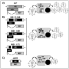
LAIV based on truncations and/or deletion of the viral NS1: Schematic representation of wild-type, WT (A), NS1 truncated (B), or NS1 deficient (C) recombinant IAV. WT NS vRNA is represented in gray boxes. WT NS1 and NEP open reading frames (ORFs) are represented as dark and light gray boxes, respectively. Modified NS segments and truncated NS1 ORFs are indicated in black boxes. White lines represent stop codons. White boxes indicate the packaging signals located at the 3´and 5´ ends of the NS vRNA. Lines at the end of the NS vRNA indicate the 3´and 5´ NCR. Expression of WT NS1 protein (A) results in inhibition of interferon (IFN) induction and efficient viral replication. NS1 1–126 (top), 1–99 (middle), or 1–73 (bottom) truncations in the NS1 ORF (B) or deletion of the entire NS1 ORF (C) result in the higher induction of IFN and reduced levels of viral replication and, therefore, virus production.
Figure 6
Figure 6
Codon deoptimization (CD) for the generation of LAIV: (A,B) Schematic representation of WT (A) and CD (B) viral NS segments: WT NS vRNA is represented in gray boxes (A). WT NS1 and NEP ORFs are represented as dark and light gray boxes, respectively (A). CD NS1 (NS1CD, top), NEP (NEPCD, middle) or both NS1 and NEP (NSCD, bottom) proteins as well as their respective NS vRNA segments are indicated with black boxes (B). After infection with a virus encoding a WT NS segment, expression of NS1 results in inhibition of IFN induction, allowing the efficient viral replication (A). Infection with viruses encoding a codon deoptimized NS1 protein (NS1CD, top; NSCD, bottom) results in reduced NS1 protein expression levels and inefficient inhibition of type I IFN responses, resulting in reduced viral replication and viral production. CD of NEP (NEPCD, middle) results in lower expression of NEP, without significantly affecting viral replication. CD of NS1 and NEP (NSCD, bottom) results in higher attenuation than viruses containing only NS1 or NEP CD ORFs, correlating with the amount of codon changes introduced in the viral segment. (C,D) Schematic representation of WT and IAV attenuated by codon-pair bias: WT PB1, HA, NP, and NA vRNA segments are indicated in gray boxes (C). Codon-pair deoptimized PB1, HA, and NP (PB1/HA/NP3F) (D, top); or, HA and NA (HA/NAMin) (D, bottom) proteins are represented in black boxes. During WT viral infection (C), vRNPs mediate viral genome replication and gene transcription, allowing efficient viral protein synthesis and viral production. Likewise, optimal expression of the viral HA and NA results in efficient production of infectious viral progeny. Infection with PB1/HA/NP3F virus (D, top), results in reduced levels of viral replication and transcription mediated by lower levels of PB1 and NP expression. The codon-pair deoptimization of HA also affects protein expression levels, contributing to the attenuation of the PB1/HA/NP3F virus in mice but with reasonable growth in vitro. Likewise, the reduction in the level of expression of the viral HA and NA during infection with the HA/NAMin virus (D, bottom) results in reduced virion formation and therefore lower infectious viral production. White boxes (AD) indicate the packaging signals that were located at the 3´and 5´ ends of each of the vRNAs. Lines at the end of each vRNA (AD) indicate the 3´and 5´ NCR.
Figure 7
Figure 7
Rearrangement of IAV genome for the generation of LAIV: (A,B) Schematic representation of WT (A) and rearranged viral segments 2 (PB1) and 8 (NS) (B): WT segments 2, 4, and 8 (A) or rearranged segment 2 (PB1 and NEP) and segment 8 (NS1) are indicated with dark gray boxes (B) The light gray box indicates the secondary H5 inserted in the NS segment. White boxes (A and B) indicate the packaging signals located at the 3´and 5´ ends of each vRNA. Black boxes (B) indicate the sequence of the foot-and-mouth disease virus (FMDV) 2A autoproteolytic cleavage site. Lines at the end of each vRNA (A and B) indicate the 3´and 5´ NCR. A white line in the NS segment (B) represents a stop codon in the NS1 resulting in a truncated (1-99 amino acids) NS1 protein (black box). Expression of NEP from a single polypeptide downstream of the modified PB1 viral segment results in a reduction on the activity of the PB1and an impaired growth of the rearranged virus (B). The expression of the H5 ORF from a modified segment 8 results in an LAIV expressing two different HA (H9, dark gray; and H5, light gray) and the induction of neutralizing antibodies against the two viral glycoproteins. (C,D) Schematic representation of WT (C) and rearranged segment 4 (HA) and segment 6 (NA) viruses (D): WT (C) and rearranged segment 4 expressing NA-HA (D) are represented in dark gray boxes. Rearranged segment 6 expressing a secondary HA is represented in a light gray box (D). White boxes indicate the packaging signals located at the 3´and 5´ ends of each vRNA (C and D). Black boxes indicate the sequence of the porcine teschovirus (PTV-1)2A autoproteolytic cleavage site (D). Lines at the end of each vRNA indicate the 3´and 5´ NCRs (C and D). While WT virus expresses the HA and NA glycoproteins from the segment 4 and 6 respectively (C), the rearranged virus expresses both the subtype 1 HA and NA glycoproteins from the modified segment 4; and the subtype 3 HA from the modified segment 6, where NA is normally located (D). This rearrangement of the viral genome results in an attenuated recombinant virus able to induce neutralizing antibodies against the two viral glycoproteins (H1 and H3) (D).
Figure 8
Figure 8
Development of LAIV based on modification of the spliced viral RNA segments 7 (M) and 8 (NS): Schematic representation of WT (A) and modified (B) viral RNA segments 7 (Ms, top), 8 (NS, middle), or both 7 and 8 (Ms/NSs, bottom) in which the overlapping ORFs of the M1 and M2 proteins (Ms, top), NS1 and NEP proteins (NSs, middle), or both (Ms/NSs, bottom) are produced from the same transcript by using the PTV-1 2A autoproteolytic cleavage site. Viral products from the M (M1 and M2) and NS (NS1 and NEP) WT (A) or modified (B) viral segments are indicated in grey boxes. M2 ORF is shown as a lighter grey box. Black boxes (B) indicate the sequence of the PTV-1 2A autoproteolytic cleavage site. The packaging signals located at the 3´and 5´ ends of each vRNA are indicated with white boxes (A,B). Lines at the end of each vRNA indicate the 3´and 5´ NCR in the M and NS viral segments (A,B). During infection with WT virus, the optimal expression of M1 and M2 proteins (M segment), as well as NS1 and NEP (NS segment) from the spliced vRNA segments, results in efficient virus replication and production. Infection with a modified M segment (B, top) results in slightly reduction of virus replication and production at 33 °C; and significant reduction of viral production at high temperatures (37 °C or 39 °C). Modification of the NS vRNA segment (B, middle) results in a slight reduction in virus replication and production that is not temperature dependent. The recombinant virus containing both modified M and NS segments (Ms/NSs) (B, bottom) results in impaired viral replication and production, similar to the recombinant virus containing the modified M segment (Ms) at non permissive temperatures (37 °C or 39 °C).
Figure 9
Figure 9
Single-cycle infectious IAV (sciIAV) as LAIV: Schematic representation of sciIAV based on deletions in the PB2 (A), PB1 (B), HA (C), or NA (D) viral proteins. SciIAV based on a modified uncleavable HA (E) or a non-functional M2, M2SR (F) are also indicated. The packaging signals located at the 3’and 5’ ends of each vRNA are indicated with white boxes (AF). Lines at the end of each vRNA indicate the 3’and 5’ NCRs (AF). Striped boxes represent an internal deletion of the PB2 (A), PB1 (B), HA (C), or NA (D) ORF. A black line represents an amino acid substitution (R325T) in the cleavage site of HA that results in an uncleavable HA protein (E). A black box represents the deletion of the M2 transmembrane domain (amino acids 25 to 53) that together with the insertion of two stop codons (black lines) downstream of M1 ORF abolish M2 expression (E). In the case of ∆PB2 (A), ∆PB1 (B), ∆HA (C), uncleavable HA (E), and M2SR (F) sciIAVs, efficient viral replication is accomplished by complementation, in trans, of the deficient (AC) or mutated (EF) viral proteins by constitutively expressing PB2 (A), PB1 (B), HA (C,E), or M2 (F) using stable cell lines. In the case of the ∆NA sciIAV (D), exogenous NA is provided in the tissue culture supernatant for the efficient release of infectious viral particles.
Figure 10
Figure 10
Generation of LAIV based on replication-incompetent viruses: (A) Introduction of premature termination codons (PTC) into the viral genome: IAV segments PB2, PB1, PA and NP containing four amber codon substitutions for the generation of replication-incompetent viruses are indicated. Packaging signals of each vRNAs located at 3’and 5’ ends are represented in white boxes. Lines at the end of each vRNA indicate the 3’and 5’ NCR. (B) Schematic representation of the orthogonal translation system: Schematic representation of ribosomal incorporation of the orthogonal unnatural amino acid (UAA) and the UAA-tRNA recruitment during the translational elongation event. An UAA is charged onto a tRNA with the required non-sense anticodon by an orthogonal tRNA synthetase. This tRNA then recognizes its corresponding mRNA non-sense codon in the ribosome, leading to the incorporation of the UAA into the protein of interest. (C) Establishment of a virion packaging system compatible with the orthogonal translation machinery: Generation of premature termination codon (PTC) viruses are characterized by replication incompetence in conventional cells (left) but efficient replication in cells that contain the cassettes for the expression of orthogonal tRNA (tRNACUA), tRNA synthase (pylRS), and a gene encoding an amber codon–containing GFP (GFP39TAG), leading to viral production (right).

Similar articles

Cited by

References

    1. Shaw M.L., Palease P. Orthomyxoviridae: The viruses and their replication. In: Knipe D.M., Howley P.M., Griffin D.E., Lamb R.A., Martin M.A., editors. Fields Virology. 5th ed. Lippincott Williams and WIlkins; Philadelphia, PA, USA: 2007.
    1. Gamblin S.J., Skehel J.J. Influenza hemagglutinin and neuraminidase membrane glycoproteins. J. Biol. Chem. 2010;285:28403–28409. doi: 10.1074/jbc.R110.129809. - DOI - PMC - PubMed
    1. Nayak D.P., Balogun R.A., Yamada H., Zhou Z.H., Barman S. Influenza virus morphogenesis and budding. Virus Res. 2009;143:147–161. doi: 10.1016/j.virusres.2009.05.010. - DOI - PMC - PubMed
    1. Samji T. Influenza A: Understanding the viral life cycle. Yale J. Biol. Med. 2009;82:153–159. - PMC - PubMed
    1. Bouvier N.M., Palese P. The biology of influenza viruses. Vaccine. 2008;26(Suppl. 4):D49–D53. doi: 10.1016/j.vaccine.2008.07.039. - DOI - PMC - PubMed

Publication types

MeSH terms

LinkOut - more resources