Comparative Study
. 2019 Jun;137(6):919-937.
doi: 10.1007/s00401-019-01979-0. Epub 2019 Feb 27.

Caspase-4 mediates cytoplasmic accumulation of TDP-43 in the primate brains

Affiliations
Comparative Study

Caspase-4 mediates cytoplasmic accumulation of TDP-43 in the primate brains

Peng Yin et al. Acta Neuropathol. 2019 Jun.

Abstract

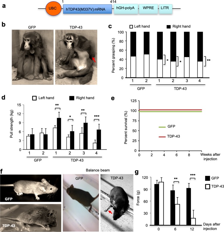
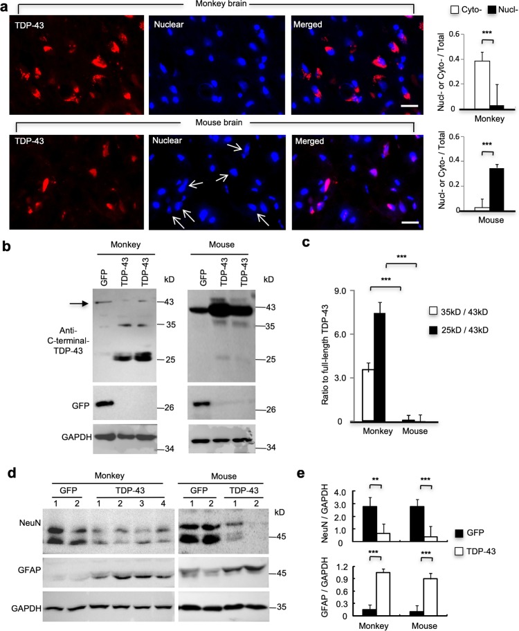
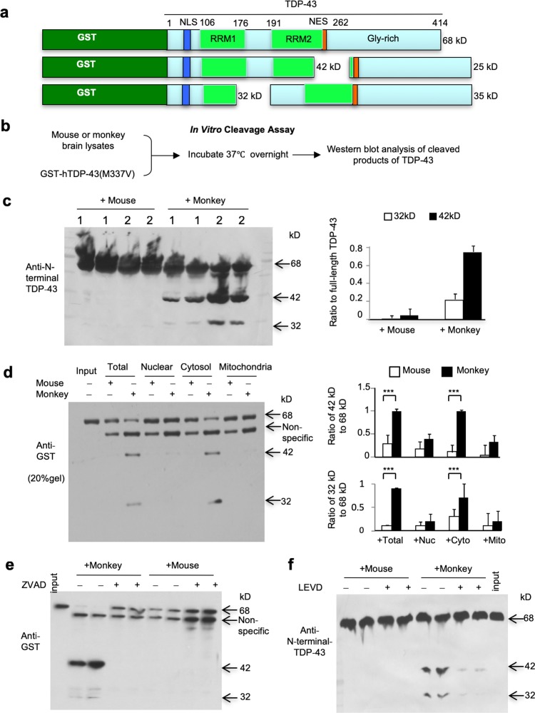
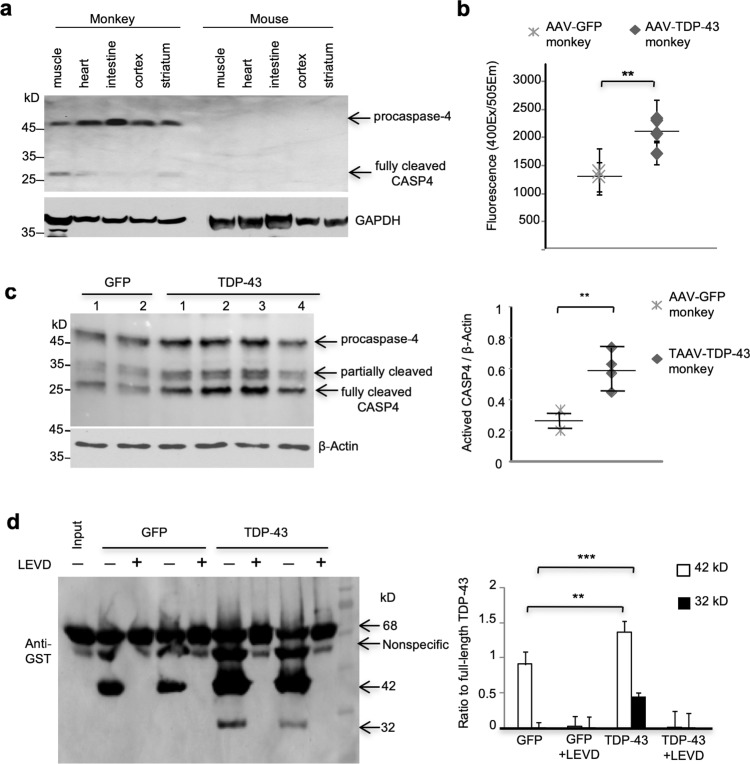
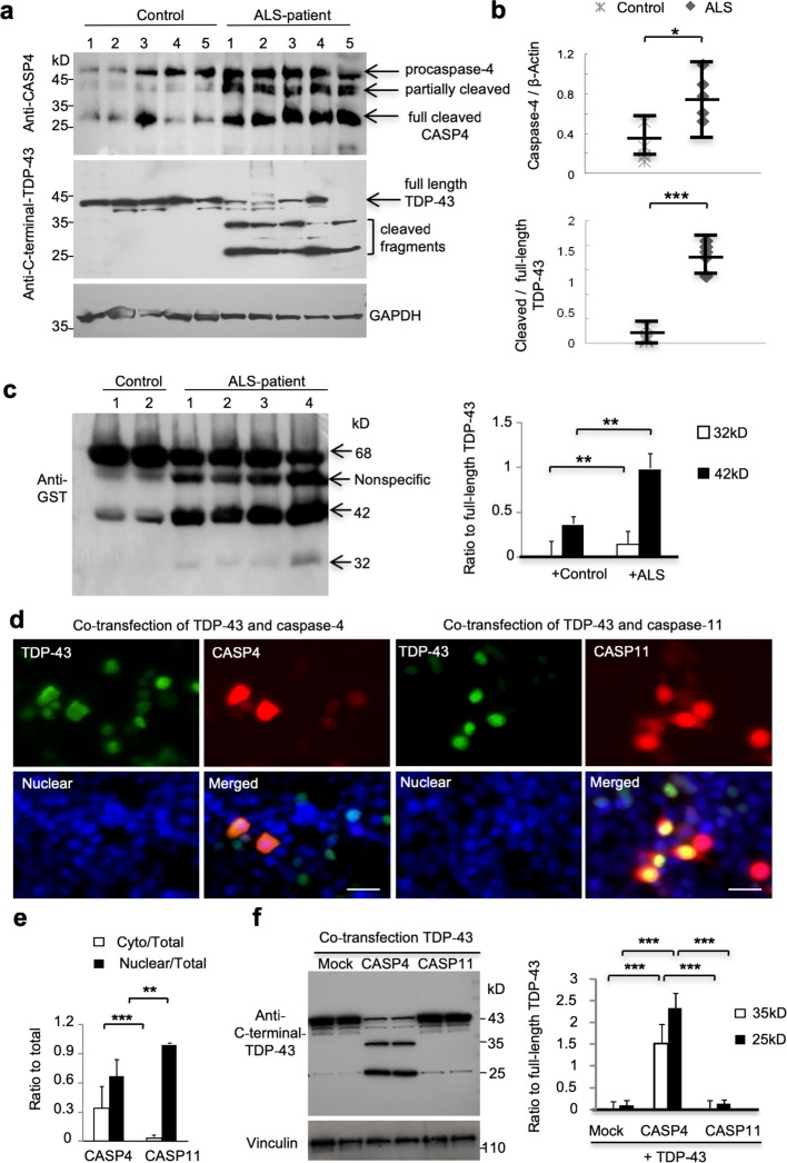
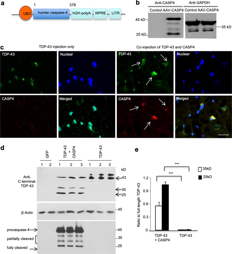
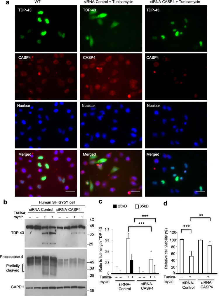
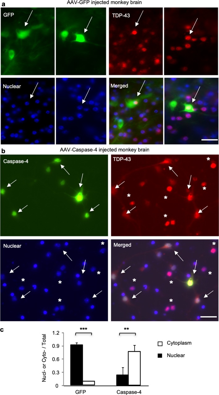
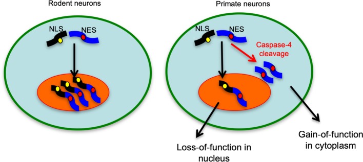
The cytoplasmic accumulation of the nuclear TAR DNA-binding protein 43 (TDP-43) is a pathologic hallmark in amyotrophic lateral sclerosis, frontotemporal lobar degeneration, and other neurological disorders. However, most transgenic TDP-43 rodent models show predominant nuclear distribution of TDP-43 in the brain. By expressing mutant TDP-43 (M337V) in the brains of rhesus monkeys and mice, we verified that mutant TDP-43 is distributed in the cytoplasm of the monkey brain and that the majority of mutant TDP-43 remains in the nuclei of the mouse brain. The primate-specific caspase-4, but not mouse homologue caspase-11, could remove the NLS-containing N-terminal domain and generate fragmented TDP-43 that accumulates in the cytoplasm. Moreover, increased expression of caspase-4 in the monkey brain promotes the cytoplasmic accumulation of endogenous TDP-43, and suppressing caspase-4 reduces the cytoplasmic distribution of endogenous TDP-43 in cultured human neural cells. Our findings suggest that primate-specific caspase-4-mediated cleavage of TDP-43 accounts for its cytoplasmic mislocalization in the primate brains and may serve as a potential therapeutic target.

Keywords: Aggregation; Caspase-4; Neurodegeneration; Non-human primate; TDP-43.


Figures

Fig. 1
Fig. 1
Phenotypes of AAV-TDP-43-injected monkeys and mice. a Adeno-associated virus vector-expressing mutant TDP-43(M337V) under the control of the human ubiquitin promoter. b Photographs of representative monkeys that were injected with AAV-GFP (control) or AAV-TDP-43 in the right side of the substantia nigra. The AAV-TDP-43-injected monkey showed the paralyzed left upper limb (arrow), whose movement is controlled by the contralateral right side of the substantia nigra. c AAV-TDP-43-injected monkeys showing decreased grasping by the left upper limb. Four AAV-TDP-43-injected and two AAV-GFP-injected monkeys were video recorded for the hand (left and right upper limbs) grasping of the ceiling fence and a test rod. Each monkey was video recorded for 30 min each time. The numbers of grasping from six recording times were used for statistical analysis (n = 6 times on the same examining day, mean ± SEM, *P < 0.05, **P < 0.01). d AAV-TDP-43-injected monkey showing decreased grip strength of the left upper limb. Monkeys in (c) were measured for the grip strength of their left and right hands by pulling a spring hand dynamometer. Each monkey was tested six times on the same examining day, and the digital score of the force (kg) on the grip strength meter was recorded for analysis (n = 6 times, mean ± SEM, *P < 0.05, **P < 0.01, ***P < 0.001). e Survival plot showing that expressing TDP-43 in the substantia nigra did not affect the monkey survival rate. The monkeys at 9–15 years of age were injected with either AAV-GFP or AAV-TDP-43 (n = 6 monkeys in each group). f Photographs of a representative mouse expressing AAV-TDP-43 (108 vg) in the right side of substantia nigra and showing the hunchback phenotype and defective balance beam movement (arrow). An AAV-GFP (108 vg)-injected mouse served as a control. g Force tests showing that injection of AAV-TDP-43 in the substantia nigra of mice caused motor function impairment, which was revealed by hind limb grip strength assay. AAV-GFP injection served as a control. Mice were 8–12 months old and injected with AAV-TDP-43 or AAV-GFP (n = 12 mice, mean ± SEM, in each group, **P < 0.01, ***P < 0.001)
Fig. 2
Fig. 2
Differential subcellular location and degradation of mutant TDP-43 (M337V) in the monkey and mouse brains. a TDP-43 immunostaining of the monkey substantia nigra showing the cytoplasmic localization of mutant TDP-43 (red, upper panel) after AAV-TDP-43 injection. The nuclei of neurons were labeled by DAPI (blue). In the mouse brain (lower panel), mutant TDP-43 was predominantly localized in the nuclei (arrows) of the injected substantia nigra. The relative numbers of cells containing nuclear (Nucl) or cytoplasmic (Cyto) TDP-43 over total DAPI-staining cells are shown in the right panel (mean ± SEM, ***P < 0.001). Scale bar 20 μm. b Western blotting analysis of the brain substantia nigra tissues of AAV-TDP-43-injected monkeys and mice. Probing with an antibody to C-terminal-TDP-43 revealed that C-terminal TDP-43 fragments (35- and 25-kD) were abundant in the monkey tissues but were weakly seen in the injected mouse brain. c Ratios of truncated TDP-43 (35- and 25-kD) to full-length TDP-43 detected by Western blotting in (b). The ratios were obtained from three independent Western blotting experiments (***P < 0.001). d Western blotting analysis of the expression of neuronal (NeuN) and astrocytic (GFAP) proteins in the AAV-GFP- or AAV-TDP-43-injected substantia nigra of monkeys and mice. Western blots were probed with antibodies to NeuN, GFAP, and GAPDH. e Ratios of NeuN/GAPDH and GFAP/GAPDH on the Western blotting in (d). The data were obtained from three independent Western blotting analysis of two AAV-GFP- and two AAV-TDP-43-injected mouse substantia nigra tissues; or two AAV-GFP- and four AAV-TDP-43-injected monkey substantia nigra tissues (**P < 0.01; ***P < 0.001)
Fig. 3
Fig. 3
Caspase-4 selectively cleaves TDP-43 in vitro. a The plasmid DNA structures for expressing GST-TDP-43 fusion protein. The cleavage of TDP-43 (35- and 25-kD) seen in the monkey brain can generate truncated GST-TDP-43 fusion proteins at 42- and 32-kD, respectively. The intact GST-TDP-43 is about 68-kD in size. RRM: RNA recognition motifs. b In vitro caspase assay of TDP-43. GST fusion proteins containing mutant TDP-43(M337V) in Sepharose beads were incubated overnight with the same quantity of mouse or monkey brain lysates. The beads were then centrifuged and analyzed by Western blotting with anti-GST. c Western blotting analysis of GST-TDP-43 that was incubated with brain homogenate lysates from monkeys or mice. The beads were centrifuged and analyzed with anti-GST. Expression levels of these fragments were minimal when incubated with mouse brain lysates. The ratios of fragmented TDP-43 to the intact full-length GST-TDP-43 (68-kD) were obtained from three independent experiments and shown in the right panel. d Western blotting analysis of GST-TDP-43(M337V) cleavage after incubation with different subcellular fractions (total homogenate, nuclear, cytosol, and mitochondria) of the mouse or monkey brains. The cytosolic fraction generated more fragmented TDP-43 (42-kD and 32-kD) than other fractions. Ratios of the 42-kD or 32-kD band to the intact GST-TDP-43 (68-kD) are presented in the right panel. The data are mean ± SEM (n = 3, ***P < 0.001). e, f Western blotting analysis of GST-mutant-TDP-43(M337V) cleavage showing that incubation of the pan-caspase inhibitor ZVAD-fmk (e) or the caspase-4 inhibitor LEVD-fmk (f) was able to block TDP-43 cleavage by the monkey brain lysates. The input lane is aliquot GST-TDP-43 (68-kD) without incubation with monkey or mice tissue lysates
Fig. 4
Fig. 4
Caspase-4 is specifically expressed in the monkey brain and its activity can be up-regulated by mutant TDP-43. a Western blotting analysis of caspase-4 (CASP4) expression in the brain and peripheral tissues of wildtype monkeys and mice. b Caspase-4 (CASP4) activity in AAV-GFP- or AAV-TDP-43(M337V)-injected monkey brain was assayed using a fluorescence method with a specific caspase-4 substrate Ac-YVAD-AFC. The data are mean ± SEM (n = 3 AAV-GFP monkeys, and n = 3 AAV-TDP-43 monkeys, **P < 0.01). c Western blotting analysis of caspase-4 expression in AAV-GFP- or AAV-TDP-43(M337V)-injected monkey brain. The activated form of caspase-4 (arrow) is more abundant in AAV-TDP-43-injected brain region than AAV-GFP-injected monkey brain. Quantitative data (ratio of activated caspase-4 to β-Actin) are shown in the right panel from three independent experiments (**P < 0.01). d Western blotting analysis of GST-TDP-43(M337V) incubated with lysates from AAV-GFP- or AAV-TDP-43-injected monkey brains. More cleaved GST-TDP-43 fragments were found in the AAV-TDP-43-injected brain lysates, and the caspase-4 inhibitor LEVD-fmk could completely block the cleavage. Quantitative data of the ratios of cleaved TDP-43 fragments to the intact form (68 kD) are shown in the right panel from three independent experiments (**P < 0.01, ***P < 0.001)
Fig. 5
Fig. 5
Caspase-4 is increased in ALS patient brains and mediates cytoplasmic localization of TDP-43. a, b Western blotting analysis of caspase-4 (CASP4) and C-terminal-TDP-43 expression in five ALS patients and five non-ALS control individuals (a). Ratios of activated caspase-4 bands (arrow) to GAPDH, or cleaved TDP-43 to full-length TDP-43 are presented right of the blots (b). *P < 0.05, ***P < 0.001. c Western blotting analysis of GST-TDP-43 (M337V) cleavage after incubation with the brain cortex lysates from ALS patients or non-ALS control individuals. Note that ALS patient brain lysates had much more abundant TDP-43 fragments (32- and 42-kD) than the control brain lysates. **P < 0.01. d, e Mouse N2A cells were transfected with pEGFP-TDP-43 (M337V) and DesRed-human caspase-4 (CASP4) or DesRed-mouse-caspase-11 (CASP11). The green fluorescent TDP-43 shows both cytoplasmic and nuclear localization in the cells co-transfected with DesRed-human-caspase-4, but not DesRed-mouse-caspase-11. The nuclei are stained by DAPI (d). Scale bars: 20 μm. Quantification of transfected N2A cells showing cytoplasmic (Cyto) or nuclear TDP43 over the total number of transfected cells when caspase-4 (CASP4) or caspase-11 (CASP11) was co-transfected. Counting the number of 120 positive cells under 40× lens in random six fields (e) (n = 3 independent experiments for each group, ** P < 0.01, *** P < 0.001). f Western blotting analysis of transfected N2A cells also showed that co-expression of caspase-4, but not caspase-11, markedly increased the generation of truncated TDP-43 (35- and 25-kD) fragments, which was also confirmed by the quantification of the ratios of truncated TDP-43 to full-length TDP-43 on the blots (right panel). n = 3 experiments, ***P < 0.001
Fig. 6
Fig. 6
Caspase-4 cleavage of mutant TDP-43 leads to the cytoplasmic localization of TDP-43 fragments in the mouse brains. a Schematic diagrams of AAV9 viral vector that expresses human caspase-4 under the control of the human ubiquitin promoter. b Expression of caspase-4 (CASP4) in mouse N2A cells infected by AAV–human-caspase-4. Caspase-4 was detected via Western blotting. c The mouse brain substantia nigra was injected with AAV-TDP-43 and AAV-caspase-4. Double immunofluorescent staining with antibodies to caspase-4 and C-terminal-TDP-43 showed the cytoplasmic localization of TDP-43 (green) in cells (arrows) that also express human caspase-4 (red). In cells expressing AAV-TDP-43 only, TDP-43 is localized in the DAPI stained nuclei. Scale bar: 40 μm. d, e Western blotting analysis of the mouse substantia nigra tissues co-injected with AAV-caspase-4 and AAV-TDP-43(M337V) using the antibody to C-terminal-TDP-43 (upper panel). The samples were also probed with anti-caspase-4 (lower panel). AAV-GFP or AAV-TDP-43(M337V) injection alone served as controls (d). Note that co-expression of human caspase-4 leads to the generation of C-terminal TDP-43 fragments of 35 and 25 kD, which is supported by the quantitative data of the ratio of cleaved TDP-43 fragments to the intact form (43 kD) (e) (n = 6, ***P < 0.001)
Fig. 7
Fig. 7
Knockdown of caspase-4 expression diminished the cytoplasmic distribution of endogenous TDP-43 in human SH-SY5Y cells. a Immunofluorescent staining of human SH-SY5Y cells transfected with caspase-4 (CASP4) siRNA or the control scrambled siRNA. The green fluorescent-labeled endogenous TDP-43 shows only nuclear localization when cells were not treated with tunicamycin. Tunicamycin treatment increased the cytoplasmic distribution of endogenous TDP-43, and this increase could be attenuated by siRNA-caspase-4 but not siRNA–control. The nuclei were stained by DAPI. Scale bars: 40 μm. b Western blotting of human SH-SY5Y cells that were transfected with caspase-4 siRNA or the scrambled control siRNA for 48 h and then treated with tunicamycin (1 μg/ml) for 12 h. The blots were probed with anti-C-terminal TDP-43 antibody. Note that tunicamycin treatment increased the level of activated caspase-4 produced from pro-caspase-4 and that siRNA-caspase-4 could inhibit this increase. c Quantitative analysis of the relative level (ratio to full-length TDP-43) of cleaved endogenous TDP-43 on Western blotting (b). The data were obtained from three independent experiments. (***P < 0.001). d Viability assay of SH-SY5Y cells transfected with siRNA-caspase-4 or its scrambled siRNA control and then treated with 1 μg/ml tunicamycin. The values (mean ± SEM) of cell viability are presented as  % of that of the control cells without tunicamycin/siRNA treatment and were obtained from three independent experiments. (**P < 0.01; ***P < 0.001)
Fig. 8
Fig. 8
Caspase-4 cleavage leads to the cytoplasmic distribution of monkey endogenous TDP-43 in vivo. The monkey brain prefrontal cortex was injected with AAV-GFP (control) (a) or AAV-caspase-4 (b). Double immunofluorescent staining with antibodies to C-terminal TDP-43 and GFP or caspase-4 showed the nuclear localization of endogenous TDP-43 (red) in cells (arrows) that express GFP (green). However, in cells expressing AAV-caspase-4, endogenous TDP-43 is localized in the cytoplasm as compared to those that do not express caspase-4. DAPI stained nuclei (star). Scale bars: 40 μm. c Quantitative analysis of the relative numbers of cells expressing nuclear (Nucl-) or cytoplasmic (Cyto-) TDP-43 over the total number of TDP-43-containing cells. The data (% of cells with nuclear or cytoplasmic TDP-43) are mean ± SEM and obtained by counting 80 cells in 6 random images (40× lens) in the injected area per sample from three monkeys (**P < 0.01, ***P < 0.001)
Fig. 9
Fig. 9
A proposed model for differential subcellular accumulation of TDP-43 in rodent and primate brains. The unique expression of caspase-4 in the primate brains causes the cleavage of full-length TDP-43 and generation of its C-terminal fragments that that can accumulate in the cytoplasm. While this cytoplasmic redistribution can reduce the level of nuclear TDP-43 to cause loss-of-function, mutations in the C-terminal region of TDP-43 could promote the misfolding and accumulation of C-terminal TDP-43 in the cytoplasm to elicit a toxic gain-of-function

Comment in

Similar articles

Cited by

References

    1. Ajroud-Driss S, Siddique T. Sporadic and hereditary amyotrophic lateral sclerosis (ALS) Biochim Biophys Acta. 2015;1852:679–684. doi: 10.1016/j.bbadis.2014.08.010. - DOI - PubMed
    1. Arai T, Hasegawa M, Akiyama H, Ikeda K, Nonaka T, Mori H, et al. TDP-43 is a component of ubiquitin-positive tau-negative inclusions in frontotemporal lobar degeneration and amyotrophic lateral sclerosis. Biochem Biophys Res Commun. 2006;351:602–611. doi: 10.1016/j.bbrc.2006.10.093. - DOI - PubMed
    1. Atkin JD, Farg MA, Walker AK, McLean C, Tomas D, Horne MK. Endoplasmic reticulum stress and induction of the unfolded protein response in human sporadic amyotrophic lateral sclerosis. Neurobiol Dis. 2008;30:400–407. doi: 10.1016/j.nbd.2008.02.009. - DOI - PubMed
    1. Bian ZM, Elner SG, Elner VM. Dual involvement of caspase-4 in inflammatory and ER stress-induced apoptotic responses in human retinal pigment epithelial cells. Invest Ophthalmol Vis Sci. 2009;50:6006–6014. doi: 10.1167/iovs.09-3628. - DOI - PMC - PubMed
    1. Cairns NJ, Neumann M, Bigio EH, Holm IE, Troost D, Hatanpaa KJ, et al. TDP-43 in familial and sporadic frontotemporal lobar degeneration with ubiquitin inclusions. Am J Pathol. 2007;171:227–240. doi: 10.2353/ajpath.2007.070182. - DOI - PMC - PubMed

Publication types

MeSH terms

LinkOut - more resources