. 2019 Apr 26;124(9):e63-e80.
doi: 10.1161/CIRCRESAHA.118.313865.

Role of SIRT1 in Modulating Acetylation of the Sarco-Endoplasmic Reticulum Ca2+-ATPase in Heart Failure

Affiliations

Role of SIRT1 in Modulating Acetylation of the Sarco-Endoplasmic Reticulum Ca2+-ATPase in Heart Failure

Przemek A Gorski et al. Circ Res. .

Erratum in

Abstract

Rationale: SERCA2a, sarco-endoplasmic reticulum Ca2+-ATPase, is a critical determinant of cardiac function. Reduced level and activity of SERCA2a are major features of heart failure. Accordingly, intensive efforts have been made to develop efficient modalities for SERCA2a activation. We showed that the activity of SERCA2a is enhanced by post-translational modification with SUMO1 (small ubiquitin-like modifier 1). However, the roles of other post-translational modifications on SERCA2a are still unknown.

Objective: In this study, we aim to assess the role of lysine acetylation on SERCA2a function and determine whether inhibition of lysine acetylation can improve cardiac function in the setting of heart failure.

Methods and results: The acetylation of SERCA2a was significantly increased in failing hearts of humans, mice, and pigs, which is associated with the reduced level of SIRT1 (sirtuin 1), a class III histone deacetylase. Downregulation of SIRT1 increased the SERCA2a acetylation, which in turn led to SERCA2a dysfunction and cardiac defects at baseline. In contrast, pharmacological activation of SIRT1 reduced the SERCA2a acetylation, which was accompanied by recovery of SERCA2a function and cardiac defects in failing hearts. Lysine 492 (K492) was of critical importance for the regulation of SERCA2a activity via acetylation. Acetylation at K492 significantly reduced the SERCA2a activity, presumably through interfering with the binding of ATP to SERCA2a. In failing hearts, acetylation at K492 appeared to be mediated by p300 (histone acetyltransferase p300), a histone acetyltransferase.

Conclusions: These results indicate that acetylation/deacetylation at K492, which is regulated by SIRT1 and p300, is critical for the regulation of SERCA2a activity in hearts. Pharmacological activation of SIRT1 can restore SERCA2a activity through deacetylation at K492. These findings might provide a novel strategy for the treatment of heart failure.

Keywords: acetylation; endoplasmic reticulum; heart failure; lysine; mice.


Figures
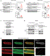
Figure 1.
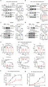
Figure 1.. SERCA2a acetylation is increased in failing hearts.
(a) SERCA2a acetylation was increased in human failing hearts. The human heart homogenates were immunoprecipitated with anti-acetyl-lysine antibody (reverse IP with anti-SERCA2a) and probed with anti-SERCA2a antibody (reverse blot with anti-acetyl-lysine). IgG was used as a negative control and glyceraldehyde-3-phosphate dehydrogenase (GAPDH) was used as a loading control. Graphs show means ± SD, with each data point representing one heart sample. Donors, n = 8; failing patients n = 8. **p < 0.01; ****p < 0.0001 vs non-failing by unpaired t-test. (b) SERCA2a acetylation was increased in TAC-induced failing mouse hearts. Graphs show means ± SD, with each data point representing one heart sample; n = 8 of Sham and n = 8 of TAC mice; **p < 0.01; ****p < 0.0001 vs Sham by unpaired t-test. (c) SERCA2a interacts with SIRT1 in adult mouse cardiomyocytes (ACMs). ACM lysates were immunoprecipitated with anti-SERCA2a or anti-SIRT1 antibodies and probed with anti-SERCA2a or anti-SIRT1 antibodies. (d) SERCA2a directly interacts with SIRT1 in HEK293 cells. Flag agarose beads or HA agarose beads were incubated with lysates of HEK293 cells transfected with either Flag-tagged SERCA2a or together with HA-tagged SIRT1. Immunoprecipitates were probed with anti-Flag or anti-HA antibodies. (e) Immunofluorescence images showing co-localization of SERCA2a and SIRT1 (merge panel, yellow) in ACMs. SERCA2a staining is shown in red, SIRT1 staining in green, and DAPI staining in blue. Pixel colocalization analyzed using the JACop plugin for Image J is shown in white. Scatter plots correspond to the colocalization between SERCA2a and SIRT1 (scale bar, 10 μm). All data shown are representative of three independent experiments.
Figure 2.
Figure 2.. SIRT1 deacetylates SERCA2a and regulates its activity.
(a) SIRT1 overexpression reduced acetylation of SERCA2a in normal ACMs. ACMs were isolated from 8 week old male C57BL/6J mice and infected with the indicated adenoviruses (50 MOI). 24 hours after adenovirus infection, ACMs were treated with trichostatin A (TSA) and nicotinamide (NAM, as SIRT1 inhibitor) for 4 hours to inhibit de-acetylation of SERCA2a. ACM lysates were used for immunoprecipitation with anti-acetyl-lysine or anti-SERCA2a antibodies. Graphs show means ± SD, with each data point representing one sample. ns, not significant; *p < 0.05 vs non-infected ACMs and #p < 0.05 vs Ad-SIRT1 by one-way ANOVA. (b) Knockout of SIRT1 elevated acetylation of SERCA2a in ACMs. ACMs were isolated from 8 weeks old male SIRT1−/− mice. SIRT1−/− ACMs were infected with the indicated adenoviruses (50 MOI) for 24 hours. Anti-acetyl-lysine or anti-SERCA2a was incubated with ACM lysates. Immunoprecipitates were probed with anti-SERCA2a or anti-acetyl-lysine antibodies. Graphs show means ± SD, with each data point representing one sample. ns, not significant; *p < 0.05 vs non-infected ACMs and ##p < 0.01 vs Ad-SIRT1 by one-way ANOVA. (c) SIRT1 overexpression reduces acetylation of SERCA2a in HEK293 cells. Anti-acetyl-lysine or anti-Flag agarose beads were incubated with lysates of HEK293 cells transfected with either Flag-tagged SERCA2a alone or together with HA-tagged SIRT1. Acetylation of SERCA2a was trigged by treatment with TSA and nicotinamide (NAM, as SIRT1 inhibitor). Immunoprecipitates were probed with anti-Flag or anti-acetyl-lysine antibody. Graphs show means ± SD, with each data point representing one sample. ns, not significant; **p < 0.01 vs Vector only by unpaired t-test. (d) Knockdown of SIRT1 elevates acetylation of SERCA2a in HEK293 cells. Lysates of HEK293 cells expressing either Flag-SERCA2a alone or together with a SIRT1-specific shRNA were immunoprecipitated with anti-acetyl-lysine agarose beads or anti-Flag beads. Immunoprecipitates were probed with anti-Flag or anti-acetyl-lysine antibody. Graphs show means ± SD, with each data point representing one sample. ns, not significant; *p < 0.05 vs sh-scrambled control by unpaired t-test. (e) SIRT1 knockout showed decreased cardiomyocyte function. ACMs were isolated from SIRT1−/− mice and infected with Ad-SIRT1 or Ad-β-gal (as a negative control). ACMs isolated from wild-type (WT) mice served as control. 24 hours after infection with adenovirus (50 MOI of each virus), the contractile response of SIRT1−/− ACMs was assessed by calcium amplitude, decay time constant (tau), peak shortening, maximal rate of contraction, and maximal rate of relaxation using a video-based edge-detection system (IonOptix, Inc. Milton, MA). 15 cardiomyocytes were measured per mouse, n = 4. Graphs show means ± SD, with each data point was represented a mean average of 15 cardiomyocytes isolated from one heart sample. ns, not significant; *p < 0.05; ***p < 0.001; ****p < 0.0001 vs WT and #p < 0.05; ##p < 0.01 vs SIRT1−/− + Ad-SIRT1 by one-way ANOVA.
Figure 3.
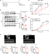
Figure 3.. Knockdown of SIRT1 elevates SERCA2a acetylation in vivo.
(a) Protocol for AAV9-sh-SIRT1-mediated SIRT1 knockdown in normal mice. (b) Knockdown of SIRT1 elevated levels of SERCA2a acetylation in normal mouse hearts. 8 week old male mice were injected with 1×10¹¹ copies of rAAV9-scrambled (Scr) or rAAV9-shRNA-SIRT1 (sh-SIRT1). 6 weeks later, hearts were harvested and homogenized. The heart homogenates were immunoprecipitated with anti-acetyl-lysine agarose beads and probed with anti-SERCA2a antibody. The representative immnoublots were presented. Graphs show means ± SD, with each data point representing one heart sample. n = 6 per each group. *p < 0.05; **p < 0.01 vs sh-scrambled control by unpaired t-test. (c-d) Knockdown of SIRT1 reduced SERCA2a activity. (c) Calcium uptake and (d) ATPase activity were measured using microsomal fractions isolated from mouse hearts injected with rAAV9- Scr or rAAV9- sh-SIRT1 (normalized to expression levels of SERCA2a). Data are represented as the mean ± SD of n = 3 hearts. * p < 0.05; *** p < 0.001 vs scrambled control by paired t-test. (e) Knockdown of SIRT1 induced cardiac dysfunction. Echocardiographic M-mode images show that mice injected with rAAV9-shRNA-SIRT1 underwent functional deterioration. Ejection fraction (EF), fractional shortening (FS), LV internal diameter during systole (LVIDs), and interventricular septal thickness at end-systole (IVSs) were determined in mice injected with rAAV9-sh-scrambled control (n = 6) and mice injected with rAAV9-shRNA-SIRT1 (n = 10). Graphs show means ± SD, with each data point representing one mouse. * p < 0.05; *** p < 0.001 vs sh-scrambled control by unpaired t-test.
Figure 4.
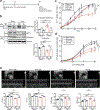
Figure 4.. β-lap treatment diminishes acetylation and restores activity of SERCA2a in a pressure-overload mouse model.
(a) Protocol for β-lap administration in TAC mice. (b) β-lap treatment reduced levels of SERCA2a acetylation in mouse hearts. 8 week old male C57BL/6J mice were subjected TAC operation. Vehicle (Veh) or 150 mg/kg/day of β-lap (βL) was administered to mice 2 weeks post sham or TAC surgery. Four weeks later, hearts were harvested, and heart homogenates were immunoprecipitated with anti-acetyl-lysine agarose beads and probed with anti-SERCA2a antibody. The representative immnoublots were presented. Graphs show means ± SD, with each data point representing one heart sample. n = 5 sham mice treated with vehicle (Sham+ Veh), n = 5 sham mice treated with β-lap (Sham+ β L), n = 5 TAC mice treated with vehicle (TAC+ Veh), and n = 5 TAC mice treated with β-lap (TAC+ βL). ns, not significant; *p < 0.05; **p < 0.01; ***p < 0.001 vs β-lap treatment and ##p < 0.01; ###p < 0.001 vs Sham+Veh by one-way ANOVA. (c-d) β-lap administration restored SERCA2a activities in TAC mouse hearts. (c) Calcium uptake and (d) ATPase activity were measured in mouse hearts (normalized to expression levels of SERCA2a). Data are represented as the mean ± SD of n = 3 experiments. *p < 0.05 vs β-lap treatment and #p < 0.05; ##p < 0.01 vs Sham+Veh by paired t-test. (e) β-lap administration restored cardiac function in TAC-operated mice. Echocardiographic M-mode images show that β-lap administration restored cardiac function in TAC-operated mice. LV chamber dimensions and LV systolic function were measured 6 weeks post sham or TAC surgery. Data are represented as the mean ± SD, with each data point representing one heart sample. Sham mice treated with vehicle (Sham+ Veh, n = 7), sham mice treated with β-lap (Sham+ βL, n = 8), TAC mice treated with vehicle (TAC+ Veh, n = 7), and TAC mice treated with β-lap (TAC+ βL, n = 10). ns, not significant; *p < 0.05; **p < 0.01; ***p < 0.001 vs β-lap treatment and ###p < 0.001; ####p < 0.0001 vs Sham+ Veh by one-way ANOVA.
Figure 5.
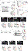
Figure 5.. β-lap treatment does not affect SERCA2a function in SIRT1 knock-down hearts.
(a) Protocol for β-lap administration in AAV9-sh-SIRT1-mediated SIRT1 knock-down mice. (b) β-lap (βL) did not restore levels of SERCA2a acetylation in SIRT1 knockdown mice. 8 week old male C57BL/6J mice were injected with 1×10¹¹ copies of rAAV9-Scr or rAAV9-sh-SIRT1. Six weeks later, vehicle or 150 mg/kg/day of β-lap was administered orally for 4 weeks. Heart homogenates were immunoprecipitated with anti-acetyl-lysine agarose beads and probed with anti-SERCA2a antibody. The representative immnoublots were presented. Graphs show means ± SD, with each data point representing one heart sample. n = 3 rAAV9-Scr injected mice treated with vehicle (rAAV9-Scr + Veh), n = 3 rAAV9-Scr injected mice treated with β-lap (rAAV9-Scr + β L), n = 6 rAAV9-sh-SIRT1 injected mice treated with vehicle (rAAV9-sh-SIRT1+ Veh), and n = 6 rAAV9-sh-SIRT1 injected mice treated with β-lap (rAAV9-sh-SIRT1+ βL). ns, not significant vs β-lap treatment and #p < 0.05; ##p < 0.01 vs rAAV9-Scr +Veh by one-way ANOVA. (c) β-lap administration did not restore SERCA2a activity in the hearts of mice treated with rAAV9-sh-SIRT1. (c) Calcium uptake and (d) ATPase activity were measured in mouse hearts. Data are represented as the mean ± SD of n = 3 experiments. ns, not significant; *p < 0.05; **p < 0.01 vs β-lap treatment and #p < 0.05; ###p < 0.001 vs rAAV9-Scr + Veh by paired t-test. (e) β-lap administration did not recover cardiac function in mice treated with rAAV9-sh-SIRT1. Echocardiographic M-mode images show that β-lap administration did not restore normal cardiac function in SIRT1 knockdown mice. LV chamber dimensions and LV systolic function were measured. Data are represented as the mean ± SD, with each data point representing one heart sample. rAAV9-Scr + Veh (n = 8), rAAV9-Scr + βL (n = 7), rAAV9-sh-SIRT1+ Veh (n = 13) and rAAV9-sh-SIRT1+ βL (n = 9). ns, not significant vs β-lap treatment and #p < 0.05; ####p < 0.0001 vs rAAV9-Scr +Veh by one-way ANOVA. (f) Protocol for β-lap administration in cardiac-specific SIRT1 knockout (SIRT1−/−) mice. (g) β-lap administration did not affect levels of SERCA2a acetylation in SIRT1−/− mice. 8 week old male SIRT1−/− mice were injected intraperitoneally with tamoxifen to induce Sirt1 gene disruption. Four weeks after tamoxifen administration vehicle or 1 mg/g/day of β-lap was administered orally for 4 weeks. Heart homogenates were immunoprecipitated with anti-SERCA2a antibody and probed with anti-acetyl-lysine antibody. The representative immnoublots were presented. Graphs show means ± SD, with each data point representing one heart sample. n=4 per each group. ns, not significant vs β-lap treatment and ##p < 0.01 vs WT+Veh by one-way ANOVA. (h) β-lap administration did not recover cardiac function in SIRT1−/− mice. Echocardiographic M-mode, LV chamber dimensions, and LV systolic function were measured. Data are represented as the mean ± SD, with each data point representing one heart sample. WT + Veh (n = 7), SIRT1−/− + Veh (n = 10) and SIRT1−/− + βL (n = 8). ns, not significant vs β-lap treatment and #p < 0.05; ##p < 0.01; ###p < 0.001; ####p < 0.0001 vs WT + Veh by one-way ANOVA.
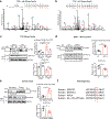
Figure 6.
Figure 6.. Acetylation of K492 on SERCA2a is increased in failing and SIRT1 knockout hearts.
(a-b) Representative MS/MS spectra of tryptic peptides of SERCA2a at K492 identified in TAC mouse hearts treated with vehicle or β-lap. (a) Acetyl-Lys peptide KACSMOXSVYCTPNKPSR (residues 492–505) was detected in heart extracts from TAC mice. (b) Lys peptide KSMSVYCTPNKPSR (residues 492–505) was detected in heart extracts from TAC mice treated with β-lap (TAC + βL). KAc: acetyl-lysine; MOx: oxidized methionine. (c) Levels of K492 acetylation of SERCA2a were elevated in TAC-induced failing mouse hearts, as determined by a specific anti-acetyl-K492 antibody. The representative immnoublots were presented. Graphs show means ± SD, with each data point representing one heart sample. n = 4 per each group. ns, not significant; *p < 0.05 vs β-lap treatment and ##p < 0.01; ###p < 0.001 vs Sham+Veh by one-way ANOVA. (d) Acetylation levels of SERCA2a at K492 were elevated in SIRT1−/− mouse hearts. The representative immnoublots were presented. Graphs show means ± SD, with each data point representing one heart sample. n = 4 per each group. ns, not significant; ***p < 0.001 vs WT by unpaired t-test. (e) Acetylation levels of SERCA2a at K492 were increased in human failing hearts. The representative immnoublots were presented. Graphs show means ± SD, with each data point representing one heart sample. n = 4 per each group. *p < 0.05; **p < 0.01 vs non-failing by unpaired t-test. (f) Amino acid sequence alignment of different members of the human P-ATPase family of proteins showing high degree of conservation of K492 (K492 and homologous lysine residues are highlighted in red).
Figure 7.
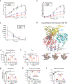
Figure 7.. Acetylation at K492 plays a critical role in regulating SERCA2a activity.
(a) The calcium uptake, (b) ATPase activity, and (c) ATP binding assays of SERCA2a in microsomal fractions isolated from HEK293 cells transfected with wild-type (WT) or mutant (K492Q or K492R) forms of SERCA2a (normalized to expression levels of SERCA2a). Data are represented as the mean ± SD of n = 3 experiments. CPM, counts per minute. *p < 0.05; ***p < 0.001 vs WT SERCA2a and #p < 0.05; ##p < 0.01 vs K492R SERCA2a by paired t-test. (d) K492 acetylation site is located in the ATP binding pocket of SERCA2a. Cartoon representation of SERCA2a based on the crystal structure of SERCA1a (PDB: 1T5S). The phosphorylation domain, nucleotide-binding domain, and actuator domain are shown in salmon, yellow, and cyan, respectively. The transmembrane domain is shown in grey. K492 (pink) is located within the nucleotide-binding domain adjacent to the ATP molecule (red). Close-up views of K492, acetylated K492 (Ac-K492), and non-acetylated K492 mutant (K492R) relative to the phosphorylation domain are shown on the bottom. (e-i) Overexpression of the acetyl-mimicking mutant (K492Q) of SERCA2a showed contractile dysfunction in adult cardiomyocytes isolated from SERCA2+/− mice. Adult ventricular cardiomyocytes were isolated from SERCA2+/− mice and infected with Ad-WT SERCA2a, Ad- K492Q SERCA2a, or Ad-β-gal (as a negative control). ACMs isolated from wild-type (WT) mice infected with Ad-β-gal served as control. The contractile response of SERCA2a restoration in SERCA2+/− ACMs was assessed by calcium amplitude (e), decay time constant (tau) (f), peak shortening (g), maximal rate of contraction (h), and maximal rate of relaxation (i) using a video-based edge-detection system (IonOptix, Inc. Milton, MA) 24 hour after infection of ACMs with adenovirus (50 MOI of each virus). n = 4. Graphs show means ± SD, with each data point was represented a mean average of 15 ACMs isolated from one heart sample. ns, not significant; *p < 0.05; **p < 0.01; ***p < 0.001; ****p < 0.0001 vs WT + β-gal and #p < 0.05; ###p < 0.001; ####p < 0.0001 vs SERCA2+/− + SERCA2a-WT by one-way ANOVA.
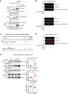
Figure 8.
Figure 8.. SERCA2a is acetylated by p300 acetyltransferase.
(a) p300 directly acetylates and interacts with SERCA2a in HEK293 cells. HEK293 cells were co-transfected with Flag-SERCA2a alone or with Myc-p300. Interaction between p300 and SERCA2a as well as acetylation levels of SERCA2a were determined by immunoprecipitation followed by probing with appropriate antibodies. The experiments shown are representative of three independent experiments. (b) SERCA2a was acetylated by p300 in vitro. Purified porcine SERCA2a protein was incubated with 10 μM acetyl-CoA in the presence or absence of human recombinant p300 (hp300) (upper panel) protein. p300-mediated acetylation of SERCA2a was prevented by treatment with C646, a selective inhibitor of p300 (lower panel). (c) K492 acetylation on SERCA2a was regulated by p300 and SIRT1. Purified porcine SERCA2a protein was incubated with 10 μM acetyl-CoA and hp300 followed by incubation in the presence or absence of recombinant SIRT1. Following incubation, the samples were subjected to analysis by mass spectrometry which showed that p300-mediated acetylation at K492 was deacetylation by SIRT1. Comparison of acetylated lysine 492 levels using MRM (multiple reaction monitoring) anaylsis. Acetyl-Lys 492 peptide KACSMSVYCTPNKPSR (residues 492–505) was acetylated by p300 and deacetylated by SIRT1. Acetyl-Lys 502 peptide SMSVYCTPNKACPSR (residues 493–505) was not perturbed in the presence of SIRT1. (d) Acetylation status of K492 in purified porcine SERCA2a protein was confirmed by immnobloting with anti-acetyl-K492 antibody. Graphs show means ± SD, with each data point representing one heart sample. n = 6 per each group. ns, not significant; *p < 0.05 vs wild-type (WT) mice by unpaired t-test.

Comment in

Similar articles

Cited by

References

    1. Kho C, Lee A and Hajjar RJ. Altered sarcoplasmic reticulum calcium cycling--targets for heart failure therapy. Nat Rev Cardiol. 2012;9:717–33. - PMC - PubMed
    1. Kawase Y and Hajjar RJ. The cardiac sarcoplasmic/endoplasmic reticulum calcium ATPase: a potent target for cardiovascular diseases. Nat Clin Pract Cardiovasc Med. 2008;5:554–65. - PubMed
    1. Sakata S, Lebeche D, Sakata N, Sakata Y, Chemaly ER, Liang LF, Takewa Y, Jeong D, Park WJ, Kawase Y and Hajjar RJ. Targeted gene transfer increases contractility and decreases oxygen cost of contractility in normal rat hearts. American journal of physiology Heart and circulatory physiology. 2007;292:H2356–63. - PubMed
    1. del Monte F, Williams E, Lebeche D, Schmidt U, Rosenzweig A, Gwathmey JK, Lewandowski ED and Hajjar RJ. Improvement in survival and cardiac metabolism after gene transfer of sarcoplasmic reticulum Ca(2+)-ATPase in a rat model of heart failure. Circulation. 2001;104:1424–9. - PMC - PubMed
    1. Miyamoto MI, del Monte F, Schmidt U, DiSalvo TS, Kang ZB, Matsui T, Guerrero JL, Gwathmey JK, Rosenzweig A and Hajjar RJ. Adenoviral gene transfer of SERCA2a improves left-ventricular function in aortic-banded rats in transition to heart failure. Proceedings of the National Academy of Sciences of the United States of America. 2000;97:793–8. - PMC - PubMed

Publication types

MeSH terms