. 2019 Feb 19;8(4):e010866.
doi: 10.1161/JAHA.118.010866.

Transcription Factor EB Activation Rescues Advanced αB-Crystallin Mutation-Induced Cardiomyopathy by Normalizing Desmin Localization

Affiliations

Transcription Factor EB Activation Rescues Advanced αB-Crystallin Mutation-Induced Cardiomyopathy by Normalizing Desmin Localization

Xiucui Ma et al. J Am Heart Assoc. .

Abstract

Background Mutations in αB-crystallin result in proteotoxic cardiomyopathy with desmin mislocalization to protein aggregates. Intermittent fasting ( IF ) is a novel approach to activate transcription factor EB (TFEB), a master regulator of the autophagy-lysosomal pathway, in the myocardium. We tested whether TFEB activation can be harnessed to treat advanced proteotoxic cardiomyopathy. Methods and Results Mice overexpressing the R120G mutant of αB-crystallin in cardiomyocytes ( Myh6-Cry ABR 120G) were subjected to IF or ad-lib feeding, or transduced with adeno-associated virus- TFEB or adeno-associated virus-green fluorescent protein after development of advanced proteotoxic cardiomyopathy. Adeno-associated virus-short hairpin RNA-mediated knockdown of TFEB and HSPB 8 was performed simultaneously with IF . Myh6-Cry ABR 120G mice demonstrated impaired autophagic flux, reduced lysosome abundance, and mammalian target of rapamycin activation in the myocardium. IF resulted in mammalian target of rapamycin inhibition and nuclear translocation of TFEB with restored lysosome abundance and autophagic flux; and reduced aggregates with normalized desmin localization. IF also attenuated left ventricular dilation and myocardial hypertrophy, increased percentage fractional shortening, and increased survival. Adeno-associated virus- TFEB transduction was sufficient to rescue cardiomyopathic manifestations, and resulted in reduced aggregates and normalized desmin localization in Myh6-Cry ABR 120G mice. Cry ABR 120G-expressing hearts demonstrated increased interaction of desmin with αB-crystallin and reduced interaction with chaperone protein, HSPB 8, compared with wild type, which was reversed by both IF and TFEB transduction. TFEB stimulated autophagic flux to remove protein aggregates and transcriptionally upregulated HSPB 8, to restore normal desmin localization in Cry ABR 120G-expressing cardiomyocytes. Short hairpin RNA-mediated knockdown of TFEB and HSPB 8 abrogated IF effects, in vivo. Conclusions IF and TFEB activation are clinically relevant therapeutic strategies to rescue advanced R120G αB-crystallin mutant-induced cardiomyopathy by normalizing desmin localization via autophagy-dependent and autophagy-independent mechanisms.

Keywords: HspB8; TFEB; cardiomyopathy; intermittent fasting; protein aggregates; αB‐crystallin.


Figures

Figure 1
Figure 1
Autophagic flux is impaired in αB‐crystallin R120G mutant transgenic mice with onset of heart failure. A through D, Representative immunoblots (A) demonstrating expression of LC3 with quantitation of LC3‐II (B), p62 (C) and αB‐crystallin (D) in Myh6‐CryABR120G transgenic mice or littermate wild‐type (WT) controls at 40 weeks of age, injected with chloroquine (40 mg/kg) or diluent to assess autophagic flux. N=4 to 6/group. E, Representative myocardial transmission electron micrographs from in Myh6‐CryABR120G transgenic mice or littermate WT controls at 40 weeks of age. Arrows point to Z‐discs, and arrowheads point to autophagic structures. Representative of N=2/group. *P<0.05.
Figure 2
Figure 2
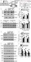
Intermittent fasting (IF) activates transcription factor EB (TFEB) and restores autophagic flux in αB‐crystallin R120G mutant transgenic mice. A, Schematic depicting experimental intervention with IF in Myh6‐CryABR120G transgenic mice. B, Survival curves in IF and ad‐lib (AL) fed Myh6‐CryABR120G transgenic mice and AL fed controls over the 6‐week experimental duration. P value depicted is by log‐rank test. C and D, Representative immunoblots (C) demonstrating expression of TFEB with quantitation (D) in the cytosolic and nuclear fractions from hearts of mice treated as in A (at 46 weeks) and subjected to biochemical fractionation. Expression of GAPDH and histone H3 is used to detect enrichment of cytosolic and nuclear proteins, respectively. N=4/group. E, Assessment of autophagic flux in mice Myh6‐CryABR120G transgenic mice subjected to IF or provided access to food AL as in A, and injected with chloroquine (40 mg/kg for 4 hours) or diluent with immunoblotting for LC3 and p62. F, Quantitation of LC3‐II and p62 in cardiac tissue from mice treated as in E. N=4/group. G and H, Representative immunoblot (G) with quantitation (H) of lysosome proteins LAMP1 and LAMP2 in total cardiac protein extracts from Myh6‐CryABR120G transgenic mice subjected to IF or AL feeding, or littermate wild type (WT) as in A. N=4/group. I and J, Representative immunoblots (I) demonstrating expression of mammalian target of rapamycin (mTOR) pathway proteins with quantitation (J) of phosphorylated mTOR (p‐mTOR), phosphorylated 70S6K (p‐S6K), and phosphorylated 4EBP1 (p‐4EBP1) in Myh6–αB‐crystallin R120G transgenic mice subjected to IF or AL feeding, and WT controls as in A. N=4/group. qOD (every other day). *P value by post hoc test after 1‐way ANOVA.
Figure 3
Figure 3
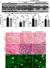
Intermittent fasting (IF) promotes myocardial protein aggregate removal in αB‐crystallin R120G mutant transgenic mice. A through C, Representative immunoblot (A) with quantitation of αB‐crystallin in the soluble (B) and insoluble (C) myocardial fractions from Myh6‐CryABR120G transgenic mice subjected to IF or ad‐lib (AL) feeding, and littermate wild‐type (WT) controls at 46 weeks of age. N=4/group. P value is by t test. “Darker” and “lighter” indicate relative exposures of the same blot. D, Representative TEM (Transmission electron microscopy) images from Myh6‐CryABR120G transgenic mice subjected to IF or AL feeding and littermate WT mice. White arrows point to protein aggregates. Black arrows point to autolysosomes. Black arrowheads point to Z‐discs, where intracellular desmin is localized, pointing to partial restoration of Z‐disc architecture in intermittently fasted Myh6‐CryABR120G transgenic mice compared with AL fed controls. Representative of N=2/group.
Figure 4
Figure 4
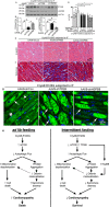
Intermittent fasting (IF) attenuates cardiomyopathy in αB‐crystallin R120G mutant transgenic mice and restores normal desmin localization. A, Representative 2‐dimensional–directed M‐mode echocardiographic images in Myh6‐CryABR120G transgenic mice subjected to IF or ad‐lib feeding for 6 weeks or ad‐lib fed littermate wild‐type (WT) mice at 46 weeks of age. B through E, Quantitation of left ventricular end‐diastolic diameter (LVEDD; B), left ventricular percentage endocardial fractional shortening (LV %FS; C), left ventricular mass (LVM; D), and heart weight normalized to tibial length (HW/TL; E) in mice treated as in A. N=5 to 7 mice/group. See Table S2 for additional echocardiographic data on these mice. F through H, Representative images demonstrating myocardial sections stained with hematoxylin‐eosin (H and E; F) and Masson's trichrome (G) and immunostained for desmin expression (H) in mice treated as in A. White arrows in F point to eosinophilic protein aggregates. In H, white arrows in groups point to Z‐discs and arrowheads indicate I lines to demonstrate desmin localization. Single long white arrows point to desmin localized in aggregates.
Figure 5
Figure 5
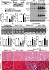
Adeno‐associated virus (AAV9)–mediated transcription factor EB (TFEB) transduction rescues cardiomyopathy in αB‐crystallin R120G mutant transgenic mice and promotes removal of protein aggregates. A, Schematic depicting experimental intervention with AAV9‐mediated TFEB transduction (or AAV9–green fluorescent protein [GFP] as control) in Myh6‐CryABR120G transgenic mice or littermate controls. B and C, Representative immunoblots demonstrating expression of TFEB in total protein extracts with quantitation (C, left) and αB‐crystallin (C, right) in hearts from Myh6‐CryABR120G transgenic mice or littermate wild‐type (WT) mice transduced with AAV9‐TFEB or AAV9‐GFP as in A, at 46 weeks of age. N=5 to 7/group. D through H, Representative 2‐dimensional–directed M‐mode echocardiograms (D) in Myh6‐CryABR120G transgenic mice or littermate WT mice transduced with AAV9‐TFEB or AAV9‐GFP as in A, at 46 weeks of age, with quantitation of left ventricular end‐diastolic diameter (LVEDD; E), left ventricular percentage endocardial fractional shortening (LV %FS; F), LV mass (G), and heart weight normalized to tibial length (HW/TL; H) in mice treated as in A. N=5 to 8/group. A total of 1 of 9 AAV9‐GFP–injected and 1 of 6 AAV9‐TFEB–injected Myh6‐CryABR120G transgenic mice died during the study. See Table S3 for additional echocardiographic data on these mice. I and J, Representative hematoxylin and eosin (H and E; I) and trichrome (J) stained myocardial sections from mice treated as in A. Arrowheads point to eosinophilic aggregates in I. *P<0.05 by post hoc test after 1‐way ANOVA.
Figure 6
Figure 6
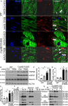
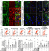
Adeno‐associated virus (AAV9)–mediated transcription factor EB (TFEB) transduction restores normal desmin localization in αB‐crystallin R120G mutant transgenic mice with upregulation of HSPB8. A, Representative images demonstrating immunolocalization of desmin (pseudocolored green) in 46‐week‐old Myh6‐CryABR120G transgenic mice or littermate controls transduced with AAV9‐TFEB or AAV9–green fluorescent protein (GFP) for 10 weeks. White arrowheads point to nuclear TFEB; white arrows in groups point to desmin associated with Z‐discs; single white arrows point to desmin associated with intercalated discs. B through E, Representative immunoblot (B) with quantitation of HSPB8 (C), HSPB1 (D), and desmin (E) in total cardiac protein extracts from mice treated as in A. N=3 to 7/group. F, Immunoblot demonstrating interaction of desmin with HSPB8 and αB‐crystallin in Myh6‐CryABR120G transgenic mice subjected to intermittent fasting (IF) or ad‐lib (AL) feeding and littermate control hearts (depicted as “‐”). Total protein (1 mg) from cardiac extracts was subjected to immunoprecipitation (IP) with anti‐desmin antibody or control IgG, followed by immunoblotting for HSPB8 and αB‐crystallin. At the exposure shown to demonstrate desmin in transgenic samples, a desmin signal was not evident in wild‐type (WT) hearts in the input lane (with 20 µg/sample) and was seen with longer exposure times (data not shown). G, Immunoblot demonstrating interaction of desmin with HSPB8 and αB‐crystallin in Myh6‐CryABR120G transgenic hearts treated with AAV9‐GFP or AAV9‐TFEB and subject to immunoprecipitation with anti‐desmin antibody (or IgG as control), as in G. For αB‐crystallin, both dark (top) and light (bottom) exposures are shown for F and G. *P<0.05 by post hoc test after 1‐way ANOVA.
Figure 7
Figure 7
Transcription factor EB (TFEB) normalizes desmin localization via HSPB8 and promotes aggregate removal independent of HSPB8 expression. A, Representative confocal images demonstrating immunolocalization of desmin and αB‐crystallin in neonatal rat cardiac myocytes (NRCMs) adenovirally transduced with TFEB or control, with and without simultaneous knockdown of HSPB8 (ie, treated with adenoviral particles coding for R120G mutant of αB‐crystallin [multiplicity of infection {MOI}=10; for 48 hours] without or with HA‐tagged TFEB [MOI=10; for 24 hours] in the presence of adenoviral particles expressing short hairpin RNA targeting rat HSPB8 [short hairpin HSPB8 {shHSPB8}; MOI=100; for 72 hours]). Adenoviral particles coding for LacZ or short hairpin LacZ were added as controls simultaneously with Ad‐TFEB or Ad–αB‐crystallin overexpression and shHSPB8, respectively, to equalize the number of viral particles. Arrows point to desmin in Z‐discs, and arrowheads point to desmin colocalized with αB‐crystallin in aggregates. Representative of n=2 experiments. B, Representative confocal images demonstrating expression of desmin, ubiquitin, and p62 in NRCMs treated as in A. Arrows point to desmin in Z‐discs, and arrowheads point to desmin colocalized with p62 and ubiquitin in aggregates. Representative of n=2 experiments. C and D, Representative flow cytometric tracings demonstrating JC‐1 fluorescence (C) and quantitation of cells predominantly expressing JC‐1 monomers (lower right quadrants in C; D) for NRCMs treated as described for A. N=7/group. E, Mitochondrial DNA content indexed to nuclear DNA in cells treated as in A. N=4/group. F, Cell death in NRCMs treated as in A. N=7 to 8/group. *P<0.05 by post hoc test after 1‐way ANOVA.
Figure 8
Figure 8
Knockdown of transcription factor EB (TFEB) and HSPB8 prevents intermittent fasting (IF)–mediated attenuation of proteotoxic cardiomyopathy with persistent desmin localization to aggregates. A through C, Representative immunoblot (A) with quantitation of TFEB expression (B) and HSPB8 abundance (C) in total cardiac protein extracts from Myh6‐CryABR120G transgenic mice transduced with adeno‐associated virus (AAV9)–short hairpin control (shCon), AAV9–short hairpin TFEB (shTFEB), or AAV9–short hairpin HSPB8 (shHSPB8) and subjected to IF, assessed at 46 weeks of age. N=4/group. *P<0.05 by post hoc test after 1‐way ANOVA. Please see schematic in Figure S12A for experimental design. D, Left ventricular percentage endocardial fractional shortening (LV %FS) in mice treated as in A and evaluated by echocardiography at baseline (4 weeks after injection of AAV9 particles and before IF, time=0 weeks) and 3 weeks and 6 weeks after initiating IF. N=4 to 5/group. *P<0.05 by post hoc test after 2‐way ANOVA. We did not observe mortality in these treatment groups. See Table S4 for additional echocardiographic data on these mice. E through G, Representative images demonstrating myocardial sections stained with hematoxylin‐eosin (H and E; E) and Masson's trichrome (F) and immunostained for desmin expression (G) in mice treated as in A. In G, white arrows in groups point to Z‐discs and arrowheads indicate intercalated discs to demonstrate desmin localization. Single long white arrows point to desmin localized in aggregates. H, Schematic depicting the mechanisms by which IF benefits advanced cardiomyopathy triggered by αB‐crystallin R120G mutation. Mutant R120G crystallin expression drives mammalian target of rapamycin (mTOR) activation with suppressed autophagic flux and aggregates sequestering desmin within the aggregates, thereby provoking mitochondrial abnormalities and cell death. IF suppresses mTOR activation, which activates TFEB to stimulate lysosome biogenesis and restores autophagic flux to remove mutant crystallin aggregates and relieve desmin sequestration. TFEB activation also stimulates HSPB8 expression, which chaperones desmin to its normal localization and restores mitochondrial quality to rescue cell death.

Comment in

Similar articles

Cited by

References

    1. Burke MA, Cook SA, Seidman JG, Seidman CE. Clinical and mechanistic insights into the genetics of cardiomyopathy. J Am Coll Cardiol. 2016;68:2871–2886. - PMC - PubMed
    1. Goldfarb LG, Dalakas MC. Tragedy in a heartbeat: malfunctioning desmin causes skeletal and cardiac muscle disease. J Clin Invest. 2009;119:1806–1813. - PMC - PubMed
    1. Bova MP, Yaron O, Huang Q, Ding L, Haley DA, Stewart PL, Horwitz J. Mutation R120G in alphaB‐crystallin, which is linked to a desmin‐related myopathy, results in an irregular structure and defective chaperone‐like function. Proc Natl Acad Sci USA. 1999;96:6137–6142. - PMC - PubMed
    1. Vicart P, Caron A, Guicheney P, Li Z, Prevost MC, Faure A, Chateau D, Chapon F, Tome F, Dupret JM, Paulin D, Fardeau M. A missense mutation in the alphaB‐crystallin chaperone gene causes a desmin‐related myopathy. Nat Genet. 1998;20:92–95. - PubMed
    1. Goldfarb LG, Park KY, Cervenakova L, Gorokhova S, Lee HS, Vasconcelos O, Nagle JW, Semino‐Mora C, Sivakumar K, Dalakas MC. Missense mutations in desmin associated with familial cardiac and skeletal myopathy. Nat Genet. 1998;19:402–403. - PubMed

Publication types

MeSH terms