. 2019 Sep 19;11(9):747-760.
doi: 10.1093/jmcb/mjz004.

MicroRNA-24 promotes pancreatic beta cells toward dedifferentiation to avoid endoplasmic reticulum stress-induced apoptosis

Affiliations

MicroRNA-24 promotes pancreatic beta cells toward dedifferentiation to avoid endoplasmic reticulum stress-induced apoptosis

Yunxia Zhu et al. J Mol Cell Biol. .

Abstract

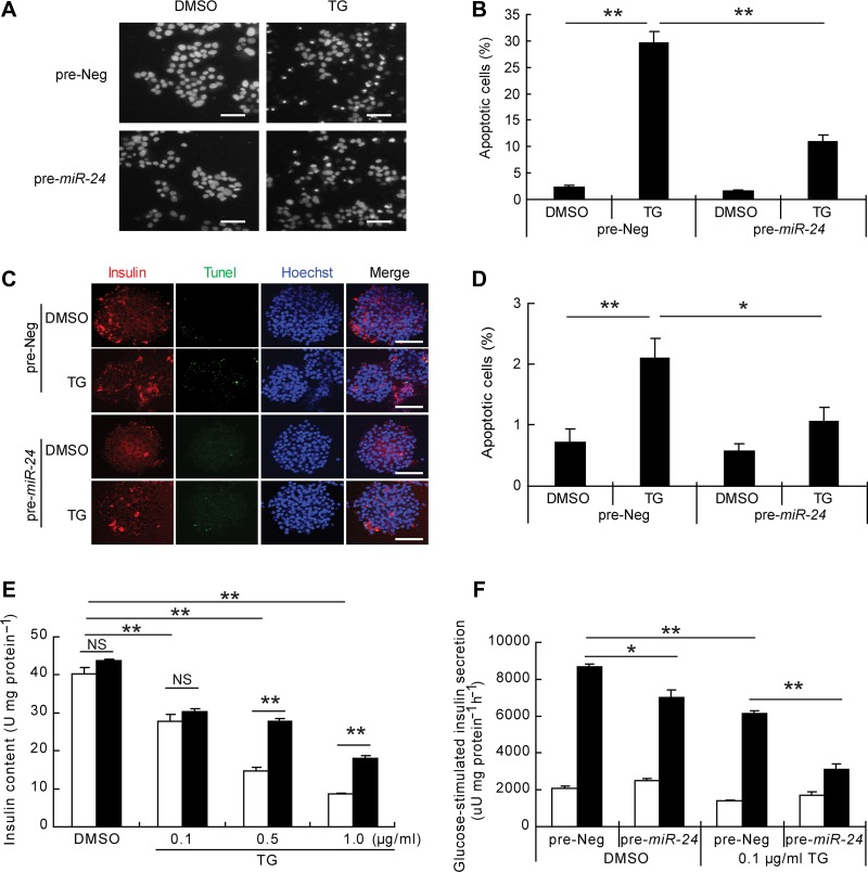
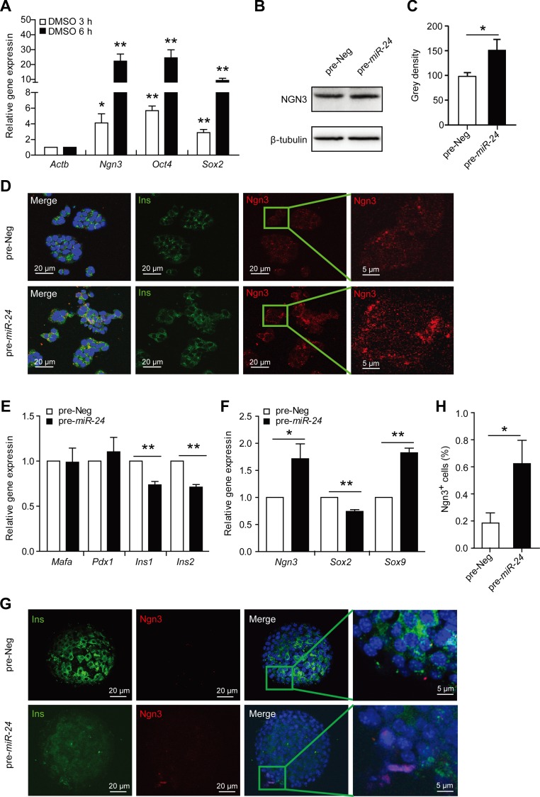
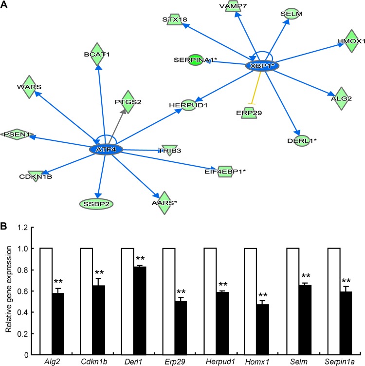
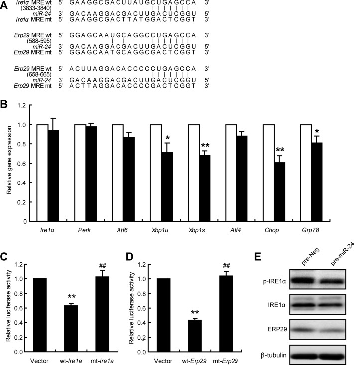
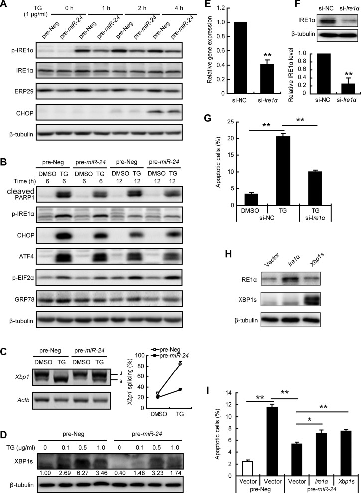
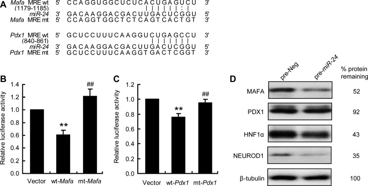
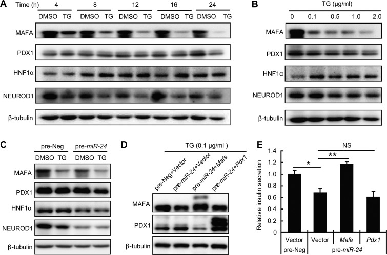
Current research indicates that beta cell loss in type 2 diabetes may be attributed to beta cell dedifferentiation rather than apoptosis; however, the mechanisms by which this occurs remain poorly understood. Our previous study demonstrated that elevation of microRNA-24 (miR-24) in a diabetic setting caused beta cell dysfunction and replicative deficiency. In this study, we focused on the role of miR-24 in beta cell apoptosis and dedifferentiation under endoplasmic reticulum (ER) stress conditions. We found that miR-24 overabundance protected beta cells from thapsigargin-induced apoptosis at the cost of accelerating the impairment of glucose-stimulated insulin secretion (GSIS) and enhancing the presence of dedifferentiation markers. Ingenuity® Pathway Analysis (IPA) revealed that elevation of miR-24 had an inhibitory effect on XBP1 and ATF4, which are downstream effectors of two key branches of ER stress, by inhibiting its direct target, Ire1α. Notably, elevated miR-24 initiated another pathway that targeted Mafa and decreased GSIS function in surviving beta cells, thus guiding their dedifferentiation under ER stress conditions. Our results demonstrated that the elevated miR-24, to the utmost extent, preserves beta cell mass by inhibiting apoptosis and inducing dedifferentiation. This study not only provides a novel mechanism by which miR-24 dominates beta cell turnover under persistent metabolic stress but also offers a therapeutic consideration for treating diabetes by inducing dedifferentiated beta cells to re-differentiation.

Keywords: ER stress; apoptosis; dedifferentiation; microRNA-24; pancreatic beta cells; type 2 diabetes.


Figures

Figure 1
Figure 1
miR-24 inhibits TG-induced apoptosis and GSIS dysfunction. (A) MIN6 cells were transfected with pre-Neg or pre-miR-24 for 48 h, treated with DMSO or TG (1 μg/ml) for 24 h, and then stained with Hoechst 33342. Scale bar, 40 μm. (B) Columns demonstrate the percentage of apoptotic cells in A. (C) Islets from 8-week-old C57BL6/J mice were transfected with pre-Neg or pre-miR-24 for 72 h and then treated with DMSO or TG (1 μg/ml) for further 24 h. Tunel combined with insulin and Hoechst 33342 staining was performed. Scale bar, 50 μm. (D) Columns show the percentage of apoptotic cells in C. (E) MIN6 cells were transfected with pre-Neg (clear bar) or pre-miR-24 (black bar) for 48 h, treated with DMSO or the indicated concentrations of TG (0.1, 0.5, or 1.0 μg/ml) for 12 h, and then the insulin content was detected. (F) The GSIS assay was performed after 48 h post-transfection and a further 12-h treatment with DMSO or 0.1 μg/ml TG. Insulin secretion in low glucose (clear bar) and high glucose (black bar) was measured. *P < 0.05 and **P < 0.01.
Figure 2
Figure 2
miR-24 increases the expressions of dedifferentiation markers. MIN6 cells were transfected with pre-Neg (control) or pre-miR-24 for 48 h, treated with DMSO for further 3 h or 6 h, and then expression levels of Ngn3, Oct4, and Sox2 were analyzed. (A) The mRNA levels of Ngn3, Oct4, and Sox2 were analyzed by qRT-PCR assay. β-actin was used as internal control. (B) The protein level of NGN3 was examined by western blot. β-tubulin was used as internal standard. (C) Grey density of B. (D) An immunofluorescence assay was used to detect protein levels and distribution of INS and NGN3. (E and F) Primary isolated islets were transfected with pre-Neg or pre-miR-24 for 72 h, and then mRNA levels of Mafa, Pdx1, Ins1, Ins2, Ngn3, Sox2, and Sox9 were detected by qRT-PCR. *P < 0.05 and **P < 0.01 vs. control. (G) Primary isolated islets were transfected with pre-Neg or pre-miR-24 for 96 h. An immunofluorescence assay was used to detect protein levels and distribution of INS and NGN3. (H) The percentage of NGN3+ cells in islets of G. *P < 0.05 vs. control.
Figure 3
Figure 3
miR-24 inhibits XBP1 and ATF4 signaling. (A) Expression levels of ER stress-associated genes were reanalyzed based on the previous mRNA microarray data by IPA. (B) MIN6 cells were transfected with pre-Neg (control, clean bar) or pre-miR-24 (black bar) for 48 h, and mRNA levels were analyzed by qRT-PCR assay. Data shown are mean ± SEMs and represent three separate experiments. β-actin was used as internal control. **P < 0.01 vs. control.
Figure 4
Figure 4
miR-24 regulates Ire1α and Erp29. (A) The 3′UTR sequences of Ire1α and Erp29 predicted to include miR-24 MREs were aligned with miR-24, and both wt and mt sequences are listed. (B) MIN6 cells were transfected with pre-Neg (control, clean bar) or pre-miR-24 (black bar) for 48 h, and mRNA levels were analyzed by qRT-PCR assay. β-actin was used as internal control. *P < 0.05 and **P < 0.01 vs. control. (C) MIN6 cells were co-transfected with pre-miR-24 and Vector, wt-Ire1α, or mt-Ire1α; after 48 h, dual luciferase reporter activities were measured. **P < 0.01 vs. Vector and ##P < 0.01 vs. wt-Ire1α. (D) MIN6 cells were co-transfected with pre-miR-24 and Vector, wt-Erp29, or mt-Erp29; after 48 h, dual luciferase reporter activities were measured. **P < 0.01 vs. Vector and ##P < 0.01 vs. wt-Erp29. (E) MIN6 cells were transfected with pre-Neg or pre-miR-24 for 48 h and protein levels were analyzed by western blot. β-tubulin was used as internal standard.
Figure 5
Figure 5
miR-24 inhibits IRE1α/XBP1s signaling. (AD) MIN6 cells were transfected with pre-Neg or pre-miR-24 for 48 h. (A and B) Transfected cells were further treated with or without 1 μg/ml TG for indicated time and protein levels were analyzed by western blot. β-tubulin was used as internal standard. (C) A semi-quantitative RT-PCR assay was performed after a 3-h treatment with or without 1 μg/ml TG, and the PCR products were separated with a 3% agarose gel. Xbp1 splicing efficiency was calculated as the ratio of the spliced Xbp1 level divided by the total Xbp1 level. (D) Transfected cells were treated with the indicated concentrations of TG for 3 h and protein level of XBP1s was determined. Relative XBP1s level was shown under the band. (EG) MIN6 cells were transfected with si-NC or si-Ire1α for 48 h. (E) mRNA level of Ire1α was analyzed by qRT-PCR assay. β-actin was used as internal control. **P < 0.01 vs. si-NC. (F) Protein level of IRE1α was analyzed by western blot. β-tubulin was used as internal standard. Grey density data are shown below. **P < 0.01 vs. si-NC. (G) Transfected cells were further treated with or without 1 μg/ml TG for 24 h, and apoptotic cells were calculated by Hoechst33342 staining. (H) MIN6 cells were transfected with Ire1α or Xbp1s for 48 h and protein levels were measured. (I) MIN6 cells were co-transfected with the indicated plasmid and miRNA for 48 h, treated with (black bar) or without (clean bar) 1 μg/ml TG for an additional 16 h, and then apoptotic cells were counted. *P < 0.05 and **P < 0.01.
Figure 6
Figure 6
miR-24 targets Mafa and Pdx1. (A) 3′UTR sequences of Mafa and Pdx1 predicted to include miR-24 MREs were aligned with miR-24, and both wt and mt sequences were listed. (B) MIN6 cells were co-transfected with pre-miR-24 and Vector, wt-Mafa, or mt-Mafa. After 48 h, dual luciferase reporter activities were measured. **P < 0.01 vs. Vector and ##P < 0.01 vs. wt-Mafa. (C) MIN6 cells were co-transfected with pre-miR-24 and Vector, wt-Pdx1, or mt-Pdx1. After 48 h, dual luciferase reporter activities were measured. **P < 0.01 vs. Vector and ##P < 0.01 vs. wt-Pdx1. (D) MIN6 cells were transfected with pre-Neg or pre-miR-24 for 48 h and protein levels were analyzed by western blot. β-tubulin was used as internal standard.
Figure 7
Figure 7
MAFA contributes to the miR-24-induced GSIS defect under ER stress condition. (A) MIN6 cells were treated with or without 0.1 μg/ml TG for the indicated times and protein levels were determined by western blot. (B) MIN6 cells were treated with the indicated concentrations of TG for 12 h, and protein levels were determined by western blot. (C) MIN6 cells were transfected with pre-Neg or pre-miR-24 for 48 h and further treated with or without 0.1 μg/ml TG for 12 h when protein levels were analyzed by western blot. (D and E) MIN6 cells were co-transfected with the indicated miRNA and plasmid for 48 h and further treated with 0.1 μg/ml TG for 12 h. (D) Protein levels were analyzed by western blot. β-tubulin was used as internal standard. (E) GSIS assay was performed and relative insulin secretion levels were normalized by the Vector and pre-Neg group. *P < 0.05 and **P < 0.01.

Similar articles

Cited by

References

    1. Allagnat F., Christulia F., Ortis F., et al. . (2010). Sustained production of spliced X-box binding protein 1 (XBP1) induces pancreatic β cell dysfunction and apoptosis. Diabetologia 53, 1120–1130. - PubMed
    1. Biden T.J., Boslem E., Chu K.Y., et al. . (2014). Lipotoxic endoplasmic reticulum stress, β cell failure, and type 2 diabetes mellitus. Trends Endocrinol. Metab. 25, 389–398. - PubMed
    1. Blum B., Roose A.N., Barrandon O., et al. . (2014). Reversal of β cell de-differentiation by a small molecule inhibitor of the TGFβ pathway. eLife 3, e02809. - PMC - PubMed
    1. Brown J.R., and Sanseau P. (2005). A computational view of microRNAs and their targets. Drug Discov. Today 10, 595–601. - PubMed
    1. Brozzi F., Gerlo S., Grieco F.A., et al. . (2014). A combined ‘omics’ approach identifies N-Myc interactor as a novel cytokine-induced regulator of IRE1 protein and c-Jun N-terminal kinase in pancreatic β cells. J. Biol. Chem. 289, 20677–20693. - PMC - PubMed

Publication types