. 2019 Oct;26(10):2046-2060.
doi: 10.1038/s41418-019-0298-5. Epub 2019 Feb 11.

Cell cycle arrest in mitosis promotes interferon-induced necroptosis

Affiliations

Cell cycle arrest in mitosis promotes interferon-induced necroptosis

Tanja Frank et al. Cell Death Differ. 2019 Oct.

Abstract

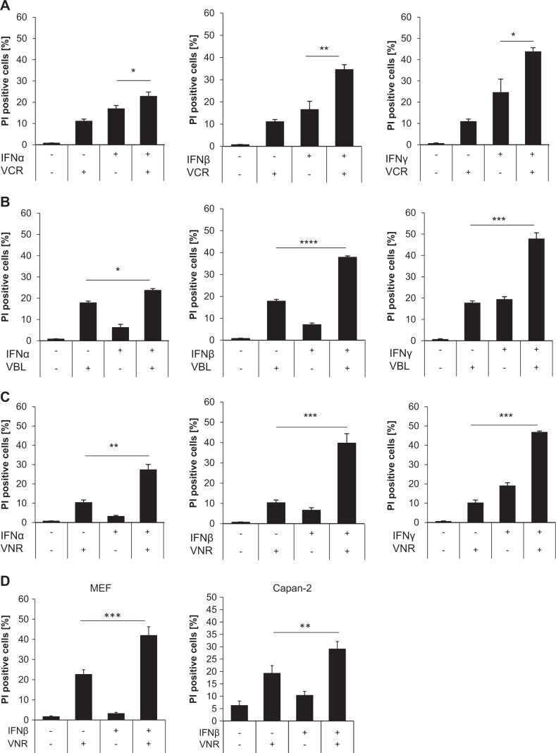
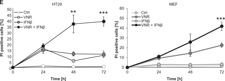
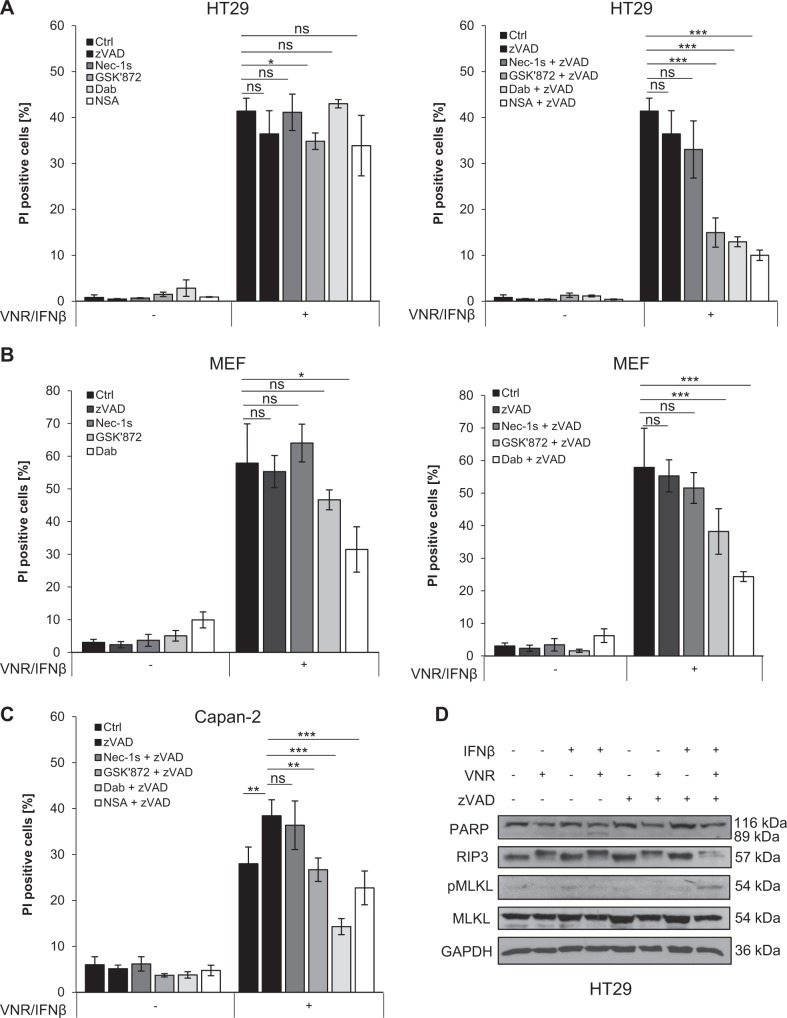
Resistance to apoptosis is a hallmark of cancer and deregulation of apoptosis often leads to chemoresistance. Therefore, new approaches to target apoptosis-resistant cancer cells are crucial for the development of directed cancer therapies. In the present study, we investigated the effect of cell cycle regulators on interferon (IFN)-induced necroptosis as an alternative cell death mechanism to overcome apoptosis resistance. Here, we report a novel combination treatment of IFNs with cell cycle arrest-inducing compounds that induce necroptosis in apoptosis-resistant cancer cells and elucidate the underlying molecular mechanisms. Combination treatment of IFNs (i.e. IFNβ) with inhibitors of the cell cycle (e.g. vinorelbine (VNR), nocodazole (Noc), polo-like kinase-1 (Plk-1) inhibitor BI 6727) co-operate to induce necroptotic cell death upon caspase inactivation. The mode of cell death was confirmed by pharmacological inhibition and siRNA-mediated downregulation of the key necroptotic factors receptor-interacting protein (RIP) kinase 3 (RIP3) and mixed-lineage kinase-like (MLKL) in various cell lines. Mechanistically, we show that necroptosis upon VNR/IFNβ/zVAD.fmk treatment is RIP1-independent but relies on IFNβ-induced gene expression of Z-DNA-binding protein 1 (ZBP1) as shown by quantitative RT-PCR and genetic knockdown experiments. Interestingly, we find that RIP3 is phosphorylated in response to compounds that trigger mitotic arrest, even in the absence of IFNβ signaling and necroptosis induction. Together, the identification of a novel combination treatment that triggers necroptosis has implications for the development of molecular-targeted therapies to circumvent apoptosis resistance and point to an underestimated role of cell cycle regulation in cell death signaling.


Conflict of interest statement

The authors declare that they have no conflict of interest.

Figures

Fig. 1
Fig. 1
Vinca alkaloids enhance IFN-induced cell death. a–c HT29 cells were treated with 10 ng/ml IFNα, IFNβ, or IFNγ and/or 100 nM VCR (a), VBL (b), or VNR (c) for 72 h. Cell death was determined by analysis of PI-stained nuclei. Mean and SD of three independent experiments performed in triplicate are shown; *P < 0.05, **P < 0.01, ***P < 0.001, ****P < 0.0001. d MEFs and Capan-2 cells were treated with 10 ng/ml IFNβ (Capan-2) or 4.5 ng/ml murine IFNβ (MEFs) and/or 100 nM VNR for 72 h (Capan-2) or 48 h (MEFs). Cell death was determined by analysis of PI-stained nuclei. Mean and SD of three independent experiments performed in triplicate are shown; **P < 0.01, ***P < 0.001. e HT29 cells and MEFs were treated with 10 ng/ml IFNβ (HT29) or 4.5 ng/ml murine IFNβ (MEFs) and/or 100 nM VNR for the indicated time points. Cell death was determined by analysis of PI-stained nuclei. Mean and SD of three independent experiments performed in triplicate are shown; **P < 0.01, ***P < 0.001
Fig. 1
Fig. 1
Vinca alkaloids enhance IFN-induced cell death. a–c HT29 cells were treated with 10 ng/ml IFNα, IFNβ, or IFNγ and/or 100 nM VCR (a), VBL (b), or VNR (c) for 72 h. Cell death was determined by analysis of PI-stained nuclei. Mean and SD of three independent experiments performed in triplicate are shown; *P < 0.05, **P < 0.01, ***P < 0.001, ****P < 0.0001. d MEFs and Capan-2 cells were treated with 10 ng/ml IFNβ (Capan-2) or 4.5 ng/ml murine IFNβ (MEFs) and/or 100 nM VNR for 72 h (Capan-2) or 48 h (MEFs). Cell death was determined by analysis of PI-stained nuclei. Mean and SD of three independent experiments performed in triplicate are shown; **P < 0.01, ***P < 0.001. e HT29 cells and MEFs were treated with 10 ng/ml IFNβ (HT29) or 4.5 ng/ml murine IFNβ (MEFs) and/or 100 nM VNR for the indicated time points. Cell death was determined by analysis of PI-stained nuclei. Mean and SD of three independent experiments performed in triplicate are shown; **P < 0.01, ***P < 0.001
Fig. 2
Fig. 2
Caspase inhibition induces a switch from apoptosis to necroptosis upon VNR and IFNβ co-treatment. a HT29 cells were treated with 10 ng/ml IFNβ, 100 nM VNR, and/or 20 µM zVAD.fmk in the presence or absence of 30 µM Nec-1s, 10 µM NSA, 20 µM GSK’872, or 20 µM Dab for 72 h. Cell death was determined by analysis of PI-stained nuclei. Mean and SD of three independent experiments performed in triplicate are shown; *P < 0.05, **P < 0.01, ***P < 0.001, ns = not significant. b MEFs were treated with 4.5 ng/ml murine IFNβ, 100 nM VNR, and/or 20 µM zVAD.fmk in the presence or absence of 30 µM Nec-1s, 10 µM GSK’872 or 10 µM Dab for 48 h. Cell death was determined by analysis of PI-stained nuclei. Mean and SD of three independent experiments performed in triplicate are shown; *P < 0.05, ***P < 0.001, ns = not significant. c Capan-2 cells were treated with 10 ng/ml IFNβ, 100 nM VNR, and/or 20 µM zVAD.fmk in the presence or absence of 30 µM Nec-1s, 10 µM NSA, 20 µM GSK’872, or 20 µM Dab for 72 h. Cell death was determined by analysis of PI-stained nuclei. Mean and SD of three independent experiments performed in triplicate are shown; **P < 0.01, ***P < 0.001. d HT29 cells were treated with 10 ng/ml IFNβ, 100 nM VNR, and/or 20 µM zVAD.fmk for 24 h. Protein expression of cleaved PARP, RIP3, pMLKL, MLKL, and GAPDH was analyzed by Western blotting. GAPDH served as loading control, ns = not significant.
Fig. 3
Fig. 3
Loss of RIP3 or MLKL inhibits VNR/IFNβ/zVAD.fmk-induced necroptosis. ad HT29 cells were transiently transfected with siRNA against RIP3 (a, b) or MLKL (c, d) or non-targeting control siRNA (siCtrl). Transfected cells were treated with 10 ng/ml IFNβ, 100 nM VNR, 20 µM zVAD.fmk, and/or 1 µM BV6 for 72 h and cell death was determined by analysis of PI-positive nuclei. Mean and SD of three independent experiments performed in triplicate are shown; *P < 0.05, **P < 0.01, ***P < 0.001 (a, c). Expression of RIP3 and MLKL was assessed by Western blotting, with GAPDH serving as loading control (b, d). eh Wt MEFs and MEFs deficient for RIP3 or MLKL were treated with 4.5 ng/ml murine IFNβ, 100 nM VNR, 20 µM zVAD.fmk, 10 ng/ml TNFα, and/or 5 µM BV6 for 72 h (IFNβ/VNR/zVAD.fmk) or 5 h (TBZ) and cell death was determined by analysis of PI-positive nuclei. Mean and SD of three independent experiments performed in triplicate are shown; *P < 0.05, ***P < 0.001 (e, g). Expression of RIP3 and MLKL was assessed by Western blotting, with GAPDH serving as loading control (f, h). i MEFs were treated with 4.5 ng/ml murine IFNβ, 100 nM VNR, and 20 µM zVAD.fmk for 72 h or with 10 ng/ml TNFα, 5 µM BV6, and 20 µM zVAD.fmk for 2 h. Lysates were fractionated on a Superose 6 3.2/300 GL column and the resulting fractions, as well as input samples, were analyzed by Western blotting (i). A schematic representation of the calibration, which is shown in detail in Supplementary Figure 4, is depicted above the Western blots
Fig. 3
Fig. 3
Loss of RIP3 or MLKL inhibits VNR/IFNβ/zVAD.fmk-induced necroptosis. ad HT29 cells were transiently transfected with siRNA against RIP3 (a, b) or MLKL (c, d) or non-targeting control siRNA (siCtrl). Transfected cells were treated with 10 ng/ml IFNβ, 100 nM VNR, 20 µM zVAD.fmk, and/or 1 µM BV6 for 72 h and cell death was determined by analysis of PI-positive nuclei. Mean and SD of three independent experiments performed in triplicate are shown; *P < 0.05, **P < 0.01, ***P < 0.001 (a, c). Expression of RIP3 and MLKL was assessed by Western blotting, with GAPDH serving as loading control (b, d). eh Wt MEFs and MEFs deficient for RIP3 or MLKL were treated with 4.5 ng/ml murine IFNβ, 100 nM VNR, 20 µM zVAD.fmk, 10 ng/ml TNFα, and/or 5 µM BV6 for 72 h (IFNβ/VNR/zVAD.fmk) or 5 h (TBZ) and cell death was determined by analysis of PI-positive nuclei. Mean and SD of three independent experiments performed in triplicate are shown; *P < 0.05, ***P < 0.001 (e, g). Expression of RIP3 and MLKL was assessed by Western blotting, with GAPDH serving as loading control (f, h). i MEFs were treated with 4.5 ng/ml murine IFNβ, 100 nM VNR, and 20 µM zVAD.fmk for 72 h or with 10 ng/ml TNFα, 5 µM BV6, and 20 µM zVAD.fmk for 2 h. Lysates were fractionated on a Superose 6 3.2/300 GL column and the resulting fractions, as well as input samples, were analyzed by Western blotting (i). A schematic representation of the calibration, which is shown in detail in Supplementary Figure 4, is depicted above the Western blots
Fig. 4
Fig. 4
RIP3 is phosphorylated during mitotic arrest upon vinca alkaloid treatment. a Fraction of cells per cell cycle phase was analyzed at 24 h after treatment with 100 nM VNR in PI-stained nuclei using FlowJow software (TreeStar Inc.). Mean and SD of three independent experiments performed in triplicate are shown. b Mitotic cells upon treatment with 100 nM VNR were quantified at 24 h by expression of mitotic marker pH3 using immunofluorescence. Mean and SD of three independent experiments performed in triplicate are shown; ****P < 0.0001. c HT29 cells were treated with 100 nM VCR, VBL, or VNR for 24 h. Protein expression of RIP3, pH3, H3, and GAPDH was analyzed by Western blotting. GAPDH served as loading control. d HT29 cells were treated with 10 ng/ml IFNβ, 100 nM VNR, and/or 20 µM zVAD for 24 h. 100 µg of each lysate was incubated with 400 U/µl λ-phosphatase for 30 min and 30 °C to remove phospho-groups. Protein expression of RIP3, pBubR1, BubR1, and β-Actin was analyzed by Western blotting. β-Actin served as loading control. e HT29 cells were treated with 10 ng/ml IFNβ, 100 nM VNR, 20 µM zVAD.fmk, and/or 20 µM GSK’872 for 24 h. Protein expression of RIP3, pMLKL, MLKL, pH3, H3, and GAPDH was analyzed by Western blotting. GAPDH served as loading control. f HT29 cells were treated with 10 ng/ml IFNβ, 100 nM VNR, 20 µM zVAD.fmk, and/or 30 µM Nec-1s for 24 h. Protein expression of RIP3 and GAPDH was analyzed by Western blotting. GAPDH served as loading control
Fig. 5
Fig. 5
Cell cycle arrest in mitosis induces RIP3 phosphorylation and enhances IFNβ-induced necroptosis. a HT29 cells were treated with 10 ng/ml IFNβ, 10 nM BI 6727, 1 µM Noc or 2 µM DME, and/or 20 µM zVAD.fmk for 24 h. Protein expression of RIP3, pMLKL, MLKL, and GAPDH was analyzed by Western blotting. GAPDH served as loading control. b HT29 cells were treated with 10 ng/ml IFNβ, 10 nM BI 6727, 1 µM Noc, and/or 2 µM DME for 72 h. Cell death was determined by analysis of PI-stained nuclei. Mean and SD of three independent experiments performed in triplicate are shown; *P < 0.05, **P < 0.01, ***P < 0.001. c HT29 cells were treated with 10 ng/ml IFNβ, 10 nM BI 6727, 1 µM Noc, and/or 2 µM DME in the presence or absence of 10 µM NSA, 20 µM GSK’872, or 20 µM Dab for 72 h. Cell death was determined by analysis of PI-stained nuclei. Mean and SD of three independent experiments performed in triplicate are shown; *P < 0.05, **P < 0.01, ***P < 0.001
Fig. 6
Fig. 6
ZBP1 upregulation after IFNβ treatment is involved in cell death induction after VNR/IFNβ/zVAD.fmk treatment. a HT29 cells were treated with 10 ng/ml IFNβ, 100 nM VNR, and/or 20 µM zVAD.fmk for 6 h. mRNA expression of MLKL, RIP1, RIP3, and ZBP1 was determined by qRT-PCR, normalized to 28S expression and is shown as x-fold mRNA expression compared with the control. Mean and SD of three independent experiments performed in triplicate are shown; *P < 0.05, **P < 0.01. b MEFs were treated with 4.5 ng/ml murine IFNβ for 24 h. Protein expression of ZBP1 was assessed by Western blotting, GAPDH served as loading control. c–g HT29 cells (c, d) or MEFs (eg) were transiently transfected with siRNA against ZBP1 or non-targeting control siRNA (siCtrl). Transfected cells were treated with 10 ng/ml IFNβ (HT29), 4.5 ng/ml murine IFNβ (MEFs), 100 nM VNR, and/or 20 µM zVAD.fmk for 72 h (HT29) or 48 h (MEFs) and cell death was determined by analysis of PI-positive nuclei. Mean and SD of three independent experiments performed in triplicate are shown; *P < 0.05, **P < 0.01. (c, e). Expression of ZBP1 was assessed by qRT-PCR, normalized to 28S (d; HT29) or murine GAPDH (f; MEFs) expression and is shown as x-fold mRNA expression compared with the control. Mean and SD of three independent experiments performed in triplicate are shown; **P < 0.01, ***P < 0.001. Protein expression of ZBP1 in MEFs was additionally shown by Western blotting, with GAPDH serving as loading control (g)

Similar articles

Cited by

References

    1. Hanahan D, Weinberg RA. Hallmarks of cancer: the next generation. Cell. 2011;144:646–74. doi: 10.1016/j.cell.2011.02.013. - DOI - PubMed
    1. Fulda S. Tumor resistance to apoptosis. Int J Cancer. 2009;124:511–5. doi: 10.1002/ijc.24064. - DOI - PubMed
    1. Vanden Berghe T, Linkermann A, Jouan-Lanhouet S, Walczak H, Vandenabeele P. Regulated necrosis: the expanding network of non-apoptotic cell death pathways. Nat Rev Mol Cell Biol. 2014;15:135–47. doi: 10.1038/nrm3737. - DOI - PubMed
    1. Pasparakis M, Vandenabeele P. Necroptosis and its role in inflammation. Nature. 2015;517:311–20. doi: 10.1038/nature14191. - DOI - PubMed
    1. Vanden Berghe T, Hassannia B, Vandenabeele P. An outline of necrosome triggers. Cell Mol Life Sci. 2016;73:2137–52. doi: 10.1007/s00018-016-2189-y. - DOI - PMC - PubMed