. 2019 Jul;15(7):1163-1181.
doi: 10.1080/15548627.2019.1580089. Epub 2019 Feb 20.

Influenza M2 protein regulates MAVS-mediated signaling pathway through interacting with MAVS and increasing ROS production

Affiliations

Influenza M2 protein regulates MAVS-mediated signaling pathway through interacting with MAVS and increasing ROS production

Ruifang Wang et al. Autophagy. 2019 Jul.

Abstract

Influenza A virus can evade host innate immune response that is involved in several viral proteins with complicated mechanisms. To date, how influenza A M2 protein modulates the host innate immunity remains unclear. Herein, we showed that M2 protein colocalized and interacted with MAVS (mitochondrial antiviral signaling protein) on mitochondria, and positively regulated MAVS-mediated innate immunity. Further studies revealed that M2 induced reactive oxygen species (ROS) production that was required for activation of macroautophagy/autophagy and enhancement of MAVS signaling pathway. Importantly, the proton channel activity of M2 protein was demonstrated to be essential for ROS production and antagonizing the autophagy pathway to control MAVS aggregation, thereby enhancing MAVS signal activity. In conclusion, our studies provided novel insights into mechanisms of M2 protein in modulating host antiviral immunity and uncovered a new mechanism into biology and pathogenicity of influenza A virus. Abbreviations: AKT/PKB: AKT serine/threonine kinase; Apo: apocynin; ATG5: autophagy related 5; BAPTA-AM: 1,2-Bis(2-aminophenoxy) ethane-N,N,N',N'-tetraacetic acid tetrakis; BECN1: beclin 1; CARD: caspase recruitment domain; CCCP: carbonyl cyanide m-chlorophenylhydrazone; CQ: chloroquine; DCF: dichlorodihyd-rofluorescein; DPI: diphenyleneiodonium; DDX58: DExD/H-box helicase 58; eGFP: enhanced green fluorescent protein; EGTA: ethylene glycol-bis(2-aminoethylether)-N,N,N',N'-tetraacetic acid; ER: endoplasmic reticulum; hpi: hours post infection; IAV: influenza A virus; IFN: interferon; IP: immunoprecipitation; IRF3: interferon regulatory factor 3; ISRE: IFN-stimulated response elements; LIR: LC3-interacting region; MAP1LC3B/LC3B: microtubule associated protein 1 light chain 3 beta; MAVS: mitochondrial antiviral signaling protein; MMP: mitochondrial membrane potential; MOI, multiplicity of infection; mRFP: monomeric red fluorescent protein; MTOR: mechanistic target of rapamycin kinase; NC: negative control; NFKB/NF-κB: nuclear factor kappa B; PI3K: class I phosphoinositide 3-kinase; RLR: RIG-I-like-receptor; ROS: reactive oxygen species; SEV: sendai virus; TM: transmembrane; TMRM: tetramethylrhodamine methylester; VSV: vesicular stomatitis virus.

Keywords: Autophagy; MAVS aggregates; influenza M2 protein; innate immunity; ion channel activity.


Figures
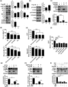
Figure 1.
Figure 1.
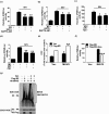
Influenza M2 protein induces autophagy through ATG5 and PI3K-AKT-MTOR pathway and cellular responses. (a and b) HEK 293T cells were transfected with indicated plasmids for 24 h, cells lysates were analyzed by western blot. * represents the indicated protein. (c) HEK 293T cells were pretreated with 10 μM LY294002 for 6 h, and then transfected with Flag-M2 for another 24 h. Cells lysates were evaluated by western blot. (d) HEK 293T cells were transfected with indicated plasmids for 12 h and treated with 5 μM amantadine. The Fluo-4 AM (Up) and Rhod-2 AM (Down) fluorescence was tested by BD FACSCalibur system after 12 h treatment. (e) HEK 293T cells were treated as in (d). Mean DCF (Up) and MitoSOX (Down) fluorescence was determined via flow cytometry. (f) HEK 293T cells were transfected with Flag-M2 and treated with 0.4 mM EGTA or 16 μM BAPTA-AM, respectively. DCF fluorescence was analyzed as (e). (g-i) Flag-M2-transfected HEK 293T cells were treated with 0.4 mM EGTA (g), or 16 μM BAPTA-AM (h), or 3 μM DPI (i), or 0.1 mM Apo (i) for 24 h. Cell lysates were analyzed by western blot. Error bars, mean ± SD of 3 experiments (*p < 0.05; **p < 0.01; ***p < 0.001).
Figure 2.
Figure 2.
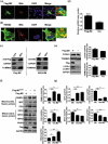
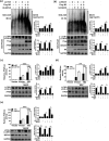
Influenza virus M2 anchors to mitochondria and disrupts the mitochondrial dynamics. (a) A549 cells were co-transfected with indicated plasmids for 24 h and analyzed for the colocalization of M2 and dsRed-mito. Scale bar: 10 μm. It was the representative of 20 cells. (b) A549 cells were transfected with dsRed-mito, and then infected with the H5N1/HM virus for 24 h and analyzed for the colocalization of M2 with dsRed-mito. Scale bar: 10 μm. It was the representative of 20 cells. (c) HEK 293T cells were transfected with Flag-M2 (left) or A549 cells were infected with the H5N1/HM virus (Right), respectively. Subcellular fractions were purified for western blot analysis (Fractions: Cyto, purified cytosolic; Mito, purified mitochondria. Organelle markers: TOMM20, mitochondria; GAPDH, cytoplasm). (d) HEK 293T cells were transfected with Flag-M2 for 24 h. Mitochondrial DNA copy number was measured by quantitative PCR and normalized to nuclear DNA levels in a ratio of mtDNA MT-COI over RNA18S rDNA. Relative mitochondrial DNA copy numbers were depicted. (e) HEK 293T cells were transfected with Flag-M2 for 24 h and analyzed by western blot. (f and g) HEK 293T cells were transfected with Flag-M2 and Flag-M2H37G, respectively. Cell lysates were evaluated by western blot. Error bars, mean ± SD of 3 independent experiments (*p < 0.05; **p < 0.01; ***p < 0.001).
Figure 3.
Figure 3.
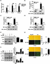
M2 protein potentiates innate immune response. (a) HEK 293T cells were transfected with Flag-M2 for 24 h and infected with SEV. The levels of IFNB, CXCL8 and CCL5 mRNAs were measured by RT-PCR at 12 hpi. (b-d) HEK 293T cells were transfected with indicated plasmids for 24 h, and then infected with SEV. Luciferase assays were performed at 12 hpi. Expression of M2 protein was confirmed by western blot. (e and f) HEK 293T cells (e) or A549 cells (f) were transfected with Flag-M2, then cells were further infected with SEV at 24 h post transfection. After 12 hpi, cells were harvested and analyzed by western blot. (g) HEK 293T cells were transfected with Flag-M2 for 24 h, and then were infected with VSV-eGFP (MOI = 0.1) for 12 h or 24 h. Replication of VSV was analyzed by microscopy and flow cytometry (BF, bright field). The presented image is a representative result from 3 independent experiments. Error bars, mean ± SD of 3 independent experiments (*p < 0.05; **p < 0.01; ***p < 0.001).
Figure 4.
Figure 4.
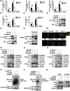
MAVS is the potential target of M2 protein. (a) HEK 293T cells were transfected with indicated plasmids. RNA was prepared for IFNB, CXCL8 and CCL5 detection by RT-PCR. (b and c) HEK 293T cells were transfected with indicated plasmids. Luciferase assays were performed at 24 h post transfection. (d) HEK 293T cells were transfected with plasmids encoding Flag-M2 and HA-MAVS. Cell lysates were subjected to IP. (e) HEK 293T cells were transfected with HA-M2 and Flag-MAVS and cell lysates were subjected to the IP. (f) Lysates of H5N1/HM-infected A549 cells were prepared, and immunoprecipitated with the anti-MAVS antibody or control IgG. (g) HeLa cells were transfected with Flag-M2 and HA-MAVS for 24 h, and then analyzed for the colocalization between M2 and MAVS. (h) HEK 293T cells were transfected with Flag-ATG5 and HA-MAVS or control vector together with Flag-M2. Cell lysates were subjected to IP. (i and j) HEK 293T cells were transfected with indicated plasmids. Cell lysates were immunoprecipitated with either anti-HA (i) or anti-GFP (j) antibody. (k and l) HEK 293T cells were transfected with indicated constructs for 24 h, cells lysates were analyzed by SDD-AGE and SDS-PAGE assays. (m) HEK 293T cells were transfected with indicated plasmids. Cell lysates were subjected to the IP. Error bars, mean ± SD of 3 independent experiments (*p < 0.05; **p < 0.01; ***p < 0.001).
Figure 5.
Figure 5.
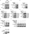
M2 His37 and the integrity of MAVS are required for the interaction between M2 and MAVS. (a) HEK 293T cells were transfected with Flag-MAVS alone or cotransfected with HA-M2-mutants (F91S, I94S and ΔLIR). Cell lysates were subjected to IP and further analyzed by western blot. (b) HEK 293T cells were transfected with Flag-M2 or Flag-M2H37G alone or along with HA-MAVS, respectively. Cell lysates were immunoprecipitated with anti-Flag and analyzed by western blot. (c) HEK 293T cells were transfected with HA-MAVS or both HA-MAVS and Flag-M2H37G. Cell lysates were immunoprecipitated with anti-HA antibody and then analyzed by western blot. (d-f) HEK 293T cells were transfected with indicated plasmids for 24 h, cell lysates were subjected to IP with respective antibodies. (g-i) HEK 293T cells were cotransfected with each HA-MAVS-mutant (Y9A, I12A, ΔLIR, 1N, 1C, ΔTM and ΔCARD) with Flag-M2. Cell lysates were immunoprecipitated with anti-Flag antibody (g) or anti-HA antibody (h and i), and then analyzed by western blot.
Figure 6.
Figure 6.
M2 protein enhances MAVS signaling through increasing ROS production. (a-d) HEK 293T cells were transfected with indicated plasmids. Cells were then pretreated with EGTA (a and b), BAPTA-AM (a and b), DPI (c and d), Apo (c and d) prior to mock-infection or infection with SEV, respectively. Luciferase assays were performed at 12 hpi. (e) HEK 293T cells were transfected with indicated plasmids for 24 h, FACS analysis were performed to determine cellular ROS production. (f) HEK 293T cells were pretreated with Apo for 4 h, and transfected with indicated plasmids. Cells were lysed to measure the IFNB induction at 24 h post transfection. (g) HEK 293T cells were pretreated with Apo for 4 h, and transfected with indicated plasmids for 24 h, cells lysates were analyzed by either SDD-AGE or SDS-PAGE assays. Error bars, mean ± SD of 3 independent experiments (*p < 0.05; **p < 0.01; ***p < 0.001).
Figure 7.
Figure 7.
M2 antagonizes the autophagy process to enhance the innate immune response. (a) After transfection with siATG5 for 6 h, the HEK 293T cells were further transfected with indicated plasmids for 24 h. The cells lysates were collected and analyzed by either SDD-AGE or SDS-PAGE assays. (b) HEK 293T cells were transfected with siBECN1 for 6 h, cells were further transfected with indicated plasmids for 24 h, and cells lysates were analyzed by either SDD-AGE or SDS-PAGE assays. (c) HEK 293T cells were transfected with siATG5 for 6 h, the cells were further transfected with indicated plasmids for 24 h, and then luciferase assays were performed. (d) HEK 293T cells were transfected with siATG5 for 6 h, the cells were further transfected with indicated plasmids for 24 h, and then mock-infected or infected with SEV for 12 h. The luciferase assays were performed. (e) HEK 293T cells were transfected with siBECN1 for 6 h, cells were further transfected with indicated plasmids. At 24 h post transfection, luciferase assays were performed. Error bars, mean ± SD of 3 independent experiments (*p < 0.05; **p < 0.01; ***p < 0.001).
Figure 8.
Figure 8.
Schematic diagram of proposed mechanisms underlying the regulation of innate immune response by influenza virus H5N1/HM M2 protein. Proton channel activity of M2 protein was essential for the increase of Ca2+ entry into cytoplasm from extracellular with a subsequent elevation in ROS production. On one hand, the increase level of ROS directly or indirectly led to the activation of ATG5 and inhibition of AKT and MTOR activity through the PI3K-AKT-MTOR pathway, thereby triggering M2-induced autophagy process. One the other hand, M2 protein anchored to the mitochondria, increased the mitochondrial number and accelerated the mitochondria fusion, which resulted in increasing ROS-dependent MAVS aggregations. The MAVS aggregates can be packed into the autophagosome and be degraded by the lysosomal enzyme in the process of autolysosome formation. LC3B and ATG5 interact with MAVS in this process, thereby attenuating MAVS-mediated antiviral signaling pathway. However, M2 physically interacted with MAVS and sequestered the MAVS-ATG5 and MAVS-LC3B complexes formation, thus leading to the release of MAVS aggregates from the ATG5-MAVS and LC3B-MAVS complexes. Furthermore, M2 protein inhibited the autolysosome formation, which suppressed the MAVS aggregates degradation. The released MAVS aggregates from ATG5-MAVS and LC3B-MAVS complexes in turn participated in the MAVS-mediated innate immune response, which resulted in subsequent amplification of IFN and inflammatory cytokine signaling by RLR signaling.

Similar articles

Cited by

References

    1. Chen C, Chi X, Bai Q, et al. Mechanisms underlying interferon-mediated host innate immunity during influenza A virus infection. Sheng Wu Gong Cheng Xue Bao. 2015;31(12):1671. - PubMed
    1. To J, Surya W, Torres J.. Targeting the channel activity of viroporins. Adv Protein Chem Struct. 2016;104:307–355. PubMed PMID: WOS:000381638000009; English. - PMC - PubMed
    1. Mould JA, Li H-C, Dudlak CS, et al. Mechanism for proton conduction of the M2 ion channel of influenza A virus. J Biol Chem. 2000. March 24;275(12):8592–8599. PubMed PMID: BCI:BCI200000349462. - PubMed
    1. Chen BJ, Leser GP, Jackson D, et al. The influenza virus M2 protein cytoplasmic tail interacts with the M1 protein and influences virus assembly at the site of virus budding. J Virol. 2008. October;82(20):10059–10070. PubMed PMID: WOS:000260109100023. - PMC - PubMed
    1. Pinto LH, Lamb RA.. Controlling influenza virus replication by inhibiting its proton channel. Mol Biosyst. 2007. January;3(1):18–23. PubMed PMID: WOS:000243429900013; English. - PubMed

Publication types

MeSH terms

Grants and funding

This work was supported by National Key Research and Development Program (2016YFD0500205 & 2016YFC1200201), Fundamental Research Funds for the Central Universities (2662017PY029), National Natural Science Foundation of China (NO. 31761133005 & 31572545 & 31772752), the Outstanding Youth Science Foundation of Hubei Province (2016CFA056).