Fast shading correction for cone-beam CT via partitioned tissue classification
- PMID: 30721886
- PMCID: PMC6571138
- DOI: 10.1088/1361-6560/ab0475
Fast shading correction for cone-beam CT via partitioned tissue classification
Abstract
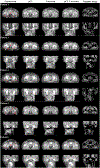
The quantitative use of cone beam computed tomography (CBCT) in radiation therapy is limited by severe shading artifacts, even with system embedded correction. We recently proposed effective shading correction methods, using planning CT (pCT) as prior information to estimate low-frequency errors in either the projection domain or image domain. In this work, we further improve the clinical practicality of our previous methods by removing the requirement of prior pCT images. Clinical CBCT images are typically composed of a limited number of tissues. By utilizing the low frequency characteristic of shading distribution, we first generate a 'shading-free' template image by enforcing uniformity on CBCT voxels of the same tissue type via a technique named partitioned tissue classification. Only a small subset of voxels in the template image are used in the correction process to generate sparse samples of shading artifacts. Local filtration, a Fourier transform based algorithm, is employed to efficiently process the sparse errors to compute a full-field distribution of shading artifacts for CBCT correction. We evaluate the method's performance using an anthropomorphic pelvis phantom and 6 pelvis patients. The proposed method improves the image quality of CBCT for both phantom and patients to a level matching that of pCT. On the pelvis phantom, the signal non-uniformity (SNU) is reduced from 12.11% to 3.11% and 8.40% to 2.21% on fat and muscle, respectively. The maximum CT number error is reduced from 70 to 10 HU and 73 to 11 HU on fat and muscle, respectively. On patients, the average SNU is reduced from 9.22% to 1.06% and 11.41% to 1.67% on fat and muscle, respectively. The maximum CT number error is reduced from 95 to 9 HU and 88 to 8 HU on fat and muscle, respectively. The typical processing time for one CBCT dataset is about 45 s on a standard PC.
Figures


Similar articles
-
Fast shading correction for cone beam CT in radiation therapy via sparse sampling on planning CT.Med Phys. 2017 May;44(5):1796-1808. doi: 10.1002/mp.12190. Epub 2017 Apr 17. Med Phys. 2017. PMID: 28261827
-
Shading correction for on-board cone-beam CT in radiation therapy using planning MDCT images.Med Phys. 2010 Oct;37(10):5395-406. doi: 10.1118/1.3483260. Med Phys. 2010. PMID: 21089775
-
Image-domain shading correction for cone-beam CT without prior patient information.J Appl Clin Med Phys. 2015 Nov 8;16(6):65-75. doi: 10.1120/jacmp.v16i6.5424. J Appl Clin Med Phys. 2015. PMID: 26699555 Free PMC article.
-
Shading correction assisted iterative cone-beam CT reconstruction.Phys Med Biol. 2017 Oct 27;62(22):8495-8520. doi: 10.1088/1361-6560/aa8e62. Phys Med Biol. 2017. PMID: 29077573
-
Planning CT-guided robust and fast cone-beam CT scatter correction using a local filtration technique.Med Phys. 2021 Nov;48(11):6832-6843. doi: 10.1002/mp.15299. Epub 2021 Oct 26. Med Phys. 2021. PMID: 34662433
Cited by
-
Single-Shot Quantitative X-ray Imaging Using a Primary Modulator and Dual-Layer Detector: Simulation and Phantom Studies.Proc SPIE Int Soc Opt Eng. 2022 Feb-Mar;12031:1203106. doi: 10.1117/12.2611591. Epub 2022 Apr 4. Proc SPIE Int Soc Opt Eng. 2022. PMID: 36560977 Free PMC article.
-
Daily dose evaluation based on corrected CBCTs for breast cancer patients: accuracy of dose and complication risk assessment.Radiat Oncol. 2022 Dec 12;17(1):205. doi: 10.1186/s13014-022-02174-4. Radiat Oncol. 2022. PMID: 36510254 Free PMC article.
-
Adaptive proton therapy.Phys Med Biol. 2021 Nov 15;66(22):10.1088/1361-6560/ac344f. doi: 10.1088/1361-6560/ac344f. Phys Med Biol. 2021. PMID: 34710858 Free PMC article. Review.
-
Obtaining dual-energy computed tomography (CT) information from a single-energy CT image for quantitative imaging analysis of living subjects by using deep learning.Pac Symp Biocomput. 2020;25:139-148. Pac Symp Biocomput. 2020. PMID: 31797593 Free PMC article.
-
Toward quantitative short-scan cone beam CT using shift-invariant filtered-backprojection with equal weighting and image domain shading correction.Proc SPIE Int Soc Opt Eng. 2019 Jun;11072:110721X. doi: 10.1117/12.2534900. Epub 2019 May 28. Proc SPIE Int Soc Opt Eng. 2019. PMID: 34248247 Free PMC article.
References
-
- Arai K et al. 2017. Feasibility of CBCT-based proton dose calculation using a histogram-matching algorithm in proton beam therapy Phys. Med 33 68–76 - PubMed
-
- Chen WJ, Giger ML and Bick U 2006. A fuzzy c-means (FCM)-based approach for computerized segmentation of breast lesions in dynamic contrast-enhanced MR images Acad. Radiol 13 63–72 - PubMed
-
- Colijn AP and Beekman FJ 2004. Accelerated simulation of cone beam x-ray scatter projections IEEE Trans. Med. Imaging 23 584–90 - PubMed
-
- Gao H, Zhu L and Fahrig R 2017. Virtual scatter modulation for x-ray CT scatter correction using primary modulator J. X-Ray Sci. Technol 25 869–85 - PubMed
Publication types
MeSH terms
Grants and funding
LinkOut - more resources
Full Text Sources
