

Coxsackievirus B infection induces the extracellular release of miR-590-5p, a proviral microRNA
- PMID: 30711774
- PMCID: PMC6382511
- DOI: 10.1016/j.virol.2019.01.025
Coxsackievirus B infection induces the extracellular release of miR-590-5p, a proviral microRNA
Abstract
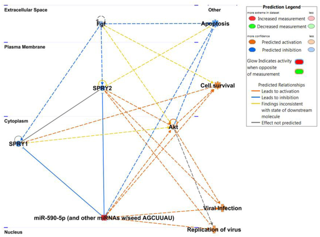
Coxsackievirus B is a significant human pathogen and is a leading cause of myocarditis. We and others have observed that certain enteroviruses including coxsackievirus B cause infected cells to shed extracellular vesicles containing infectious virus. Recent reports have shown that vesicle-bound virus can infect more efficiently than free virus. Though microRNAs are differentially regulated in cells following infection, few have been associated with the vesicles shed from infected cells. Here we report exclusive trafficking of specific microRNAs into viral vesicles compared to vesicles from non-infected cells. We found that the most highly-expressed unique microRNA in viral vesicles was miR-590-5p, which facilitates prolonged viral replication by blocking apoptotic factors. Cells over-expressing this miR were significantly more susceptible to infection. This may be a mechanism by which coxsackievirus B boosts subsequent rounds of infection by co-packaging virus and a select set of pro-viral microRNAs in extracellular vesicles.
Keywords: Apoptosis; Coxsackievirus; MicroRNA; Vesicles.
Copyright © 2019 The Authors. Published by Elsevier Inc. All rights reserved.
Figures


Similar articles
-
TBK1 and GABARAP family members suppress Coxsackievirus B infection by limiting viral production and promoting autophagic degradation of viral extracellular vesicles.PLoS Pathog. 2022 Aug 31;18(8):e1010350. doi: 10.1371/journal.ppat.1010350. eCollection 2022 Aug. PLoS Pathog. 2022. PMID: 36044516 Free PMC article.
-
[The suppressive effect of MiR-490 on coxsackievirus B3 replication].Bing Du Xue Bao. 2014 Nov;30(6):619-23. Bing Du Xue Bao. 2014. PMID: 25868275 Chinese.
-
Protein kinase B/Akt regulates coxsackievirus B3 replication through a mechanism which is not caspase dependent.J Virol. 2004 Apr;78(8):4289-98. doi: 10.1128/jvi.78.8.4289-4298.2004. J Virol. 2004. PMID: 15047842 Free PMC article.
-
Coxsackievirus B transmission and possible new roles for extracellular vesicles.Biochem Soc Trans. 2013 Feb 1;41(1):299-302. doi: 10.1042/BST20120272. Biochem Soc Trans. 2013. PMID: 23356301 Review.
-
CVB infection and mechanisms of viral cardiomyopathy.Curr Top Microbiol Immunol. 2008;323:315-35. doi: 10.1007/978-3-540-75546-3_15. Curr Top Microbiol Immunol. 2008. PMID: 18357777 Review.
Cited by
-
Coxsackievirus B3 infects and disrupts human induced-pluripotent stem cell derived brain-like endothelial cells.Front Cell Infect Microbiol. 2023 Apr 17;13:1171275. doi: 10.3389/fcimb.2023.1171275. eCollection 2023. Front Cell Infect Microbiol. 2023. PMID: 37139492 Free PMC article.
-
Dysregulated CD4+ T Cells and microRNAs in Myocarditis.Front Immunol. 2020 Mar 25;11:539. doi: 10.3389/fimmu.2020.00539. eCollection 2020. Front Immunol. 2020. PMID: 32269577 Free PMC article. Review.
-
The Expression Levels of MicroRNAs Differentially Expressed in Sudden Sensorineural Hearing Loss Patients' Serum Are Unchanged for up to 12 Months after Hearing Loss Onset.Int J Mol Sci. 2023 Apr 15;24(8):7307. doi: 10.3390/ijms24087307. Int J Mol Sci. 2023. PMID: 37108470 Free PMC article.
-
The Role of MicroRNA in the Pathophysiology and Diagnosis of Viral Myocarditis.Int J Mol Sci. 2024 Oct 11;25(20):10933. doi: 10.3390/ijms252010933. Int J Mol Sci. 2024. PMID: 39456716 Free PMC article. Review.
-
TBK1 and GABARAP family members suppress Coxsackievirus B infection by limiting viral production and promoting autophagic degradation of viral extracellular vesicles.PLoS Pathog. 2022 Aug 31;18(8):e1010350. doi: 10.1371/journal.ppat.1010350. eCollection 2022 Aug. PLoS Pathog. 2022. PMID: 36044516 Free PMC article.
References
-
- Liu PP, Mason JW. Advances in the understanding of myocarditis. Circulation. 2001;104(9):1076–82. - PubMed
-
- Pons F, Lupon J, Urrutia A, Gonzalez B, Crespo E, Diez C, et al. Mortality and cause of death in patients with heart failure: findings at a specialist multidisciplinary heart failure unit. Rev Esp Cardiol. 2010;63(3):303–14. - PubMed
Publication types
MeSH terms
Substances
Grants and funding
LinkOut - more resources
Full Text Sources
