

Review
doi: 10.1016/j.ceb.2018.12.006.
Epub 2019 Jan 29.
Spatial encoding of GPCR signaling in the nervous system
Affiliations
- PMID: 30708280
- PMCID: PMC6462236
- DOI: 10.1016/j.ceb.2018.12.006
Item in Clipboard
Review
Spatial encoding of GPCR signaling in the nervous system
Curr Opin Cell Biol.
2019 Apr.
Abstract
Several GPCRs, including receptors previously thought to signal primarily from the cell surface, have been recently shown to signal from many intracellular compartments. This raises the idea that signaling by any given receptor is spatially encoded in the cell, with distinct sites of signal origin dictating distinct downstream consequences. We will discuss recent developments that address this novel facet of GPCR physiology, focusing on the spatial segregation of signaling from the cell surface, endosomes, and the Golgi by receptors relevant to the nervous system.
Copyright © 2019 Elsevier Ltd. All rights reserved.
Conflict of interest statement
Disclosure:
The authors declare no conflicts of interest.
Figures
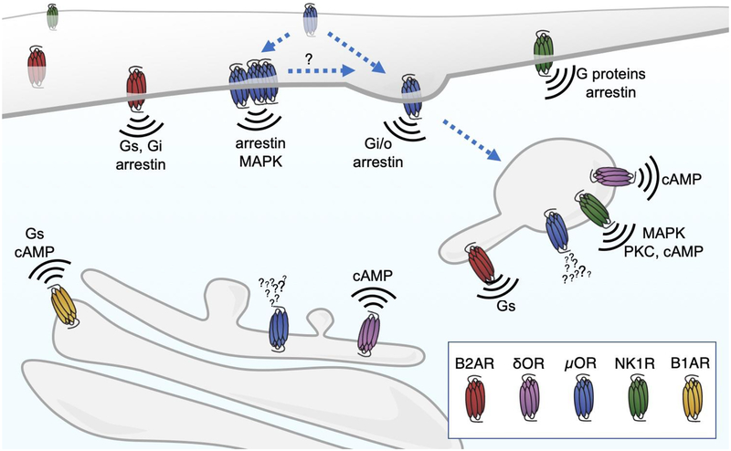
At the plasma membrane GPCRs can signal through both G proteins and arrestins. Receptors like NK1R and B2AR can couple to different G proteins depending on the circumstances, although how this selectivity is regulated is not well understood. Most GPCRs change their diffusion, clustering, and localization to endocytic domains in response to agonist. The rates of these changes are different between different agonists, which could in turn dictate effector coupling and signaling as has been described for μOR. Many GPCRs also signal from endosomes once internalized. Some receptors like NK1R could couple directly to different effectors in endosomes compared to the plasma membrane to cause different physiological effects. B2AR couples to Gs in both locations but cause divergent transcriptional outputs depending on where it is activated. GPCR signaling from the Golgi has recently been described for B1AR and δOR, two GPCRs with steady-state Golgi localization. The physiological consequences and in vivo relevance of receptors signaling at the Golgi are still being explored. Exciting future work on the mechanisms and signaling of receptors at different sites in their trafficking itinerary may allow us to develop therapeutic strategies that spatially restrict receptor activation in subcellular locations and therefore fine-tune the consequences of receptor signaling.
Similar articles
-
Compartmentalized GPCR Signaling from Intracellular Membranes.J Membr Biol. 2021 Jun;254(3):259-271. doi: 10.1007/s00232-020-00158-7. Epub 2020 Nov 24. J Membr Biol. 2021. PMID: 33231722 Free PMC article. Review.
-
Spatial encryption of G protein-coupled receptor signaling in endosomes; Mechanisms and applications.Biochem Pharmacol. 2017 Nov 1;143:1-9. doi: 10.1016/j.bcp.2017.04.028. Epub 2017 Apr 27. Biochem Pharmacol. 2017. PMID: 28456515 Review.
-
Advances in Membrane Trafficking and Endosomal Signaling of G Protein-Coupled Receptors.Int Rev Cell Mol Biol. 2018;339:93-131. doi: 10.1016/bs.ircmb.2018.03.001. Epub 2018 Apr 30. Int Rev Cell Mol Biol. 2018. PMID: 29776606 Review.
-
Hardwiring wire-less networks: spatially encoded GPCR signaling in endocrine systems.Curr Opin Cell Biol. 2019 Apr;57:77-82. doi: 10.1016/j.ceb.2018.12.009. Epub 2019 Jan 23. Curr Opin Cell Biol. 2019. PMID: 30682696 Review.
-
Internalized TSH receptors en route to the TGN induce local Gs-protein signaling and gene transcription.Nat Commun. 2017 Sep 5;8(1):443. doi: 10.1038/s41467-017-00357-2. Nat Commun. 2017. PMID: 28874659 Free PMC article.
Cited by
-
Specific motifs mediate post-synaptic and surface transport of G protein-coupled receptors.iScience. 2021 Dec 18;25(1):103643. doi: 10.1016/j.isci.2021.103643. eCollection 2022 Jan 21. iScience. 2021. PMID: 35024582 Free PMC article.
-
Opioid Research: Past and Future.Mol Pharmacol. 2020 Oct;98(4):389-391. doi: 10.1124/molpharm.120.000093. Epub 2020 Jul 13. Mol Pharmacol. 2020. PMID: 32660966 Free PMC article.
-
Rab43 GTPase directs postsynaptic trafficking and neuron-specific sorting of G protein-coupled receptors.J Biol Chem. 2021 Jan-Jun;296:100517. doi: 10.1016/j.jbc.2021.100517. Epub 2021 Mar 4. J Biol Chem. 2021. PMID: 33676895 Free PMC article.
-
Heterotrimeric G protein signaling without GPCRs: The Gα-binding-and-activating (GBA) motif.J Biol Chem. 2024 Mar;300(3):105756. doi: 10.1016/j.jbc.2024.105756. Epub 2024 Feb 15. J Biol Chem. 2024. PMID: 38364891 Free PMC article. Review.
-
The Kinase Specificity of Protein Kinase Inhibitor Peptide.Front Pharmacol. 2021 Jan 29;12:632815. doi: 10.3389/fphar.2021.632815. eCollection 2021. Front Pharmacol. 2021. PMID: 33584320 Free PMC article.
References
-
- Pierce KL, Premont RT, Lefkowitz RJ: Seven-transmembrane receptors. Nat Rev Mol Cell Biol 2002, 3:639–650. - PubMed
-
- Zastrow von M, Kobilka BK: Antagonist-dependent and -independent steps in the mechanism of adrenergic receptor internalization. Journal of Biological Chemistry 1994, 269:18448–18452. - PubMed
-
- Hanyaloglu AC, Zastrow MV: Regulation of GPCRs by Endocytic Membrane Trafficking and Its Potential Implications. Annu. Rev. Pharmacol. Toxicol 2008, 48:537–568. - PubMed
-
- Hausdorff WP, Caron MG, Lefkowitz RJ: Turning off the signal: desensitization of beta-adrenergic receptor function. FASEB J. 1990, 4:2881–2889. - PubMed
Publication types
MeSH terms
Substances
Grants and funding
LinkOut - more resources
Full Text Sources
