. 2019 Apr 12;124(8):1214-1227.
doi: 10.1161/CIRCRESAHA.118.314438.

Protective Effects of Activated Myofibroblasts in the Pressure-Overloaded Myocardium Are Mediated Through Smad-Dependent Activation of a Matrix-Preserving Program

Affiliations

Protective Effects of Activated Myofibroblasts in the Pressure-Overloaded Myocardium Are Mediated Through Smad-Dependent Activation of a Matrix-Preserving Program

Ilaria Russo et al. Circ Res. .

Abstract

Rationale: The heart contains abundant interstitial and perivascular fibroblasts. Traditional views suggest that, under conditions of mechanical stress, cytokines, growth factors, and neurohumoral mediators stimulate fibroblast activation, inducing ECM (extracellular matrix) protein synthesis and promoting fibrosis and diastolic dysfunction. Members of the TGF (transforming growth factor)-β family are upregulated and activated in the remodeling myocardium and modulate phenotype and function of all myocardial cell types through activation of intracellular effector molecules, the Smads (small mothers against decapentaplegic), and through Smad-independent pathways.

Objectives: To examine the role of fibroblast-specific TGF-β/Smad3 signaling in the remodeling pressure-overloaded myocardium.

Methods and results: We examined the effects of cell-specific Smad3 loss in activated periostin-expressing myofibroblasts using a mouse model of cardiac pressure overload, induced through transverse aortic constriction. Surprisingly, FS3KO (myofibroblast-specific Smad3 knockout) mice exhibited accelerated systolic dysfunction after pressure overload, evidenced by an early 40% reduction in ejection fraction after 7 days of transverse aortic constriction. Accelerated systolic dysfunction in pressure-overloaded FS3KO mice was associated with accentuated matrix degradation and generation of collagen-derived matrikines, accompanied by cardiomyocyte myofibrillar loss and apoptosis, and by enhanced macrophage-driven inflammation. In vitro, TGF-β1, TGF-β2, and TGF-β3 stimulated a Smad3-dependent matrix-preserving phenotype in cardiac fibroblasts, suppressing MMP (matrix metalloproteinase)-3 and MMP-8 synthesis and inducing TIMP (tissue inhibitor of metalloproteinases)-1. In vivo, administration of an MMP-8 inhibitor attenuated early systolic dysfunction in pressure-overloaded FS3KO mice, suggesting that the protective effects of activated cardiac myofibroblasts in the pressure-overloaded myocardium are, at least in part, because of suppression of MMPs and activation of a matrix-preserving program. MMP-8 stimulation induces a proinflammatory phenotype in isolated macrophages.

Conclusions: In the pressure-overloaded myocardium, TGF-β/Smad3-activated cardiac fibroblasts play an important protective role, preserving the ECM network, suppressing macrophage-driven inflammation, and attenuating cardiomyocyte injury. The protective actions of the myofibroblasts are mediated, at least in part, through Smad-dependent suppression of matrix-degrading proteases.

Keywords: extracellular matrix; fibroblasts; inflammation; macrophages; matrix metalloproteinases.


Figures

Figure 1:
Figure 1:. All three TGF-β isoforms, but not angiotensin II, BMP2, BMP4 and BMP7, activate Smad3 in cardiac fibroblasts.
A. The specificity of the antibodies to p-Smad3 and Smad3 was validated using fibroblasts from WT and global Smad3 KO hearts (S3KO). B. Representative western blotting experiment demonstrates that TGF-β1 (10ng/ml), -β2 (10ng/ml) and -β3 (10ng/ml) increase Smad3 phosphorylation at the S423/S425 sites after 30 min of stimulation. In contrast, BMP2 (50ng/ml), BMP4 (50ng/ml), BMP7 (50ng/ml) and angiotensin II (50ng/ml) do not activate Smad3. C-E: Quantitative analysis shows that TGF-βs significantly increase p-Smad3 levels (C) and the pSmad3:Smad3 ratio (D), without affecting Smad3 expression (E). (*p<0.05, **p<0.01, ***p<0.001, ****p<0.0001 vs. control, n=4).
Figure 2:
Figure 2:. Fibroblast-specific Smad3 loss accelerates systolic dysfunction following cardiac pressure overload.
Mice with loss of Smad3 in activated myofibroblasts (FS3KO) were generated using the periostin-Cre driver. A-D: Documentation of cell-specific Smad3 knockdown in FS3KO mice. A: Cardiac fibroblasts isolated from pressure-overloaded hearts after 7 days of TAC exhibited markedly lower Smad3 mRNA expression, when compared with Smad3 fl/fl animals (**p<0.01, n=5/group). In contrast, pressure-overloaded FS3KO mice and corresponding Smad3 fl/fl animals had comparable levels of Smad3 in the spleen (B). C: Western blotting showed markedly reduced Smad3 protein in fibroblasts harvested from pressure-overloaded FS3KO hearts, when compared with fibroblasts from Smad3 fl/fl hearts after 7 days of TAC (*p<0.05, n=3/group). Specificity of the Smad3 antibody was validated using cells from mice with global Smad3 loss (S3KO). E-L: Echocardiography was used to assess the effects of fibroblast-specific Smad3 loss on cardiac remodeling following pressure overload. E, I: FS3KO mice and Smad3 fl/fl had no statistically significant differences in LVEDV. E-L: After 7-28 days of pressure overload, control Smad3 fl/fl animals exhibited left ventricular hypertrophy in the absence of significant systolic dysfunction. In contrast, FS3KO mice exhibited a marked >40% reduction in ejection fraction after 7 days of TAC (****p<0.0001 vs. Smad3 fl/fl), suggesting accelerated systolic dysfunction (G, K). After 28 days of TAC, FS3KO and not Smad3 fl/fl mice exhibited depressed systolic function (^^p<0.01 vs. corresponding baseline); however there was no significant difference in ejection fraction between FS3KO and Smad3 fl/fl, reflecting late dysfunction in Smad3 fl/fl mice. E, L: The increase in LV mass was not significantly different between Smad3 fl/fl and FS3KO mice after 7-28 days of TAC (^p<0.05, ^^p<0.01, ^^^^p<0.0001 vs. corresponding baseline) (n=12-21 mice/group).
Figure 3:
Figure 3:. Fibroblast-specific Smad3 loss accentuates matrix degradation in the pressure-overloaded heart, promoting release of collagen-derived matrikines.
A-J: Sirius red staining was used to identify the collagen network in pressure-overloaded hearts. A-F: In the TAC model, cardiac pressure overload is typically associated with interstitial and perivascular fibrosis (arrows), in the absence of replacement fibrosis. FS3KO mice exhibited foci of replacement fibrosis (arrowheads); in contrast Smad3 fl/fl animals had predominantly interstitial and perivascular collagen deposition (arrows). G. Quantitative analysis of replacement fibrosis areas showed that after 7 days of TAC, 8 of 9 FS3KO mice had >2.5% of the left ventricular myocardium replaced by scar (p=0.015 vs. Smad3 fl/fl mice, n=8-9/group). H: The difference in the area of replacement fibrosis between pressure-overloaded FS3KO and Smad3 fl/fl mice did not reach statistical significance (p=0.09). I: Quantitative analysis of interstitial fibrosis was performed in areas with no evidence of replacement fibrosis (black rectangle, B), and showed no significant differences between Smad3 fl/fl and FS3KO animals. J: FS3KO and Smad3 fl/fl mice did not have a statistically significant difference in the mean ratio of peri-adventitial collagen-stained area:medial area, an indicator of perivascular fibrosis (p=0.06). K-M: Fibroblast-specific Smad3 loss accentuates matrix degradation. In order to study the effects of fibroblast-specific Smad3 loss on collagen fragmentation and denaturation we used two distinct methods: a) fluorescent staining with collagen hybridizing peptide (CHP), which binds only to unfolded denatured collagen fibers (K, arrows), and b) mass spectrometry to assess levels of the collagen-derived matrikine Pro-Gly-Pro (PGP). FS3KO mice had extensive collagen denaturation, evidenced by a marked increase in the CHP-stained area in the pressure-overloaded cardiac interstitium after 7 days of TAC (K, arrows; M, ****p<0.0001, n=7-8/group). M: Mass spectrometry of serum samples after 7 days of TAC showed that FS3KO mice had a 2-fold increase in generation of PGP (*p<0.05, n=5/group).
Figure 4:
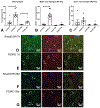
Figure 4:. Fibroblast-specific Smad3 loss is associated with increased cardiomyocyte injury after 7 days of TAC.
A-B: Hematoxylin-eosin staining demonstrated that many cardiomyocytes in pressure-overloaded FS3KO mice exhibited significant myofibrillar loss (myocytolysis, arrows). C: Quantitative analysis showed that FS3KO animals had a significantly higher myocytolysis index (*p<0.05, n=8–9). D-E: TUNEL staining was used to identify apoptotic cells in pressure overloaded hearts (arrows). Quantitative analysis showed that after 7 days of TAC, FS3KO mice had a significantly higher density of apoptotic cells (F), when compared with corresponding Smad3 fl/fl mice.
Figure 5:
Figure 5:. Fibroblast-specific Smad3 loss accentuates macrophage-driven inflammation in the pressure-overloaded myocardium.
A. Mac2 immunofluorescence was used to identify macrophages in the remodeling myocardium. FS3KO mice exhibited a marked increase in macrophage density after 7-28 days of TAC (*p<0.05 vs. Smad3 fl/fl, n=6-7/group). B. Dual immunofluorescence for Mac2 and iNOS was used to identify pro-inflammatory M1-like macrophages. The density of iNOS+ macrophages was significantly higher in FS3KO mice after 7 days of TAC (*p<0.05 vs. Smad3 fl/fl). The number of pro-inflammatory macrophages in FS3KO mice was significantly reduced after 28 days of TAC when compared to the 7-day timepoint (^p<0.05 vs. FS3KO 7d, n=7/group). C. Dual immunofluorescence for Mac2 and Arginase-1 (Arg1) was used to identify anti-inflammatory M2-like macrophages. The density of Arg1+ macrophages was comparable between groups. D-G: Representative images show Mac2/iNOS dual fluorescence (arrows) in Smad3 fl/fl and FS3KO mice after 7 and 28 days of TAC.
Figure 6:
Figure 6:. Smad3 signaling mediates the matrix-preserving effects of TGF-β in cardiac fibroblasts.
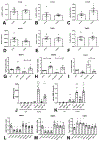
Fibroblasts harvested from FS3KO and Smad3 fl/fl hearts after 7 days of TAC had comparable MMP2 (A) and MMP3 (B) mRNA expression levels, but exhibited markedly elevated MMP8 expression (C, *p<0.05, n=5/group). MMP9 (D), TIMP1 (E) and TIMP2 (F) expression levels were comparable between Smad3 fl/fl and FS3KO fibroblasts. G-I: Effects of TGF-β on MMP and TIMP expression in cardiac fibroblasts cultured in collagen pads are dependent on Smad3. In WT cells, TGF-β1 stimulation suppressed synthesis of MMP3 (G) and MMP8 (H) and induced TIMP-1 (I) expression (**p<0.01 vs. control, n=6/group). In contrast, TGF-β1 had no significant effects on MMP3, MMP8 and TIMP1 expression in Smad3 KO fibroblasts (^p<0.05, ^^p<0.01 vs. corresponding control). J-K: WT cardiac fibroblasts had low levels of MMP8 protein and activity in the supernatant, in the presence or absence of TGF-β. In contrast, Smad3 KO cells exhibited marked release of MMP8 protein and activity 30 min and 12h after TGF-β1 stimulation (****p<0.0001 vs. corresponding WT cells; ^p<0.05, ^^p<0.01, ^^^^p<0.0001 vs. unstimulated cells, n=4/group). The findings suggested that Smad3 signaling restrains release of MMP8 protein and activity by cardiac fibroblasts. L-N: Effects of Smad3 activating members of the TGF-β superfamily on expression of MMP3, MMP8 and TIMP1. Only TGF-β1, β2 and β3, and not activin-A, activin-B, myostatin and GDF-11 suppressed MMP3 (L) and MMP8 (M) expression, and induced TIMP-1 synthesis (N) in fibroblasts cultured in collagen pads (**p<0.01 vs. control, n=5/group).
Figure 7:
Figure 7:. MMP8 inhibition attenuates systolic dysfunction in pressure-overloaded FS3KO mice.
A-B. FS3KO mice exhibited significant early reduction in ejection fraction after 7 days of TAC (*p<0.05 vs. baseline). Administration of an MMP8 inhibitor, but not vehicle attenuated systolic dysfunction in FS3KO mice. Surprisingly, MMP8 inhibition caused reduction of ejection fraction in Smad3 fl/fl mice undergoing TAC protocols, suggesting that both excessive MMP8 (in FS3KO mice) and very low MMP8 activity (in control mice treated with inhibitor) have detrimental effects. C-D: MMP8 inhibition also attenuated the increase in LVESV noted in pressure-overloaded FS3KO mice. E-F: There were no significant effects of MMP8 inhibition on LVEDV. G-H: MMP8 inhibition did not significantly affect the effects of fibroblast-specific Smad3 loss on left ventricular mass (LVM). (*p<0.05, **p<0.01, n=9-13/group).
Figure 8:
Figure 8:. MMP8 promotes a pro-inflammatory macrophage phenotype in vitro and in vivo.
A-C: Treatment with the MMP8 inhibitor did not improve the myocytolysis score in FS3KO mice (A) and did not affect the density of apoptotic cells (B) in the pressure-overloaded myocardium after 7 days of TAC. Although the effects of MMP8 inhibition on the density of iNOS+ M1-like macrophages (C) and Arg1+ M2-like macrophages (D) did not reach statistical significance, administration of the inhibitor increased the ratio of M2 like to M1 like cells (E, *p<0.05). In vitro, recombinant MMP8 (50ng/ml) markedly reduced levels of active TGF-β1 (F) and increased levels of the pro-inflammatory chemokine CCL2 (G) in the supernatant of isolated bone marrow macrophages (*p<0.05, n=5/group). H: Schematic cartoon illustrating the protective effects of TGF-β/Smad3-activated fibroblasts in the pressure-overloaded myocardium. Mechanical stress stimulates angiotensin II (Ang II) and generates reactive oxygen species (ROS), activating proteases. TGF-β-mediated stimulation of fibroblasts promotes a matrix-preserving program, suppressing MMP3 and MMP8 expression and inducing TIMP1 synthesis. Reduced matrix degradation decreases release of collagen-derived matrikines and attenuates macrophage-driven inflammation protecting cardiomyocytes from death and dysfunction. Moreover, restrained MMP activity may inhibit inflammation by decreasing formation of bioactive cytokine and chemokine molecules. The cartoon was designed using Servier Medical Art (https:smart.servier.com).

Comment in

Similar articles

Cited by

References

    1. Pinto AR, Ilinykh A, Ivey MJ, Kuwabara JT, D’Antoni ML, Debuque R, Chandran A, Wang L, Arora K, Rosenthal NA and Tallquist MD. Revisiting Cardiac Cellular Composition. Circ Res. 2016;118:400–9. - PMC - PubMed
    1. Tallquist MD and Molkentin JD. Redefining the identity of cardiac fibroblasts. Nat Rev Cardiol. 2017;14:484–491. - PMC - PubMed
    1. Souders CA, Bowers SL and Baudino TA. Cardiac fibroblast: the renaissance cell. Circ Res. 2009;105:1164–76. - PMC - PubMed
    1. Cleutjens JP, Verluyten MJ, Smiths JF and Daemen MJ. Collagen remodeling after myocardial infarction in the rat heart. Am J Pathol. 1995;147:325–38. - PMC - PubMed
    1. Kanisicak O, Khalil H, Ivey MJ, Karch J, Maliken BD, Correll RN, Brody MJ, SC JL, Aronow BJ, Tallquist MD and Molkentin JD. Genetic lineage tracing defines myofibroblast origin and function in the injured heart. Nature communications. 2016;7:12260. - PMC - PubMed

Publication types

MeSH terms

Substances