. 2019 Mar;179(3):1111-1131.
doi: 10.1104/pp.18.01434. Epub 2019 Jan 18.

Targeted Endoplasmic Reticulum Localization of Storage Protein mRNAs Requires the RNA-Binding Protein RBP-L

Affiliations

Targeted Endoplasmic Reticulum Localization of Storage Protein mRNAs Requires the RNA-Binding Protein RBP-L

Li Tian et al. Plant Physiol. 2019 Mar.

Abstract

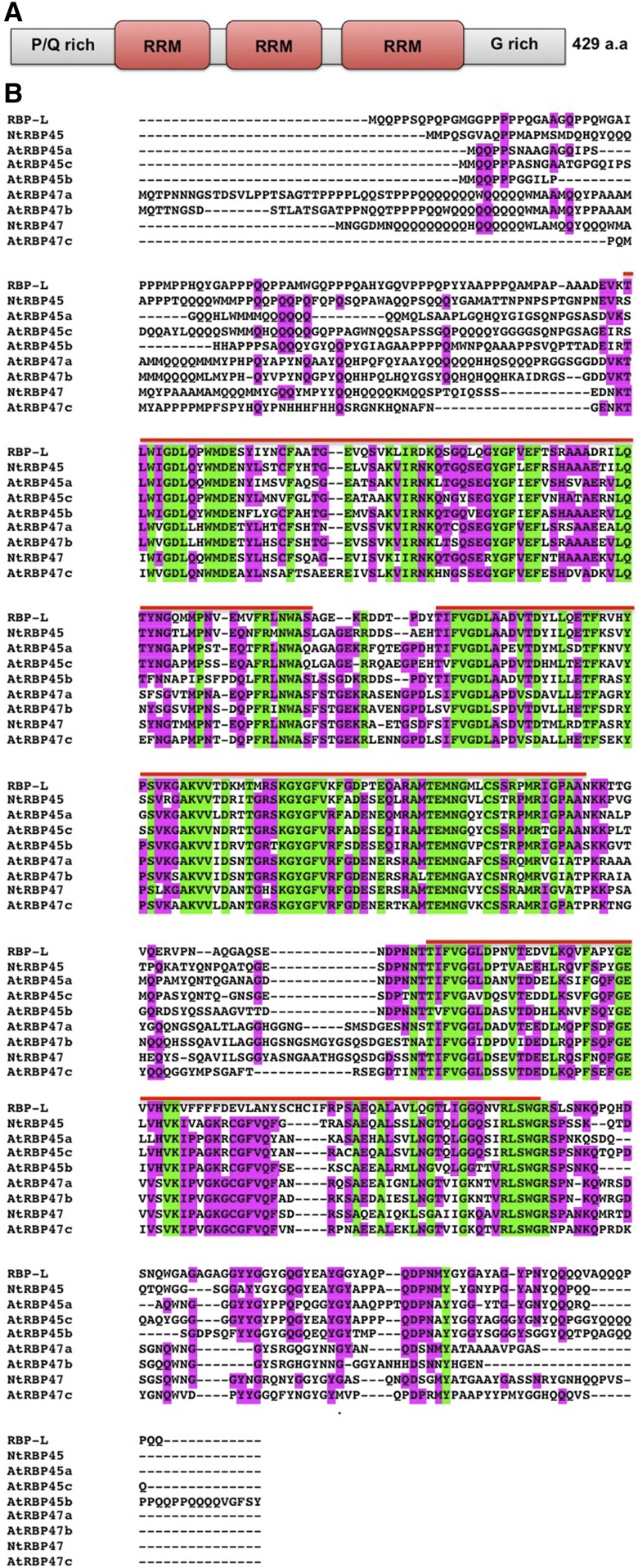
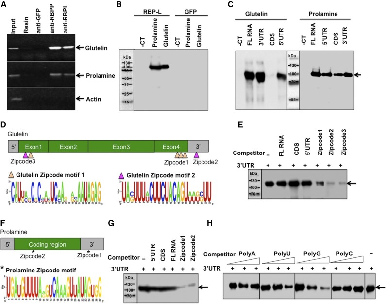
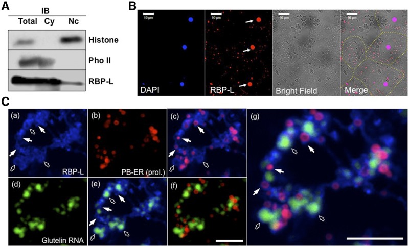
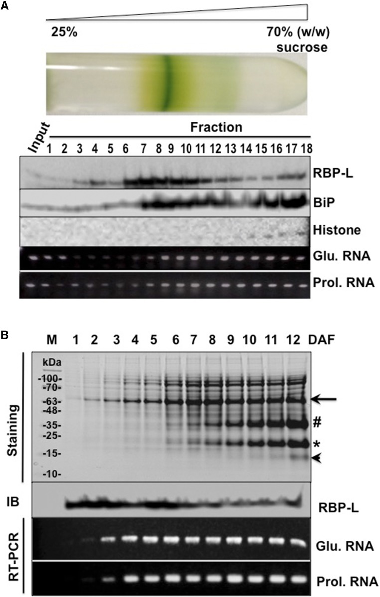
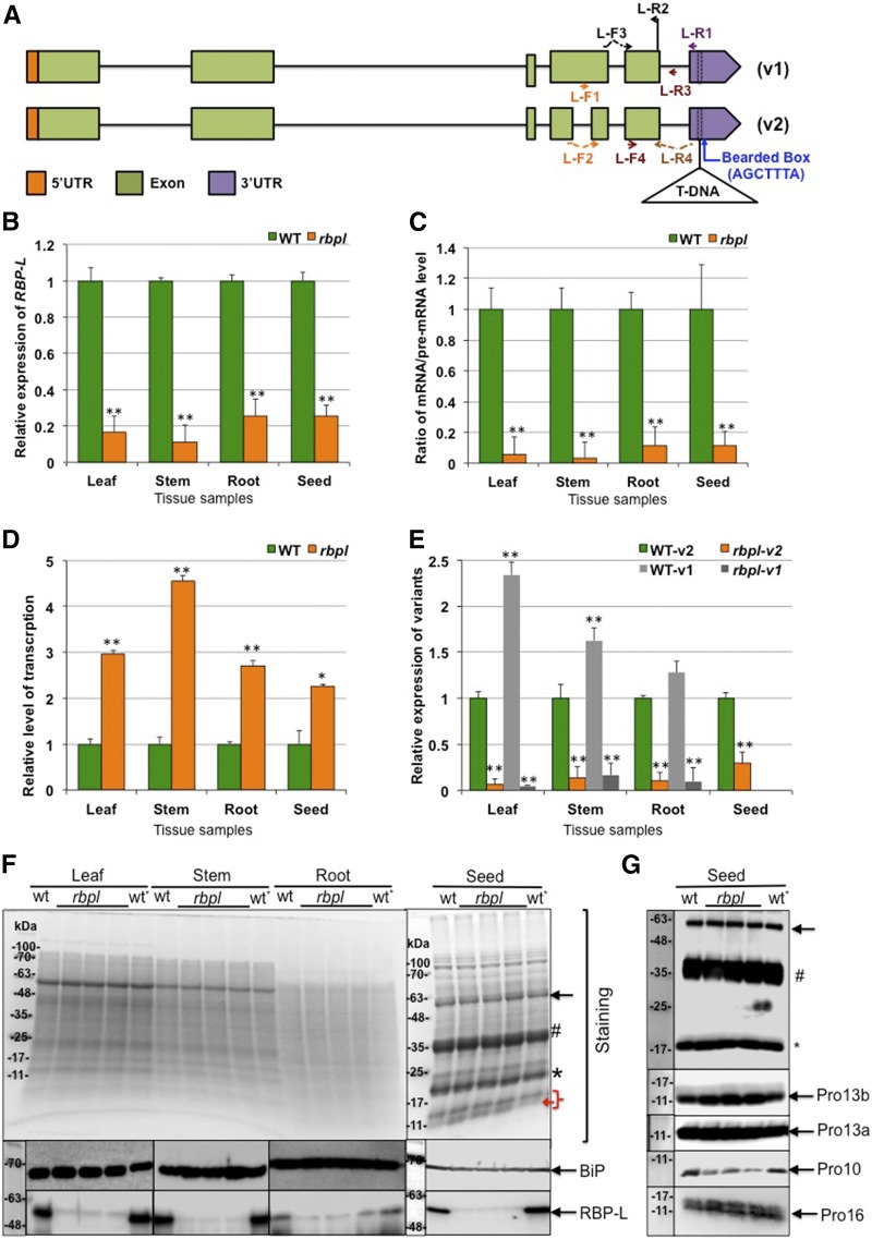
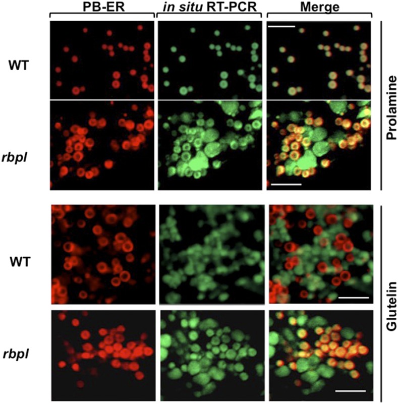
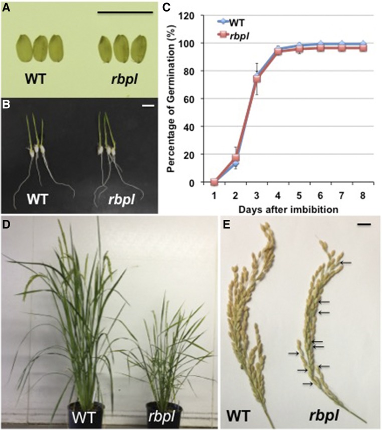
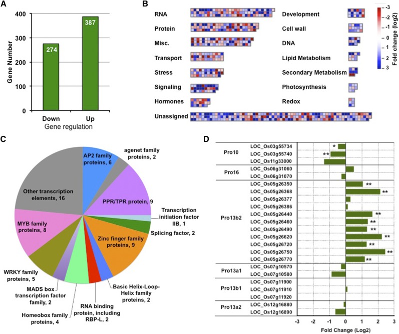
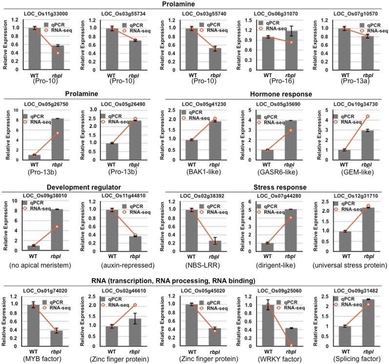
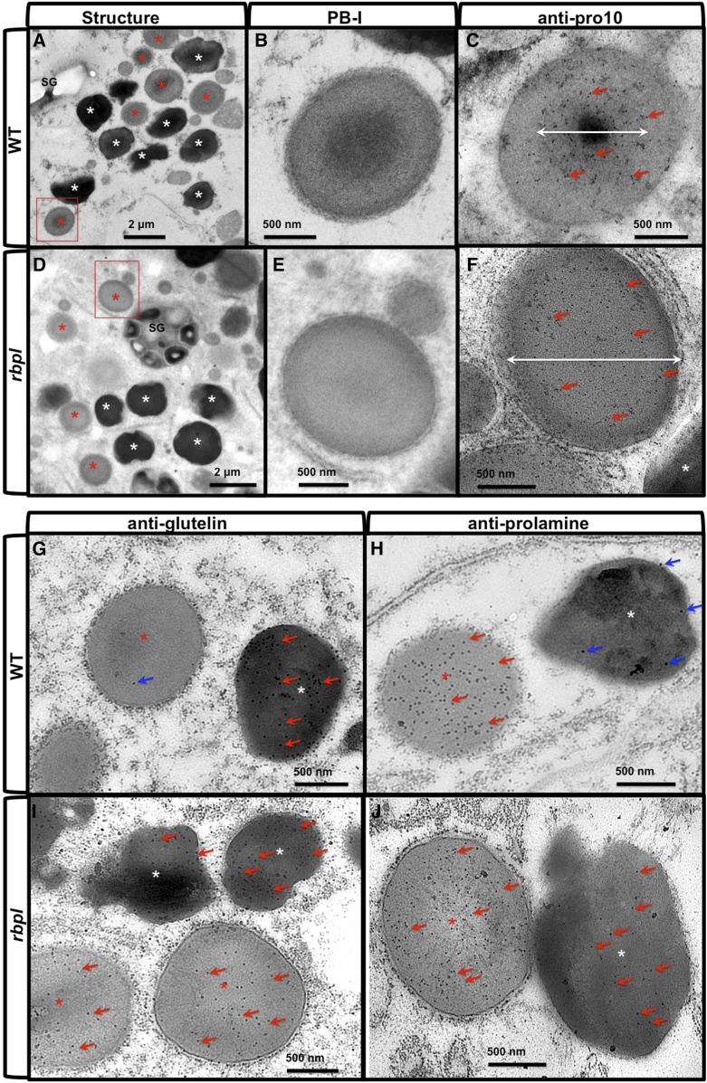
The transport and targeting of glutelin and prolamine mRNAs to distinct subdomains of the cortical endoplasmic reticulum is a model for mRNA localization in plants. This process requires a number of RNA-binding proteins (RBPs) that recognize and bind to mRNA cis-localization (zipcode) elements to form messenger ribonucleoprotein complexes, which then transport the RNAs to their destination sites at the cortical endoplasmic reticulum. Here, we present evidence that the rice (Oryza sativa) RNA-binding protein, RBP-L, like its interacting RBP-P partner, specifically binds to glutelin and prolamine zipcode RNA sequences and is required for proper mRNA localization in rice endosperm cells. A transfer DNA insertion in the 3' untranslated region resulted in reduced expression of the RBP-L gene to 10% to 25% of that in the wild-type. Reduced amounts of RBP-L caused partial mislocalization of glutelin and prolamine RNAs and conferred other general growth defects, including dwarfism, late flowering, and smaller seeds. Transcriptome analysis showed that RBP-L knockdown greatly affected the expression of prolamine family genes and several classes of transcription factors. Collectively, these results indicate that RBP-L, like RBP-P, is a key RBP involved in mRNA localization in rice endosperm cells. Moreover, distinct from RBP-P, RBP-L exhibits additional regulatory roles in development, either directly through its binding to corresponding RNAs or indirectly through its effect on transcription factors.


Figures

Figure 1.
Figure 1.
Sequence information of RBP-L. A, Schematic structure of RBP-L. B, Protein sequence alignment of RBP-L to RBP45/47 family members from Arabidopsis and tobacco. The three RRMs are indicated by the red line above the sequences. The conserved (shared by all protein sequences) and semiconserved (shared by six out of nine protein sequences) amino acids are highlighted in green and magenta, respectively. P/Q rich, Pro- and Gln-rich domain; G-rich, Gly-rich domain; a.a., amino acids.
Figure 2.
Figure 2.
RBP-L binds to glutelin and prolamine mRNAs, especially to their zipcode RNA sequences. A, RNA-IP analysis showing that RBP-L is associated with glutelin and prolamine mRNAs in vivo. The RNAs extracted from IPs generated by anti-RBP-P, anti-RBP-L, anti-GFP, and empty resin were subjected to RT-PCR to detect glutelin, prolamine, and actin gene sequences. The figure depicts an agarose gel with resolved PCR products. Anti-GFP and empty resin were used as negative controls. Input, PCR using cDNA synthesized from RNAs isolated from IPs. B, Binding activity of RBP-L to glutelin and prolamine mRNAs compared to RNA control (for detailed information, see “Materials and Methods”). The figure depicts an immunoblot where anti-DIG antibody was used to detect bound DIG-labeled RNAs (see “Materials and Methods” for detailed experimental details). GFP protein was used as negative control for the binding test. C, Binding activities of RBP-L to different region of glutelin and prolamine RNAs. Arrow indicates the location of recombinant RBP-L. D, The glutelin RNA zipcode elements, which consist of two types of motifs, zipcode motif 1 (orange triangle) and zipcode motif 2 (magenta triangle). (Top) Location and components of three glutelin zipcode elements; (bottom) consensus zipcode sequences generated by WebLogo (http://weblogo.berkeley.edu). E, Competition assays using equimolar amounts of DIG-labeled glutelin 3′UTR and the indicated unlabeled competitor RNAs. Arrows indicates the location of recombinant RBP-L. F, The prolamine zipcode elements, which consist of only a single zipcode motif (*). (Top) Location of the two zipcode elements within prolamine mRNA; (bottom) consensus sequence of prolamine zipcode. G, Competition assays using equimolar amounts of DIG-labeled prolamine 3′UTR and indicated unlabeled competitor RNAs. Arrows indicate the location of recombinant RBP-L. H, Binding specificity of RBP-L to different homoribopolymers. Unlabeled poly(U), poly(A), poly(C), and poly(G) were added at 5-, 10-, and 20-fold mole excess over DIG-labeled glutelin 3′ UTR RNA. Arrows indicates the location of recombinant RBP-L. -CT, pBlueScript KS vector sequence; 5′, 5′UTR; 3′, 3′UTR; -, no competitor.
Figure 3.
Figure 3.
Subcellular localization of RBP-L in rice seed endosperm cells. A, The distribution of RBP-L in the nuclear and cytoplasmic subcellular fractions as revealed by immunoblot analysis. Histone H3 and starch phosphorylase II were used as marker proteins of the nuclear and cytoplasmic fractions. B, Immunofluorescence analysis to locate RBP-L (red) in rice endosperm cells using its specific antibody. The labeling was performed on 5-μm-thin sections from LR-white embedded rice grain tissue. Nuclei stained by 4′,6-diamidino-2-phenylindole (blue) are indicated by arrows and the different cells shown in the figure are distinguished by dashed yellow line. Scale bar = 10 μm. C, Triple labeling for detecting the distribution of RBP-L (blue, a), PB-ER (red, b), and glutelin RNAs (green, d). Fresh-frozen 20-μm cryosections of rice seed tissues were initially subjected to in situ RT-PCR in the presence of glutelin primers and Alexa Fluor 488-UTP (green, d) to visualize the distribution of glutelin mRNAs. The sections were then treated by immunofluorescence analysis for detection of RBP-L and then poststained with rhodamine B to visualize PB-ER (red, b), the ER membranes where prolamine mRNAs are localized. C, (e) to (g), are the merged images of (a) and (b), (a) and (d), (b) and (d), and the combined (a), (b) and (d) panels, respectively. White arrows denote colocalized RBP-Lwith rhodamine-B–labeled PB-ER membranes and open arrows indicate distribution of RBP-L protein surrounding glutelin mRNA patches. Scale bar = 20 μm. IB, immunoblot analysis; Cy, cytoplasmic; Nc, nuclear; DAPI, 4′,6-diamidino-2-phenylindole; Pho II, phosphorylase II; prol., prolamine.
Figure 4.
Figure 4.
The association of RBP-L with glutelin and prolamine mRNAs in developing rice grains. A, The distribution of RBP-L, glutelin, and prolamine mRNAs, histone, and BiP as resolved by Suc density gradient centrifugation. Samples from the Suc gradient were collected from the top (25% [w/w] Suc) to bottom (70% [w/w] Suc) and subjected to SDS-PAGE, immunoblot analyses, and RT-PCR. Input, rice seed lysate sample before centrifugation. Histone H3 and BiP were used as nuclear and the ER markers, respectively. Glutelin and prolamine mRNA distribution was assessed by RT-PCR using the total RNA isolated from each fraction. B, Expression of RBP-L protein and glutelin/prolamine RNAs during rice seed development. Total proteins and RNAs extracted from developing rice seeds, collected daily from 1 to 12 DAF, were subject to SDS-PAGE (Coomassie brilliant-blue–stained gel) followed by immunoblot analysis or by RT-PCR using glutelin- and prolamine-specific primers (RT-PCR), respectively. Black arrow, Glutelin precursor; #, acidic subunit; *, basic subunit; arrowhead, prolamine polypeptide; Glu. mRNA, Glutelin RNA; Prol. mRNA, prolamine RNA; IB, immunoblot analysis.
Figure 5.
Figure 5.
Knockdown of RBP-L by a T-DNA insertion within its 3′UTR. A, Gene structure and T-DNA insertion site of the RBP-L gene. Two splicing variants are labeled as “v1” and “v2,” respectively. T-DNA insertion site is bordered by dashed lines in the 3′UTR. Arrows indicate the location and direction of the corresponding primers. B, Relative expression level of the RBP-L gene revealed by RT-qPCR using primer set of L-F3 and L-R2. C, The relative apparent splicing efficiency as denoted by the ratio of RBP-L mRNA/premRNA levels. RT of total RNA was performed using primer L-R1, and mRNA and premRNA levels amplified by primer sets of L-F3 + L-R2, and L-F4 + L-R3, respectively. D, Total transcription level of the RBP-L gene expression was determined by RBP-L gene transcripts (spliced and unspliced) amplified by primers L-F4 and L-R2. E, Relative expression of the two splicing variants of the RBP-L gene amplified by L-R4 with L-F1 or L-F2. All relative expression levels of RBP-L gene transcripts in rbpl line shown in (B) to (E) are normalized to that of wild type. **P value < 0.01. The expression level of the splicing variant v1 in developing seeds is not shown due to its low expression in developing rice grains. F, Expression level of RBP-L protein revealed by immunoblot using anti-RBP-L antibody. BiP levels as assessed by immunoblot analysis was used as a loading control. (Top) Coomassie brilliant-blue–stained SDS-PAGE; (center and bottom) the immunoblot results for BiP and RBP-L, respectively. The three lanes between “wt” and “wt*” present total protein samples from three individual plants of rbpl line. G, The differential expression of prolamine family proteins during grain development. Pro-10 polypeptides levels are depressed whereas Pro-13b polypeptides amounts are elevated in rbpl line. Black arrow, Glutelin precursor; #, acidic subunit; *, basic subunit; red bracket, prolamine polypeptide; red arrow, possible up-regulation of 13 kD prolamine polypeptides; wt, wild type; wt*, wild-type genotype segregated from heterozygous rbpl line (for detailed information, see “Materials and Methods”).
Figure 6.
Figure 6.
Knockdown of RBP-L results in the partial mislocalization of glutelin and prolamine mRNAs on the cortical-ER as revealed by in situ RT-PCR. In situ RT-PCR was performed directly on developing rice grain sections in the presence of Alexa-488-UTP (green) and specific primers to label prolamine and glutelin mRNAs. PB-ER was stained using Rhodamine B dye (red). Note that prolamine and glutelin mRNAs are localized to the PB-ER (red) and cisternal-ER, respectively, in wild type. In the rbpl line, glutelin and prolamine mRNAs are distributed on both the PB-ER and cisternal-ER. Scale bar = 5 μm. WT, wild type.
Figure 7.
Figure 7.
Knockdown of RBP-L confers several growth defects in rbpl line in comparison to wild type. A, Seed morphology of wild-type and rbpl line. Scale bar = 1 cm. B, Germinated wild-type and rbpl seeds after 6 d of imbibition. Scale bar = 1 cm. C, Germination rate of wild-type and rbpl seeds. Seeds were considered germinated when the radicle was extended >1 cm or more. D, The rbpl line showed late flowering and dwarfism. When rbpl line plants were blooming, the grains from wild-type plants were approaching maturity. E, The rbpl lines showed lower spikelet fertility than wild type. Some empty glumes on the rbpl tassel are indicated by arrows. WT, wild type.
Figure 8.
Figure 8.
Transcriptome changes mediated by knockdown of RBP-L expression. A, Chart showing the numbers of down- and up-regulated genes (log 2 fold change > 1, P value < 0.01) in rbpl developing seeds compared to that in wild-type. B, Distribution of molecular function and biological pathways that were enriched by the 661 DEGs in the rbpl line. C, Composition of the DEGs involved in RNA metabolic pathways. D, Fold changes on gene expression of prolamine family members. Prolamine genes with relative low RPKM (<1) were not analyzed and are not shown in the chart. *P value of two-tail t test < 0.05; **P value of two-tail t test < 0.01, based on RNA-seq data. Detailed information shown in this figure can be found in Supplemental Tables S2–S6.
Figure 9.
Figure 9.
Validation of DEGs in rbpl by RT-qPCR. Analysis of selected genes from prolamine family, hormone signaling pathway, development, stress response, and RNA metabolism confirmed the differentially expressed patterns determined by RNA-seq. Gray columns indicate the fold-changes based on qPCR results, whereas orange lines show the data obtained from RNA-seq. y axis, fold changes relative to that of wild-type. Error bars shown on gray column represent ses of the mean fold changes calculated from three qPCR replicates.
Figure 10.
Figure 10.
PB-I structure and localization of glutelin and prolamine proteins in wild-type and rbpl line endosperm cells. A to F, Ultrastructure of PB-I in wild-type (A–C) and rbpl line (D–F) endosperm cells. B and E, Enlarged picture of the areas indicated by the red boxes in (A) and (D). C and F, Immunolabeling patterns using anti-prolamine (Pro-10) antibody and 10-nm gold particle-conjugated secondary antibody in wild type (C) and rbpl (F) PB-I, respectively. White double-headed arrows indicate the primary distribution area of Pro-10 proteins within PB-I. Scale bar = 2 μm (A and D) or 500 nm (B, C, E, and F). Red and white asterisks in (A) and (D) denote PSV and PB-I, respectively. G to J, Immunolabeling of glutelin (G, I) and prolamine (H, J) proteins using monospecific antibodies and 15-nm gold particles-conjugated secondary antibodies. G and H, wild-type. I and J, rbpl line. Red arrows denote positive gold particle labeling, and blue arrows denote slight background nonspecific labeling. Scale bar = 500 nm. WT, wild type.
Figure 11.
Figure 11.
A proposed working model to show the critical roles of RBP-L in rice glutelin and prolamine mRNA localization and gene expression. (1) Through posttranscriptional control of genes that encode regulation on transcription factors, RBP-L indirectly regulates the transcription of prolamine genes, particularly Pro-10 and Pro-13b genes. This regulatory mechanism can also be extended to other genes that are regulated by RBP-L. (2) Through its direct binding affinity to glutelin and prolamine zipcode RNAs, RBP-L together with RBP-P (Tian et al., 2018) and other factors drive storage protein mRNAs to their destination on subdomains of the cortical-ER. (3) RBP-L may also regulate glutelin and prolamine mRNA localization through its regulation on the expression of other RBPs (orange oval) that are components of the mRNP complex.

Similar articles

Cited by

References

    1. Ambawat S, Sharma P, Yadav NR, Yadav RC (2013) MYB transcription factor genes as regulators for plant responses: An overview. Physiol Mol Biol Plants 19: 307–321 - PMC - PubMed
    1. Arora R, Agarwal P, Ray S, Singh AK, Singh VP, Tyagi AK, Kapoor S (2007) MADS-box gene family in rice: Genome-wide identification, organization and expression profiling during reproductive development and stress. BMC Genomics 8: 242. - PMC - PubMed
    1. Baron KN, Schroeder DF, Stasolla C (2014) GEm-Related 5 (GER5), an ABA and stress-responsive GRAM domain protein regulating seed development and inflorescence architecture. Plant Sci 223: 153–166 - PubMed
    1. Belkhadir Y, Jaillais Y (2015) The molecular circuitry of brassinosteroid signaling. New Phytol 206: 522–540 - PubMed
    1. Bilenoglu O, Basak AN, Russell JE (2002) A 3'UTR mutation affects beta-globin expression without altering the stability of its fully processed mRNA. Br J Haematol 119: 1106–1114 - PubMed

Publication types