

miR-142-5p suppresses proliferation and promotes apoptosis of human osteosarcoma cell line, HOS, by targeting PLA2G16 through the ERK1/2 signaling pathway
- PMID: 30655907
- PMCID: PMC6312951
- DOI: 10.3892/ol.2018.9712
miR-142-5p suppresses proliferation and promotes apoptosis of human osteosarcoma cell line, HOS, by targeting PLA2G16 through the ERK1/2 signaling pathway
Abstract
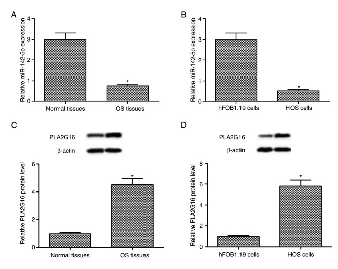
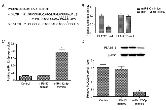
Previous studies have revealed that miR-142-5p serves a critical role in human cancer progression. However, the biological function of miR-142-5p in osteosarcoma (OS) development remains unclear. In the present study, the role of miR-142-5p in human OS HOS cells was determined, and the underlying mechanism involved was examined. Compared with the adjacent healthy tissues, the expression level of miR-142-5p was downregulated and the expression level of group XVI phospholipase A2 (PLA2G16) protein was upregulated in human OS tissues. The aforementioned results were also indicated in human OS HOS cells when compared with human fetal osteoblastic hFOB1.19 cells. Additionally, the results demonstrated that PLA2G16 was a direct target of miR-142-5p. miR-142-5p transfection upregulated the expression level of miR-142-5p and suppressed the expression level of PLA2G16 protein in HOS cells. MTT assays indicated a time-dependent decrease by miR-142-5p transfection in the proliferation of HOS cells. 5-bromo-2'-deoxyuridine incorporation assays confirmed that miR-142-5p transfection inhibited DNA synthesis in HOS cells. In addition, miR-142-5p transfection increased the Caspase-3 (CASP3) activity and apoptotic rate. Western blot analysis indicated that miR-142-5p transfection reduced BCL2, apoptosis regulator expression and upregulated the expression of CASP3 and BCL2 associated X, apoptosis regulator in HOS cells. Furthermore, miR-142-5p transfection decreased the expression levels of phosphorylated (p)-proto-oncogene, serine/threonine kinase, p-mitogen-activated protein kinase kinase, and p-extracellular signal-regulated kinase (ERK) 1/2 proteins in HOS cells. PLA2G16 overexpression restored the expression level of p-ERK 1/2 protein, which was reduced by miR-142-5p overexpression. MTT and CASP3 activity assays indicated that restoration of PLA2G16 reversed the tumour-suppressive role of miR-142-5p transfection in HOS cells. In conclusion, the results of the present study indicated that miR-142-5p suppressed proliferation and promoted apoptosis in human OS HOS cells by targeting PLA2G16 through ERK1/2 signaling pathway.
Keywords: Group XVI phospholipase A2; apoptosis; miR-142-5p; osteosarcoma; proliferation.
Figures


Similar articles
-
[LncRNA LINC-PINT regulating proliferation and apoptosis of osteosarcoma cells by targeting miR-524-5p].Zhonghua Zhong Liu Za Zhi. 2020 Apr 23;42(4):325-330. doi: 10.3760/cma.j.cn112152-20190726-00471. Zhonghua Zhong Liu Za Zhi. 2020. PMID: 32375449 Chinese.
-
LncRNA FER1L4 induces apoptosis and suppresses EMT and the activation of PI3K/AKT pathway in osteosarcoma cells via inhibiting miR-18a-5p to promote SOCS5.Gene. 2019 Dec 30;721:144093. doi: 10.1016/j.gene.2019.144093. Epub 2019 Aug 29. Gene. 2019. PMID: 31473323
-
MicroRNA-340-5p suppresses osteosarcoma development by down-regulating the Wnt/β-catenin signaling pathway via targeting the STAT3 gene.Eur Rev Med Pharmacol Sci. 2019 Feb;23(3):982-991. doi: 10.26355/eurrev_201902_16985. Eur Rev Med Pharmacol Sci. 2019. PMID: 30779064
-
[microRNA-16-5p targeted tetraspanin 15 gene to inhibit the proliferation, migration and invasion of osteosarcoma cell through phospoinositide 3-kinase/protein kinase B signaling pathway].Zhonghua Yi Xue Za Zhi. 2020 Jun 2;100(21):1668-1675. doi: 10.3760/cma.j.cn112137-20191101-02376. Zhonghua Yi Xue Za Zhi. 2020. PMID: 32486604 Chinese.
-
miR-491-5p inhibits osteosarcoma cell proliferation by targeting PKM2.Oncol Lett. 2018 Nov;16(5):6472-6478. doi: 10.3892/ol.2018.9451. Epub 2018 Sep 18. Oncol Lett. 2018. PMID: 30405785 Free PMC article.
Cited by
-
Disruption of Cancer Metabolic SREBP1/miR-142-5p Suppresses Epithelial-Mesenchymal Transition and Stemness in Esophageal Carcinoma.Cells. 2019 Dec 18;9(1):7. doi: 10.3390/cells9010007. Cells. 2019. PMID: 31861383 Free PMC article.
-
miRNome Profiling Detects miR-101-3p and miR-142-5p as Putative Blood Biomarkers of Frailty Syndrome.Genes (Basel). 2022 Jan 26;13(2):231. doi: 10.3390/genes13020231. Genes (Basel). 2022. PMID: 35205276 Free PMC article.
-
METTL3 stabilizes HDAC5 mRNA in an m6A-dependent manner to facilitate malignant proliferation of osteosarcoma cells.Cell Death Discov. 2022 Apr 8;8(1):179. doi: 10.1038/s41420-022-00926-5. Cell Death Discov. 2022. PMID: 35396379 Free PMC article.
-
A novel lncRNA ROPM-mediated lipid metabolism governs breast cancer stem cell properties.J Hematol Oncol. 2021 Oct 29;14(1):178. doi: 10.1186/s13045-021-01194-z. J Hematol Oncol. 2021. PMID: 34715882 Free PMC article.
-
Long Non-Coding RNA FEZF1-AS1 Modulates CXCR4 to Promote Cell Proliferation, Warburg Effect and Suppress Cell Apoptosis in Osteosarcoma by Sponging miR-144.Onco Targets Ther. 2020 Apr 5;13:2899-2910. doi: 10.2147/OTT.S235970. eCollection 2020. Onco Targets Ther. 2020. PMID: 32308422 Free PMC article.
References
LinkOut - more resources
Full Text Sources
Research Materials
Miscellaneous