. 2019 Feb 15;14(2):266-275.
doi: 10.1021/acschembio.8b00972. Epub 2019 Jan 24.

Crystal Structures of Fumarate Hydratases from Leishmania major in a Complex with Inhibitor 2-Thiomalate

Affiliations

Crystal Structures of Fumarate Hydratases from Leishmania major in a Complex with Inhibitor 2-Thiomalate

Patricia R Feliciano et al. ACS Chem Biol. .

Abstract

Leishmaniases affect the poorest people on earth and have no effective drug therapy. Here, we present the crystal structure of the mitochondrial isoform of class I fumarate hydratase (FH) from Leishmania major and compare it to the previously determined cytosolic Leishmania major isoform. We further describe the mechanism of action of the first class-specific FH inhibitor, 2-thiomalate, through X-ray crystallography and inhibition assays. Our crystal structures of both FH isoforms with inhibitor bound at 2.05 Å resolution and 1.60 Å resolution show high structural similarity. These structures further reveal that the selectivity of 2-thiomalate for class I FHs is due to direct coordination of the inhibitor to the unique Fe of the catalytic [4Fe-4S] cluster that is found in class I parasitic FHs but is absent from class II human FH. These studies provide the structural scaffold in order to exploit class I FHs as potential drug targets against leishmaniases as well as Chagas diseases, sleeping sickness, and malaria.


Conflict of interest statement

The authors declare no competing financial interest.

Figures
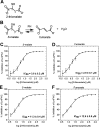
Figure 1
Figure 1
Inhibition of LmFH isoforms by 2-thiomalate. (A) Structure of 2-thiomalate molecule. (B) FH catalytic reversible reaction of S-malate to fumarate. (C) Dose–response curve of the inhibition of mitochondrial LmFH-1 by RS-2-thiomalate against S-malate (3 mM). (D) Dose–response curve of the inhibition of mitochondrial LmFH-1 by RS-2-thiomalate against fumarate (3 mM). (E) Dose–response curve of the inhibition of cytosolic LmFH-2 by RS-2-thiomalate against S-malate (3 mM). (F) Dose–response curve of the inhibition of cytosolic LmFH-2 by RS-2-thiomalate against fumarate (6 mM). Error bars represent three independent measurements.
Figure 2
Figure 2
Crystal structures of LmFH isoforms in a complex with 2-thiomalate. (A) Overall structure of cytosolic LmFH-2 functional dimer. Monomers are shown in blue (chain A) and pink (chain B) and are comprised of two domains: N-terminal domain (darker blue and pink) and C-terminal domain (lighter blue and pink) connected by a flexible linker. (B) Overall structure of mitochondrial LmFH-1 functional dimer. Monomers are shown in purple (chain A) and green (chain B). N-terminal domain is shown in darker purple and green, and C-terminal domain is shown in lighter purple and green. The domain linker is shown in dark blue. (C) Ribbon diagram of the superposition of LmFHs monomers (chain A). The N-terminal domain is divided into two nonsequential subdomains 1 (purple in LmFH-1 and blue in LmFH-2) and 2 (cyan in LmFH-1 and light cyan in LmFH-2), connected to C-terminal domain (light purple in LmFH-1 and light blue in LmFH-2) by a linker (green in LmFH-1 and black arrow in LmFH-2 to where linker would be if it was ordered). Linear schematic indicates subdomain order. (D) LmFH-2 in a complex with S-2-thiomalate. The left panel shows the FoFc difference electron density map contoured at 3.0 rmsd (green mesh) for S-2-thiomalate (green) suggesting its double conformation. The center panel shows the sulfur anomalous difference electron density map contoured at 3.0 rmsd (purple mesh) supporting the assignment of S-2-thiomalate (green) double conformation. The C2-thiol groups (yellow) are coordinated to the unique Fe (labeled in white) of the [4Fe-4S] cluster. The right panel shows the final 2FoFc electron density map contoured at 1.5 rmsd (blue mesh) for S-2-thiomalate (green) and [4Fe-4S] cluster. (E) LmFH-1 in a complex with S-2-thiomalate. The left panel shows the FoFc difference electron density map contoured at 3.0 rmsd (green mesh) for S-2-thiomalate (cyan) in chains A and B, consistent with a double conformation. The right panel shows the FoFc difference electron density map contoured at 3.0 rmsd (green mesh) for S-2-thiomalate (cyan) in chains C and D, consistent with a single conformation. The catalytic [4Fe-4S] clusters are shown in orange (Fe) and yellow (S) spheres.
Figure 3
Figure 3
Sequence alignment of LmFH isoforms. LmFH-1 and LmFH-2 are the mitochondrial and cytosolic isoforms of Leishmania major, respectively. The conserved residues are indicated in the blue boxes. The conserved active site residues among class I FHs that coordinate to inhibitor S-2-thiomalate are indicated in the pink boxes. The three conserved cysteine residues, which are shown to bind a [4Fe-4S] cluster, are indicated in the yellow boxes. Secondary structures of LmFH-1 and LmFH-2 are shown on top and at the bottom of sequence alignment, respectively. The dimer interface residues of LmFH-1 are indicated in green stars. The alignment was performed using MULTALIN and graphically displayed using ESPript.
Figure 4
Figure 4
Electrostatic surface potential of LmFH isoforms. The upper and lower panels represent two orthogonal views of the electrostatic surface potential representation of LmFH-2 (A) and LmFH-1 (B).
Figure 5
Figure 5
Positively charged cavity on the top of LmFHs. (A) This solvent exposed “top” cavity is the entrance of the positively charged tunnel that goes through the entire length of the proteins LmFH-2 (left panel) and LmFH-1 (right panel). (B) The left and right panels show the “top” cavities of LmFH-2 and LmFH-1, respectively. The N-terminal loop residues are shown in magenta (LmFH-2) and green (LmFH-1) sticks.
Figure 6
Figure 6
LmFHs active sites in a complex with S-2-thiomalate and S-malate. (A) Double conformation of S-2-thiomalate (green) in the active site of LmFH-2 from chain B. The residues of N- and C-terminal domains are shown in pink and light pink, respectively. (B) Double conformation of S-2-thiomalate (cyan) in the active site of LmFH-1 from chain B. The residues of N- and C-terminal domains are shown in green and light green, respectively. (C) Single conformation (canonical) of S-2-thiomalate (cyan) in the active site of LmFH-1 from chain C. The residues of N- and C-terminal domains are shown in green and light green, respectively. The [4Fe-4S] cluster is shown in orange (Fe) and yellow (S) spheres. The water molecule is shown in red sphere. (D) 2D representation of the interactions between S-2-thiomalate (purple; canonical conformation) and the active site residues in LmFH-2. (E) 2D representation of the interactions between S-2-thiomalate (purple; alternative conformation) and the active site residues in LmFH-2. (F) 2D representation of the interactions between S-malate (dark blue) and the active site residues in LmFH-2 (PDB code 5L2R). The double conformation of the residues Asp135 and Thr467 are labeled in parts A and B. The water molecule, C, N, O, Fe, and S atoms are shown in cyan, black, blue, red, orange, and yellow, respectively. The hydrogen bonds are shown as green dashed lines. Image created with LigPlot+.

Similar articles

Cited by

References

    1. Aagaard-Hansen J.; Nombela N.; Alvar J. (2010) Population movement: a key factor in the epidemiology of neglected tropical diseases. Trop. Med. Int. Health 15, 1281–1288. 10.1111/j.1365-3156.2010.02629.x. - DOI - PubMed
    1. Argaw D.; Mulugeta A.; Herrero M.; Nombela N.; Teklu T.; Tefera T.; Belew Z.; Alvar J.; Bern C. (2013) Risk Factors for Visceral Leishmaniasis among Residents and Migrants in Kafta-Humera, Ethiopia. PLoS Neglected Trop. Dis. 7, e2543.10.1371/journal.pntd.0002543. - DOI - PMC - PubMed
    1. Field M. C.; Horn D.; Fairlamb A. H.; Ferguson M. A. J.; Gray D. W.; Read K. D.; De Rycker M.; Torrie L. S.; Wyatt P. G.; Wyllie S.; Gilbert I. H. (2017) Anti-trypanosomatid drug discovery: an ongoing challenge and a continuing need. Nat. Rev. Microbiol. 15, 217.10.1038/nrmicro.2016.193. - DOI - PMC - PubMed
    1. Coustou V.; Besteiro S.; Riviere L.; Biran M.; Biteau N.; Franconi J. M.; Boshart M.; Baltz T.; Bringaud F. (2005) A mitochondrial NADH-dependent fumarate reductase involved in the production of succinate excreted by procyclic Trypanosoma brucei. J. Biol. Chem. 280, 16559–16570. 10.1074/jbc.M500343200. - DOI - PubMed
    1. Yogev O.; Yogev O.; Singer E.; Shaulian E.; Goldberg M.; Fox T. D.; Pines O. (2010) Fumarase: A Mitochondrial Metabolic Enzyme and a Cytosolic/Nuclear Component of the DNA Damage Response. PLoS Biol. 8, e1000328.10.1371/journal.pbio.1000328. - DOI - PMC - PubMed

Publication types

MeSH terms