. 2019 Feb;51(2):285-295.
doi: 10.1038/s41588-018-0305-7. Epub 2019 Jan 14.

Nuclear positioning and pairing of X-chromosome inactivation centers are not primary determinants during initiation of random X-inactivation

Affiliations

Nuclear positioning and pairing of X-chromosome inactivation centers are not primary determinants during initiation of random X-inactivation

Tim Pollex et al. Nat Genet. 2019 Feb.

Abstract

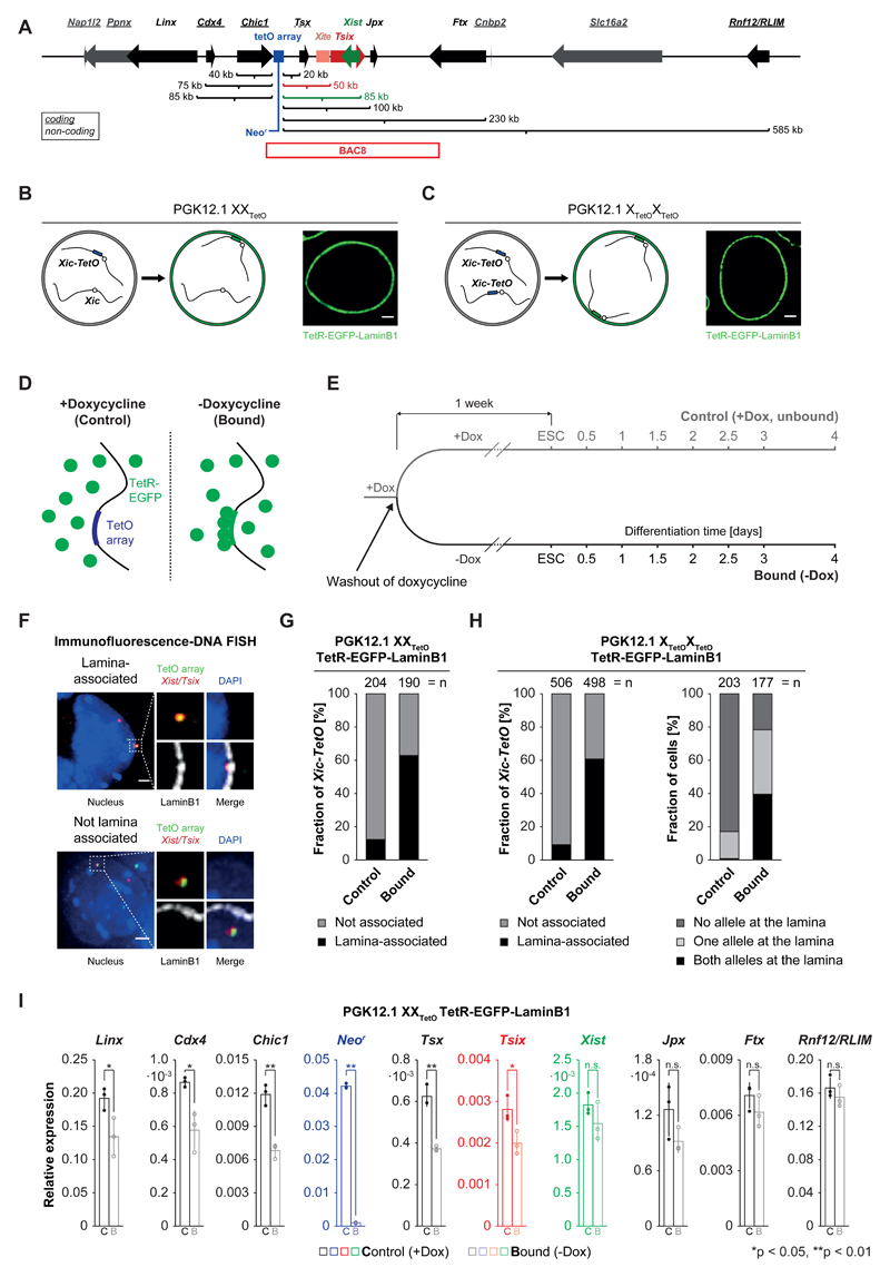
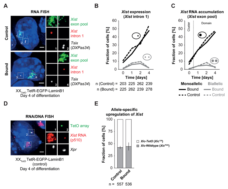
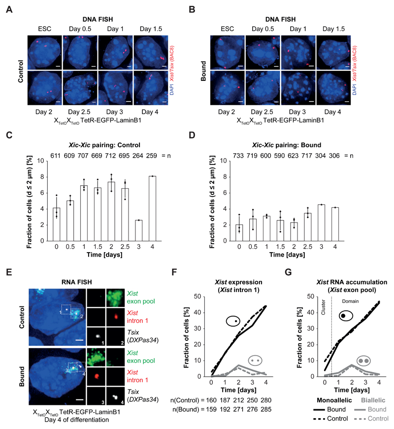
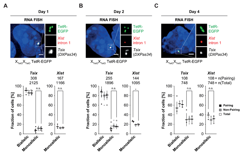
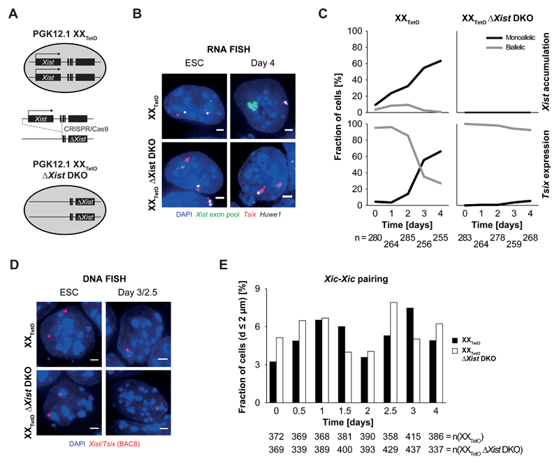
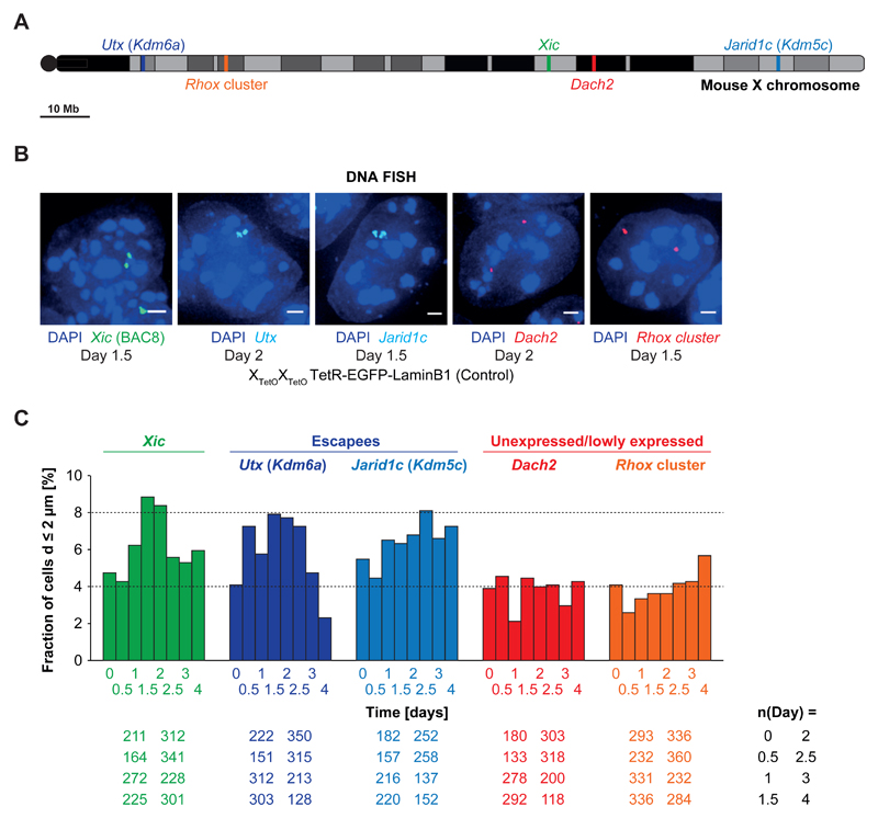
During X-chromosome inactivation (XCI), one of the two X-inactivation centers (Xics) upregulates the noncoding RNA Xist to initiate chromosomal silencing in cis. How one Xic is chosen to upregulate Xist remains unclear. Models proposed include localization of one Xic at the nuclear envelope or transient homologous Xic pairing followed by asymmetric transcription factor distribution at Xist's antisense Xite/Tsix locus. Here, we use a TetO/TetR system that can inducibly relocate one or both Xics to the nuclear lamina in differentiating mouse embryonic stem cells. We find that neither nuclear lamina localization nor reduction of Xic homologous pairing influences monoallelic Xist upregulation or choice-making. We also show that transient pairing is associated with biallelic expression, not only at Xist/Tsix but also at other X-linked loci that can escape XCI. Finally, we show that Xic pairing occurs in wavelike patterns, coinciding with genome dynamics and the onset of global regulatory programs during early differentiation.


Conflict of interest statement

Competing interests

The authors declare no competing financial interest.

Figures

Figure 1
Figure 1. Expression of TetR-EGFP-LaminB1 in PGK12.1 XXTetO/XTetOXTetO cells induces relocalization of the Xic-TetO and gene repression in the relocalized Xic
A) Schematic representation of the murine X-inactivation center (Xic) with an insertion of a TetO array (blue box, 224 repeats, 11.2 kb). Indicated are the linear genomic distances of the transcriptional start sites of genes in the Xic with respect to the insertion site of the TetO array. Genes giving rise to non-coding transcripts are underlined. The position of BAC8 is indicated below the scheme. B) Schematic representation of the experimental approach for the relocalization of the single Xic-TetO to the nuclear lamina upon expression of a TetR-EGFP-LaminB1 fusion protein in heterozygous PGK12.1 XXTetO cells. The EGFP fluorescence and localization of the fusion protein are detectable in PGK12.1 XXTetO stably expressing the transgene. C) Like B) for PGK12.1 XTetOXTetO cells. D) Schematic representation of binding of TetR fusion proteins to the TetO array in the absence (bound) or presence (control) of doxycycline. E) Scheme of the experimental set up. One population of ‘control’ cells (+Dox) was separated into one population of ‘control’ cells and one population of ‘bound’ cells one week prior to initiation of differentiation. F) Immunofluorescence (IF) DNA fluorescence in situ hybridization (FISH) for the nuclear lamina (anti-LaminB1 IF) and for the TetO array locus and the Tsix/Xist region in the Xic (DNA FISH). Depicted are two representative cells with association (upper panel) or no association (lower panel) of the TetO array locus (Xic-TetO) with the nuclear lamina. Depicted is the entire nucleus as a projection (without the lamina signal) and the region of interest magnified on the right of the nucleus (with the lamina signal). Scored as association was only a direct overlap between the TetO array locus signal and the signal of the nuclear lamina as depicted in the example. Depicted are representative cells for each condition. G) Quantification of association of the single Xic-TetO with the nuclear lamina in PGK12.1 XXTetO ESCs based on IF DNA FISH as depicted in D). (n = number of scored Xic-TetO) H) Quantification of the association of the two Xic-TetO in homozygous PGK12.1 XTetOXTetO for all the Xic-TetO in a population of cells (left panel) and quantification of the association of the two Xic-TetO in single cells (right panel). (n (left) = number of scored Xic-TetO, n(right) = number of cells). I) Mean relative expression of Xic-linked genes in control (empty bars) and bound (empty bars, lighter shade) PGK12.1 XXTetO TetR-EGFP-LaminB1 cells (n = 3), assessed by qRT-PCR relative to the expression level of the Arp0 gene. Individual data points are depicted as filled and empty circles. Significant differences in gene expression are marked (asterisk: p < 0.05; double asterisk: p < 0.01; t-test (unpaired, two-tailed); error bars indicate standard deviation; p-values for genes with p<0.05: Linx 0.037, Cdx4 0.015, Chic1 0.0016, Neor 9x10⁻⁸, Tsx: 0.0011, Tsix: 0.028).
Figure 2
Figure 2. Xist upregulation from the Xic-TetO remains unaffected upon relocalization to the nuclear lamina
A) RNA FISH for Xist (red) and Tsix (white) nascent transcripts as well as for processed, accumulating Xist RNA (green) in control and bound PGK12.1 XXTetO TetR-EGFP-LaminB1 cells on day 4 of differentiation by LIF withdrawal. Depicted are representative cells for each condition. B) Quantification of Xist expression by presence of nascent Xist RNA as focal signal as depicted in A) in control and bound PGK12.1 XXTetO TetR-EGFP-LaminB1 cells in ESCs and throughout differentiation by LIF withdrawal (n = number of cells). C) Quantification of Xist RNA accumulation by presence of Xist RNA clouds/domains as depicted in A) in control and bound PGK12.1 XXTetO TetR-EGFP-LaminB1 cells in ESCs and throughout differentiation by LIF withdrawal. In ESCs no Xist RNA domains could be detected and only the presence of small clusters of Xist RNA was scored (n = number of cells). D) Simultaneous RNA/DNA FISH for Xist RNA (red) and Xpr (X pairing region, BAC 5 (474E04), white) and the TetO array locus (green) performed on control PGK12.1 XXTetO TetR-EGFP-LaminB1 cells on day 4 of differentiation by LIF withdrawal. Xist RNA is stable during denaturation and can be detected after performing DNA FISH. Depicted is a representative example in which Xist RNA became up-regulated from the wildtype Xic and started coating the wildtype X chromosome. E) Quantification of the distribution of up-regulation of Xist from the Xic-TetO or the wildtype Xic in control and bound PGK12.1 XXTetO TetR-EGFP-LaminB1 cells on day 4 of differentiation by LIF withdrawal based on simultaneous RNA/DNA FISH as depicted in D) (n = total number of cells; mean results of three experiments; error bars indicate standard deviation; for B,C: one representative experiment is depicted in the quantification).
Figure 3
Figure 3. Relocalization and tethering of both Xic in PGK12.1 XTetOXTetO reduces the relative number of Xic trans associations but has no impact on initiation of XCI
A) + B) DNA FISH for the Xist/Tsix region (red, BAC 8) in control (A) and bound (B) differentiating PGK12.1 XTetOXTetO TetR-EGFP-LaminB1 cells. Depicted are representative cells for each condition. C) + D) Quantification of the mean relative amount of cells with Xic at pairing distance of d < 2 µm in populations of differentiating control (C) and bound (D) PGK12.1 XTetOXTetO TetR-EGFP-LaminB1 cells. The distance between the centers of mass of intensity of the DNA FISH signals was determined in 3D. The displayed relative amount represents the ratio of cells with d < 2 µm and cells with d > 2 µm (n = number of total cells analyzed; mean pairing frequency of three independent experiments for day 0 – day 2.5 of differentiation; error bars indicate standard deviation; one experiment was performed for day 3 and day 4 of differentiation; individual data points are depicted as filled circles). E) RNA FISH for nascent Tsix (white) and Xist (red) transcripts as well as for processed, accumulating Xist RNA (green) in control and bound PGK12.1 XTetOXTetO TetR-EGFP-LaminB1 cells at day 4 of differentiation by LIF withdrawal. Depicted are representative cells for each condition. F) Quantification of Xist expression by presence of nascent Xist RNA as focal signal as depicted in E) in control and bound PGK12.1 XTetOXTetO TetR-EGFP-LaminB1 cells in ESCs and throughout differentiation by LIF withdrawal (n = number of cells). G) Quantification of Xist RNA accumulation by presence of Xist RNA clouds/domains as depicted in E) in control and bound PGK12.1 XTetOXTetO TetR-EGFP-LaminB1 cells in ESCs and throughout differentiation by LIF withdrawal. In ESCs no Xist RNA domains could be detected and only the presence of small clusters of Xist RNA was scored (n = number of cells; for F,G: one representative experiment is depicted in the quantification).
Figure 4
Figure 4. Pairing of the Xic is associated with biallelic Tsix expression or expression of Tsix and Xist
A) + B) + C) RNA FISH for nascent Xist (red) and Tsix (white) transcripts in differentiating PGK12.1 XTetOXTetO TetR-EGFP cells at day 1 (A), day 2 (B) and day 4 (C) of differentiation. Binding of TetR-EGFP can be detected after RNA FISH as focal green fluorescent signal. Two focal signals per allele indicate the presence of sister chromatids after replication of the Xic. Depicted are three examples of cells with pairing Xic (d < 2 µm) at day 1, day 2 and day 4 after LIF withdrawal. Note the different expression patterns of Tsix and Xist in these cells, demonstrating that no single expression pattern is associated with homologous Xic pairing. The bottom part of panels A), B) and C) depicts the quantification of Tsix and Xist expression in the total population of cells (empty circles), the fraction of cells with non-pairing Xic (grey circles) and cells with pairing Xic (black circles) at day 1, day 2 and day 4 of differentiation based on the presence of focal RNA FISH signals (lines indicate the median). (n = number of cells; * = p < 0.05; t-test (unpaired, two-tailed); scale bar = 2 µm; error bars indicate standard deviation of at least three independent experiments)
Figure 5
Figure 5. Homologous Xic pairing can occur in the absence of biallelic Tsix expression
A) Schematic representation of a female ESC line with a doxycycline-inducible promoter regulating the expression of one endogenous Xist allele (TX1072). B) Schematic representation of the initiation of differentiation and induction of Xist expression by addition of doxycycline in TX1072 cells. C) RNA FISH for nascent Xist (red, red arrowheads) and Tsix (white, white arrowheads) transcripts as well as processed and accumulating Xist RNA (green) in undifferentiated TX1072 or TX1072 cells differentiated for 2 days by LIF withdrawal either in the presence or absence of doxycycline in the culture medium. Depicted are representative cells for each condition. D) Quantification of the fraction of cells with monoallelic Tsix expression and monoallelic Xist (intron 1) expression in differentiating TX1072 either not induced with doxycycline or with Xist expression induced one day prior to the initiation of differentiation. E) DNA FISH for the Xist/Tsix region (red, BAC 8) in induced and uninduced TX1072 ESCs and at day 1.5 of differentiation after LIF withdrawal. Depicted are representative cells for each condition. F) Fraction of cells with Xic at pairing distance in differentiating induced and uninduced TX1072 cells. 3D distance was determined after DNA FISH for the Xist/Tsix region (BAC 8) as depicted in D). (n = number of cells, scale bar = 2 µm)
Figure 6
Figure 6. Xist expression is not necessary for homologous Xic pairing
A) Schematic representation of the derivation of a homozygous Xist double knock-out (DKO) cell line in PGK12.1 XXTetO cells by CRISPR/Cas9-mediated genetic engineering. B) RNA FISH for nascent Tsix (red) and Huwe1 (white) transcripts as well as processed and accumulating Xist RNA (green) performed in PGK12.1 XXTetO (control) and PGK12.1 XXTetO ΔXist DKO in ESCs and at day 4 of differentiation after LIF withdrawal (depicted are representative cells for each condition). C) Quantification of monoallelic and biallelic Xist RNA accumulation as well as monoallelic and biallelic Tsix expression determined by presence of Xist RNA domains/clusters as well as focal signals for Tsix in RNA FISH. D) DNA FISH for the Xist/Tsix region (BAC 8, red) performed in PGK12.1 XXTetO (control) and PGK12.1 XXTetO ΔXist DKO in ESCs and at day 4 of differentiation after LIF withdrawal. Depicted are representative cells for each condition. E) Fraction of cells with Xic at pairing distance in differentiating PGK12.1 XXTetO (control) and PGK12.1 XXTetO ΔXist DKO cells. 3D distance was determined after DNA FISH for the Xist/Tsix region (BAC 8) as depicted in D). (n = number of cells, scale bar = 2 µm)
Figure 7
Figure 7. Other biallelically active loci on the X chromosome show homologous pairing
A) Schematic overview of the position of the analyzed X-linked loci Xic (green), Utx (dark blue), Jarid1c (light blue), Dach2 (red) and the Rhox cluster (orange) on the X chromosome. Dach2 and Rhox7/8 (spanned by the BACs used for the FISH analysis) expression is very low but detectable in undifferentiated female ESCs. Rhox6 and Rhox9 (also spanned by the BAC used for FISH analysis) are expressed at higher levels than Rhox 7/8 (comparable to the lowly expressed Xic gene Chic1). Utx and Jarid1c are well expressed at levels similar to the house-keeping genes Dhfr and G6pdx. B) DNA FISH for the Xic (BAC 8), Utx, Jarid1c, Dach2 and the Rhox cluster in differentiating PGK12.1 XTetOXTetO TetR-EGFP-LaminB1 (control) cells. Depicted are representative cells for each condition. C) Fraction of cells with Xic, Utx, Jarid1c, Dach2 or Rhox loci at pairing distance in differentiating PGK12.1 XTetOXTetO TetR-EGFP-LaminB1 (control) cells. 3D distance was determined after DNA FISH (n = number of cells, scale bar = 2 µm).

Similar articles

Cited by

References

    1. Rastan S, Robertson EJ. X-chromosome deletions in embryo-derived (EK) cell lines associated with lack of X-chromosome inactivation. J Embryol Exp Morphol. 1985;90:379–388. - PubMed
    1. Augui S, Nora EP, Heard E. Regulation of X-chromosome inactivation by the X-inactivation centre. Nat Rev Genet. 2011;12:429–442. - PubMed
    1. Galupa R, Heard E. X-chromosome inactivation: new insights into cis and trans regulation. Curr Opin Genet Dev. 2015;31:57–66. - PubMed
    1. Pollex T, Heard E. Recent advances in X-chromosome inactivation research. Curr Opin Cell Biol. 2012;24:825–832. - PubMed
    1. Borsani G, et al. Characterization of a murine gene expressed from the inactive X chromosome. Nature. 1991;351:325–329. - PubMed

Publication types

LinkOut - more resources