

Isotype Specific Assembly of B Cell Antigen Receptors and Synergism With Chemokine Receptor CXCR4
- PMID: 30619343
- PMCID: PMC6305424
- DOI: 10.3389/fimmu.2018.02988
Isotype Specific Assembly of B Cell Antigen Receptors and Synergism With Chemokine Receptor CXCR4
Abstract
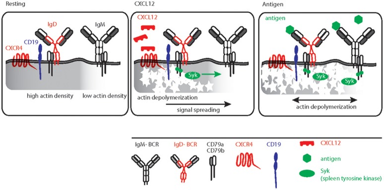
Expression of the membrane-bound form of the immunoglobulin (Ig) as part of the antigen receptor is indispensable for both the development and the effector function of B cells. Among five known isotypes, IgM and IgD are the common B cell antigen receptors (BCRs) that are co-expressed in naïve B cells. Despite having identical antigen specificity and being associated with the same signaling heterodimer Igα/Igβ (CD79a/CD79b), IgM and IgD-BCR isotypes functionally differ from each other in the manner of antigen binding, the formation of isolated nanoclusters and in their interaction with co-receptors such as CD19 and CXCR4 on the plasma membrane. With recent developments in experimental techniques, it is now possible to investigate the nanoscale organization of the BCR and better understand early events of BCR engagement. Interestingly, the cytoskeleton network beneath the membrane controls the BCR isotype-specific organization and its interaction with co-receptors. BCR triggering results in reorganization of the cytoskeleton network, which is further modulated by isotype-specific signals from co-receptors. For instance, IgD-BCR is closely associated with CXCR4 on mature B cells and this close proximity allows CXCR4 to employ the BCR machinery as signaling hub. In this review, we discuss the functional specificity and nanocluster assembly of BCR isotypes and the consequences of cross-talk between CXCR4 and IgD-BCR. Furthermore, given the role of BCR and CXCR4 signaling in the development and survival of leukemic B cells, we discuss the consequences of the cross-talk between CXCR4 and the BCR for controlling the growth of transformed B cells.
Keywords: B cell antigen receptor (BCR); B cell malignancies; Chemokine receptor 4 (CXCR4); Cytoskeleton; Nanoclusters.
Figures

Similar articles
-
CXCR4 signaling and function require the expression of the IgD-class B-cell antigen receptor.Proc Natl Acad Sci U S A. 2017 May 16;114(20):5231-5236. doi: 10.1073/pnas.1621512114. Epub 2017 May 1. Proc Natl Acad Sci U S A. 2017. PMID: 28461496 Free PMC article.
-
The importance of B cell receptor isotypes and stereotypes in chronic lymphocytic leukemia.Leukemia. 2019 Feb;33(2):287-298. doi: 10.1038/s41375-018-0303-x. Epub 2018 Dec 16. Leukemia. 2019. PMID: 30555163 Free PMC article. Review.
-
Control of B Cell Responsiveness by Isotype and Structural Elements of the Antigen Receptor.Trends Immunol. 2016 May;37(5):310-320. doi: 10.1016/j.it.2016.03.004. Epub 2016 Apr 1. Trends Immunol. 2016. PMID: 27052149 Review.
-
The actin and tetraspanin networks organize receptor nanoclusters to regulate B cell receptor-mediated signaling.Immunity. 2013 Mar 21;38(3):461-74. doi: 10.1016/j.immuni.2012.11.019. Epub 2013 Mar 14. Immunity. 2013. PMID: 23499492
-
IgD shapes the pre-immune naïve B cell compartment in humans.Front Immunol. 2023 Jan 26;14:1096019. doi: 10.3389/fimmu.2023.1096019. eCollection 2023. Front Immunol. 2023. PMID: 36776874 Free PMC article.
Cited by
-
B-Cell Receptor Signaling and Beyond: The Role of Igα (CD79a)/Igβ (CD79b) in Normal and Malignant B Cells.Int J Mol Sci. 2023 Dec 19;25(1):10. doi: 10.3390/ijms25010010. Int J Mol Sci. 2023. PMID: 38203179 Free PMC article. Review.
-
P66Shc: A Pleiotropic Regulator of B Cell Trafficking and a Gatekeeper in Chronic Lymphocytic Leukemia.Cancers (Basel). 2020 Apr 19;12(4):1006. doi: 10.3390/cancers12041006. Cancers (Basel). 2020. PMID: 32325830 Free PMC article. Review.
-
Cyclosporine A Modulates LSP1 Protein Levels in Human B Cells to Attenuate B Cell Migration at Low O2 Levels.Life (Basel). 2022 Aug 22;12(8):1284. doi: 10.3390/life12081284. Life (Basel). 2022. PMID: 36013463 Free PMC article.
-
Peripheral Blood B-Cell Subsets Frequency and Distribution and the BSF-2(IL-6) to CSIF:TGIF(IL-10) Ratio as Severity-Associated Signatures in Primary Open-Angle Glaucoma: A Case-Controlled Study.Biomedicines. 2024 Feb 21;12(3):485. doi: 10.3390/biomedicines12030485. Biomedicines. 2024. PMID: 38540099 Free PMC article.
References
Publication types
MeSH terms
Substances
Grants and funding
LinkOut - more resources
Full Text Sources
Miscellaneous